Pantoscopic Ophthalmoscope
Ask for Price$0.00
Shipped from abroad
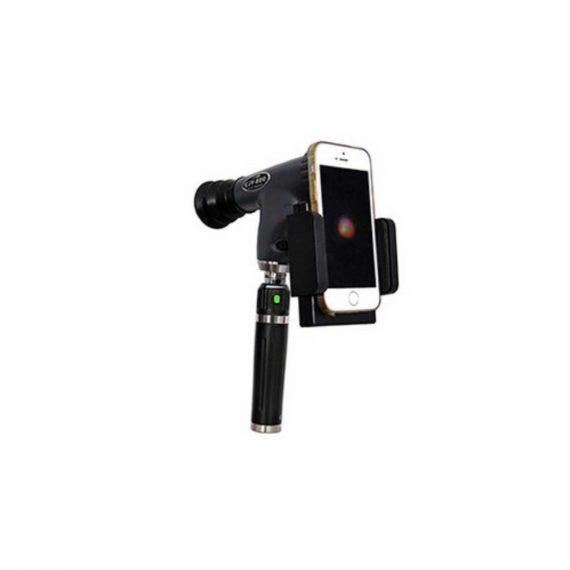
The brand-new Pantoscopic Ophthalmoscope is a portable digital imaging device which makes it possible to view and take pictures of the eyes.
Delivery & Availability:
Typically 14 working days – excluding furniture and heavy/bulky equipment. Please contact us for further information.
Description
The brand-new Pantoscopic Ophthalmoscope is a portable digital imaging device which makes it possible to view and take pictures of the eyes. The optical access of the Pantoscopic Ophthalmoscope is aligned to the visual axis of the smartphone camera by the adaptor which allows to you take pictures of the fundus and retinal nerve in high resolution. You could save pictures for each patient or email and print as needed. The Pantoscopic Ophthalmoscope provides a 5X larger view of the fundus compared with the standard ophthalmoscope. It has a wider view field of 230. Without dilating the pupil, the fundus imagines could be captured at any time and places.
Features:
- Rapid capture of fundus images on your cellphone.
- Get eyeground documentation in an easy and low-cost way.
- Send images to your peers across the continum of care instantly.
- Easy carry to capture images at any clinical circumstance and work efficiently.
- Make patient education easier.
- 5X larger view of the fundus vs. standard ophthalmoscopes in an undilated eye.
- Halogen lamp provides bright, white light.
- Apertures and Filters: Looking at the pupil through an add-on corneal viewing lens, the practitioner can detect lens opacities easily.
Quick Comparison
| Settings | Pantoscopic Ophthalmoscope remove | DrGem Ceiling Mounted Digital X-ray remove | DrGem Diamond All-In-One Digital X-ray Machine remove | DrGem Floor Mounted Analogue X-ray remove | Sonoscape P10 Ultrasound Machine remove | ASPEL Stress ECG with Treadmill and Software remove |
|---|---|---|---|---|---|---|
| Name | Pantoscopic Ophthalmoscope remove | DrGem Ceiling Mounted Digital X-ray remove | DrGem Diamond All-In-One Digital X-ray Machine remove | DrGem Floor Mounted Analogue X-ray remove | Sonoscape P10 Ultrasound Machine remove | ASPEL Stress ECG with Treadmill and Software remove |
| Image |  |  |  |  |  |  |
| SKU | SF1033560107-3 | SF1033560074-4 | SF1033560074-3 | SF1033560074-6 | SF1033560012-7 | SF1033560075-2 |
| Rating | ||||||
| Price | Ask for Price | $68,468.00 | Ask for Price | $26,924.00 | Ask for Price | Ask for Price |
| Stock | ||||||
| Availability | ||||||
| Add to cart | ||||||
| Description | Shipped from abroad
The brand-new Pantoscopic Ophthalmoscope is a portable digital imaging device which makes it possible to view and take pictures of the eyes.
| In Stock The GXR-SD is a diagnostic digital radiography system that provides reliable high quality digital radiographic images with a reduced dose. The GXR-SD DR systems offer comprehensive digital solutions to all radiography needs, featuring ACQUIDR digital imaging system with stationary or portable digital flat-panel detectors as well as reliable high-frequency x-ray generators that are known worldwide for their excellent performance, lifetime and stability. Patient tables and wall stands are also offered. Delivery & Availability: Typically 21 working days – excluding furniture and heavy/bulky equipment. Please contact us for further information. | Shipped from Abroad DrGem Diamond All-In-One Digital X-ray Machine is a fully automatic digital radiography system providing state-of-the-art image quality, image processing and user interface. With a wide selection of anatomical studies on the imaging software, DIAMOND automatically sets up the x-ray generator’s preprogrammed exposure technique settings, motorized radiographic stand positioning, x-ray collimation and post-image processing for the selected study. Specifically designed to increase workflow, this fully digital system offers convenient auto-positioning and advanced image processing to achieve big performance with little effort. Delivery & Availability: Typically 21 working days – excluding furniture and heavy/bulky equipment. Please contact us for further information. | In Stock GXR Analogue X-ray system matches with a radiographic room which perfectly fits your workow and can be easily upgraded to DR system with the help of DR interface and PC interface in GXR generator as well as Bucky suitable to Flat Panel Detector. GXR X-ray system is equipped with a high frequency X-ray generator which consistently produces high quality radiograph in favor of high quality X-ray output with a very small kV ripple and accurate mA and mAs. GXR X-ray system is designed to provide convenience to operator and comfort to patient. Delivery & Availability: Typically 21 working days – excluding furniture and heavy/bulky equipment. Please contact us for further information. | Shipped from Abroad The P10 color Doppler ultrasound system is a new generation product from SonoScape. It is designed to give high quality images, rich probe configurations, various clinical tools and automatic analysis software to provide you with comprehensive solutions for your growing demand for clinical applications. Delivery & Availability: Typically 5-7 working days – excluding furniture and heavy/bulky equipment. Please contact us for further information. | Shipped from Abroad It is a system with professional tool dedicated to exercise and resting ECG examination. Treadmill has 12 lead ECG modules. With ECG Analyzing Software. Delivery & Availability: Typically 21 working days – excluding furniture and heavy/bulky equipment. Please contact us for further information. |
| Content | The brand-new Pantoscopic Ophthalmoscope is a portable digital imaging device which makes it possible to view and take pictures of the eyes. The optical access of the Pantoscopic Ophthalmoscope is aligned to the visual axis of the smartphone camera by the adaptor which allows to you take pictures of the fundus and retinal nerve in high resolution. You could save pictures for each patient or email and print as needed. The Pantoscopic Ophthalmoscope provides a 5X larger view of the fundus compared with the standard ophthalmoscope. It has a wider view field of 230. Without dilating the pupil, the fundus imagines could be captured at any time and places.
Features:
| DrGem Ceiling Mounted Digital X-ray is a diagnostic digital radiography system that provides reliable high quality digital radiographic images with a reduced dose. The GXR-SD DR systems offer comprehensive digital solutions to all radiography needs, featuring ACQUIDR digital imaging system with stationary or portable digital flat-panel detectors as well as reliable high-frequency x-ray generators that are known worldwide for their excellent performance, lifetime and stability. Patient tables and wall stands are also offered.
Features:
Click Here To Download Catalogue | DrGem Diamond All-In-One Digital X-ray Machine is a fully automatic digital radiography system providing state-of-the-art image quality, image processing and user interface. With a wide selection of anatomical studies on the imaging software, DIAMOND automatically sets up the x-ray generator’s pre-programmed exposure technique settings, motorized radiographic stand positioning, x-ray collimation and post-image processing for the selected study. Specifically designed to increase workflow, this fully digital system offers convenient auto-positioning and advanced image processing to achieve big performance with little effort.
Features of DrGem Diamond All-In-One Digital X-ray Machine:
Outstanding Image Quality -
Digital radiography via at panel detector improves your workflow, exam speed and comfort with efficiency. Digital at panel detector with Csl screen provides excellent spatial resolution, MTF, DQE and stability based on ne pixel pitch. A 3-field ion-chamber is provided for AEC function.
Automatic Collimation –
Automatic x-ray eld size control of the motorized collimator corresponds to dierent SIDs. Includes user adjustable lamp timer with on/oswitch.
Automatic Positioning –
Click Here To Download Catalogue | DrGem GXR Floor Mounted Analogue X-ray system matches with a radiographic room which perfectly fits your workflow and can be easily upgraded to DR system with the help of DR interface and PC interface in GXR generator as well as Bucky suitable to Flat Panel Detector. GXR (Analogue X-ray)system is equipped with a high frequency X-ray generator which consistently produces high quality radiograph in favor of high quality X-ray output with a very small kV ripple and accurate mA and mAs. GXR (Analogue X-ray) system is designed to provide convenience to operator and comfort to patient.
Features of DrGem GXR Floor Mounted Analogue X-ray:
Click Here To Download Catalogue | DETAILS
B + Compound
B + Compound utilizes several lines of sight for optimal contrast resolution, speckle reduction and border detection, with which P10 is ideal for superficial and abdominal imaging with better clarity and improved continuity of structures.
μ-Scan
The new generation μ-Scan imaging technology gives you better image quality by reducing noise, improving signal strength and improving visualization.
P10 offers a comprehensive selection of electronic probes to maximize its capabilities to meet a wide range of applications including abdomen, pediatric, OB/GYN, cardiovascular, musculoskeletal, etc. The advanced probe technologies also effectively enhance the image quality and confidence in reaching clinical diagnoses, even in difficult patients.
Convex Probe 3C-A
Ideal for an abundant of application such as abdomen, gynecology, obstetrics, urology and even abdomen biopsy.
Linear Probe L741
This linear probe is designed to satisfy vascular, breast, thyroid, and other small parts diagnosis, and its adjustable parameters could also present users a clear view of MSK and deep vessels.
Phase Array Probe 3P-A
For the purpose of adult and pediatric cardiology and emergency, the phase array probe provides elaborate presets for different exam modes, even for difficult patients.
Intracavitary Probe 6V1
Intracavitary probe could face application of gynecology, urology, prostate, and its temperature detection technology not only protects the patient but also extends the service life.
Click Here To Download Catalogue | It is a system with professional tool dedicated to exercise and resting ECG examination. Treadmill has 12 lead ECG modules. With ECG Analyzing Software.
Technical Specification:
Click Here To Download Catalogue |
| Weight | N/A | N/A | N/A | N/A | N/A | N/A |
| Dimensions | N/A | N/A | N/A | N/A | N/A | N/A |
| Additional information |


Reviews
There are no reviews yet.