

Anke Anatom 16 Slice CT Scan
$0.00
Shipped from Abroad
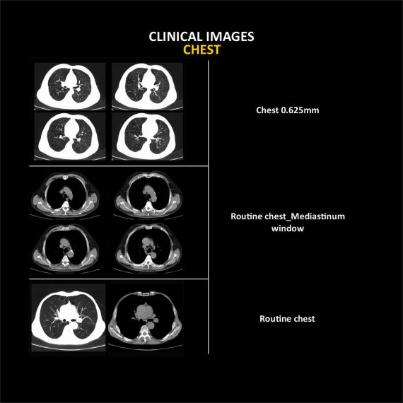
ANATOM16 HD, is a tool of precision medicine in diagnosis imaging. Via the breakthrough designs in precise hardware, software and imaging technologies, ANATOM 16 HD can provide precise diagnosis information and early detection for small lesions.
Delivery & Availability:
Typically 90 working days – excluding furniture and heavy/bulky equipment. Please contact us for further information.
Description
ANATOM16 HD, is a tool of precision medicine in diagnosis imaging. Via the breakthrough designs in precise hardware, software and imaging technologies, ANATOM 16 HD can provide precise diagnosis information and early detection for small lesions.
Features:
- Seamlessly upgrade to meet your future needs: Anke takes full consideration of the increasing clinical requirements of your business in today’s rapidly changing medical environment.
- Precise hardware, Precise technology, Precise imaging: OptiWave detector, High precision gantry control, Dual-mode gantry tilt, Admir3D iterative technology, Dual-energy head imaging, 1024 x1024 matrix imaging technology, High-definition imaging of targeted organs, Low dose platform, 3D enhanced VR
- Precision Technology Platform: ANATOM precision technology platform is equipped with advanced imaging technologies, and adopts OptiWave detector, Ahead dual-energy imaging, Admir3D iterative reconstruction technology and AccuTilt dual-mode tilt gantry technology to provide powerful support for accurate diagnosis
- Admir3D iterative reconstruction technology: Admir applies mathematical and physics models to accurately construct and describe the signal’s quantum characteristics. Iterative operations are performed in the three domains of raw data, projection and image, to greatly reduce the image noise and achieve optimal image quality with low dose
- Ahead-Head dual-energy head imaging technology: Ahead creatively uses 140kV and 80kV dual energy switching scan mode for brain imaging. By careful analyzing the high and low energy characteristics, images can show more valuable information about the brain tissues
- AccuTilt dual-mode gantry tilt technology: The system provides digital and mechanical tilt to accommodate different user habits and clinical needs. Real-time collision preventing system is available for the patients’ safety
- AccuOrgan-Targeted organ imaging: To achieve high precision imaging of each part of human body at low dose and low energy consumption
- AccuDose-Comprehensive low dose imaging: Pediatric Scan Protocol, Individual Dose Monitoring, AccuShape Filter, Efficient Detector, Adose Dose Modulation, Ahead – Head Dual-energy Imaging, Iterative Reconstruction, Amast, Contrast Agent Tracking Technology
- AccuScan-Enjoy ease: Convenient and efficient operation process greatly improve work efficiency to achieve high volume of patients
- Clinical Applications: Fast, precise and low-dose imaging technologies provide a full range of clinical solutions to meet the current and future clinical diagnostic needs
- Service Innovation creating maximum value for customers: Service Support within 24 Hours, Local Service Partners, On-line Service Support, After-sales Maintenance Stations
- AccuSaving Green & Energy-saving: AccuSaving is an innovative energy saving technology. The system will enter the “dormant”, which is a low carbon mode, after a certain idle time or per user’s request. To bring the system back to working status is as easy as pushing a button. The system will also remind the user to perform necessary warm-up and calibration procedures, which are fully automated processes. AccuSaving technology can reduce operation and standby power consumption and save the electricity cost by 30% by adopting different operation modes in working and off hours
Technical Specifications:
| No. | Technical feature | Description |
| 1 | Gantry | |
| 1.01 | Gantry type | Low voltage slip-ring with
AccuSlip-ring technology |
| 1.02 | Gantry driven type | Strap-driven |
| 1.03 | Patient opening | 70cm |
| 1.04 | Gantry tilt mode | Dual-mode gantry tilt |
| 1.05 | Mechanical tilt capability | ±30° |
| 1.06 | Digital tilt capability | ±50° |
| 1.07 | Gantry remote-Control | Provided |
| 1.08 | Detector type | OptiWave rare-earth ceramic detector |
| 1.09 | Numbers of detector rows | 32 |
| 1.10 | Width of Z-axle detector | 20mm |
| 1.11 | Detector columns of channels per row | 912 |
| 1.12 | Numbers of detector columns | 29184 |
| 1.13 | Data-transfer type | RF,optical fiber communication |
| 1.14 | 3D laser orientation | Provided |
| 1.15 | External X-ray enable | Interface for Foot-Pedal Provided |
| 1.16 | Automatic exposure control(mA Modulation) | Provided |
| 1.17 | Auto-voice manager | Breath Graphical Display
Hold Message (Record/Playback) Breath Message(Record/Playback) |
| 1.18 | ANKE energy conservation management | Provided |
| 1.19 | Acquisition mode | 16 × 0.625mm, 16 × 1.25mm |
| 2 | Scan parameter | |
| 2.01 | Shortest 360 degree rotation time | 0.5s |
| 2.02 | Allowed rotation times | 0.5s,0.8s,1.0s,1.5s,2.0s |
| 2.03 | Slice numbers per rotation | 16 |
| 2.04 | Minimum slice thickness of scan | 0.625mm |
| 2.05 | Minimum slice thickness of reconstruction | 0.625mm |
| 2.06 | Maximum slice thickness of scan | 10mm |
| 2.07 | Nominal reconstruction slice thickness | 0.625mm,1.25mm,2.5mm,5.0mm,
7.5mm,10mm |
| 2.08 | Speed of image reconstruction(512×512) | 65 frames/s |
| 2.09 | Scan FOV | 52cm |
| 2.10 | Image reconstruction matrix | 512×512,1024×1024 |
| 2.11 | Image display matrix | 512×512,1024×1024 |
| 2.12 | Maximum continuous scan duration | 120s |
| 2.13 | Maximum continuous scan length | 180cm |
| 2.14 | Direction of TOPO | Front-back,Left-right |
| 2.15 | Max. length of TOPO | 180cm |
| 2.16 | Range of pitch | 0.5~1.5 |
| 2.17 | Scan mode | Scout scan
Axial scan Helical scan Cine scan |
| 3 | HVPS and Tube | |
| 3.01 | Maximum continuous output of HV generator | 50kW |
| 3.02 | Tube kV selections | 80kV,100 kV,120 kV,140 kV |
| 3.03 | Tube mA range | 10~420mA |
| 3.04 | Tube anode heat capacity | 5.0MHU |
| 3.05 | Heat dissipation rate | 815kHU/min |
| 3.06 | Type of cooling | Oil cooling + Air cooling |
| 3.07 | Tube focus | Large:1.0 mm×1.0mm
Small:0.5mm×1.0mm |
| 3.08 | Dynamic flying focal spot technology | Provided |
| 4 | Patient table | |
| 4.01 | Maximum horizontal-movable range | 1850mm |
| 4.02 | Table horizontal-scannable range | 1800mm |
| 4.03 | Table horizontal-position repeatability | ±0.25mm |
| 4.04 | Maximum vertical-movable range | 500mm |
| 4.05 | Maximum speed of vertical movement | 20mm/s |
| 4.06 | Maximum speed of horizontal movement | 150mm/s |
| 4.07 | Maximum patient weight | 205kg |
| 4.08 | Foot pedal of patient table control | Provided |
| 5 | Image Quality | |
| 5.01 | High contrast resolution | 21lp/cm@0%MTF |
| 5.02 | Low contrast resolution | 2.0mm@0.30% |
| 5.03 | Isotropic imaging resolution | 0.625mm |
| 5.04 | Range of CT numbers | -32767~32768 |
| 5.05 | Image noise | ≤0.25@28mGy |
| 6 | Computer subsystem | |
| 6.01 | CPU | 3.5GHz |
| 6.02 | Memory | 16GB×4 |
| 6.03 | Storage of hard-disk | 1T×2 |
| 6.04 | Monitor | 24’’ LCD Monitor |
| 6.05 | Resolution of monitor | 1920×1200 |
| 6.06 | Image-data external storage type | CD/DVD/USB |
| 6.07 | Time of image reconstruction(512×512) | 15.4ms/frame |
| 6.08 | DICOM 3.0 interface | Provided |
| 6.09 | Printer DICOM 3.0 interface | Provided |
| 6.10 | Auto filming | Provided |
| 6.11 | Worklist function | Provided |
| 7 | Advanced application | |
| 7.01 | Multi-Planar Reconstruction(MPR) | Provided |
| 7.02 | Curve Multi-Planar Reconstruction(CPR) | Provided |
| 7.03 | Surface Shaded Display(SSD) | Provided |
| 7.04 | Volume Rendering(VR) | Provided |
| 7.05 | Maximum Intensity Projection(MIP) | Provided |
| 7.06 | Minimum Intensity Projection(MinIP) | Provided |
| 7.07 | Virtual Endoscopy(VE) | Provided |
| 7.08 | CT angiography(CTA) | Provided |
| 7.09 | Tissue segmentation | Provided |
| 7.10 | One click bone remove | Provided |
| 7.11 | One click patient table remove | Provided |
| 7.12 | Bolus-tracking Technology | Provided |
| 7.13 | Spiral auto start | Provided |
| 7.14 | Cine display | Provided |
| 7.15 | AbastTM bone artifact suppression technology | Provided |
| 7.16 | AmastTM metal artifact suppression technology | Provided |
| 7.17 | Admir3D fulll-domain iterative reconstruction | Provided |
| 7.18 | Low-dose pediatric scan technology | Provided |
| 7.19 | Low-dose lung scan technology | Provided |
| 7.20 | AccuHead grey-white matter enhanced
technology |
Provided |
| 7.21 | AccuLung high resolution scan technology | Provided |
| 7.22 | AccuOtica inner-ear high resolution scan
technology |
Provided |
| 7.23 | AccuBody high resolution scan technology | Provided |
| 7.24 | AccuBone high resolution scan technology | Provided |
Click Here To Download Catalogue
Additional information
| Model | Advanced, Advanced Plus, Basic, Smart |
|---|
Review(1)
Quick Comparison
| Anke Anatom 16 Slice CT Scan remove | ASPEL AsCARD Green B/W ECG Machine remove | Sonoscape P10 Ultrasound Machine remove | ASPEL Ambulatory BP Machine remove | DrGem Ceiling Analogue X-ray Machine remove | Sonoscape P15 Ultrasound Machine With Four Probes remove | |||||||||||||||||||||||||||||||||||||||||||||||||||||||||||||||||||||||||||||||||||||||||||||||||||||||||||||||||||||||||||||||||||||||||||||||||||||||||||||||||||||||||||||||||||||||||||||||||||||||||||||||||||||||||||||||||||||||||||||||||||||||||||||||||||||||||||||||||||||||||||||||||||||||||||||
|---|---|---|---|---|---|---|---|---|---|---|---|---|---|---|---|---|---|---|---|---|---|---|---|---|---|---|---|---|---|---|---|---|---|---|---|---|---|---|---|---|---|---|---|---|---|---|---|---|---|---|---|---|---|---|---|---|---|---|---|---|---|---|---|---|---|---|---|---|---|---|---|---|---|---|---|---|---|---|---|---|---|---|---|---|---|---|---|---|---|---|---|---|---|---|---|---|---|---|---|---|---|---|---|---|---|---|---|---|---|---|---|---|---|---|---|---|---|---|---|---|---|---|---|---|---|---|---|---|---|---|---|---|---|---|---|---|---|---|---|---|---|---|---|---|---|---|---|---|---|---|---|---|---|---|---|---|---|---|---|---|---|---|---|---|---|---|---|---|---|---|---|---|---|---|---|---|---|---|---|---|---|---|---|---|---|---|---|---|---|---|---|---|---|---|---|---|---|---|---|---|---|---|---|---|---|---|---|---|---|---|---|---|---|---|---|---|---|---|---|---|---|---|---|---|---|---|---|---|---|---|---|---|---|---|---|---|---|---|---|---|---|---|---|---|---|---|---|---|---|---|---|---|---|---|---|---|---|---|---|---|---|---|---|---|---|---|---|---|---|---|---|---|---|---|---|---|---|---|---|---|---|---|---|---|---|---|---|---|---|---|---|---|---|---|---|---|---|---|---|---|---|---|---|---|---|---|
| Name | Anke Anatom 16 Slice CT Scan remove | ASPEL AsCARD Green B/W ECG Machine remove | Sonoscape P10 Ultrasound Machine remove | ASPEL Ambulatory BP Machine remove | DrGem Ceiling Analogue X-ray Machine remove | Sonoscape P15 Ultrasound Machine With Four Probes remove | ||||||||||||||||||||||||||||||||||||||||||||||||||||||||||||||||||||||||||||||||||||||||||||||||||||||||||||||||||||||||||||||||||||||||||||||||||||||||||||||||||||||||||||||||||||||||||||||||||||||||||||||||||||||||||||||||||||||||||||||||||||||||||||||||||||||||||||||||||||||||||||||||||||||||||||
| Image | 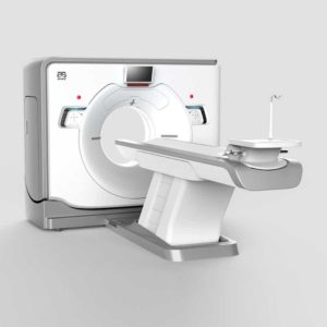 | 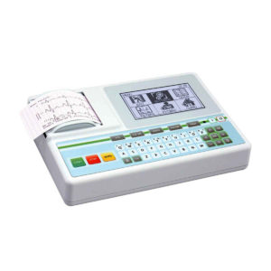 | 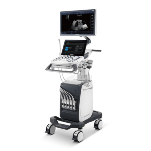 | 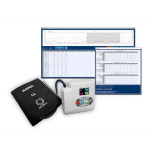 | 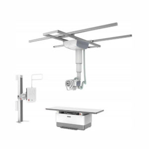 | 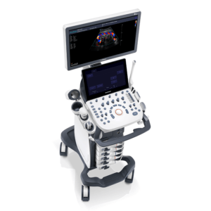 | ||||||||||||||||||||||||||||||||||||||||||||||||||||||||||||||||||||||||||||||||||||||||||||||||||||||||||||||||||||||||||||||||||||||||||||||||||||||||||||||||||||||||||||||||||||||||||||||||||||||||||||||||||||||||||||||||||||||||||||||||||||||||||||||||||||||||||||||||||||||||||||||||||||||||||||
| SKU | SF1033560092-5 | SF1033560075-8 | SF1033560012-7 | SF1033560075-13 | SF1033560074-7 | SF1033560012-8 | ||||||||||||||||||||||||||||||||||||||||||||||||||||||||||||||||||||||||||||||||||||||||||||||||||||||||||||||||||||||||||||||||||||||||||||||||||||||||||||||||||||||||||||||||||||||||||||||||||||||||||||||||||||||||||||||||||||||||||||||||||||||||||||||||||||||||||||||||||||||||||||||||||||||||||||
| Rating | ||||||||||||||||||||||||||||||||||||||||||||||||||||||||||||||||||||||||||||||||||||||||||||||||||||||||||||||||||||||||||||||||||||||||||||||||||||||||||||||||||||||||||||||||||||||||||||||||||||||||||||||||||||||||||||||||||||||||||||||||||||||||||||||||||||||||||||||||||||||||||||||||||||||||||||||||||
| Price |
|
|
|
|
|
| ||||||||||||||||||||||||||||||||||||||||||||||||||||||||||||||||||||||||||||||||||||||||||||||||||||||||||||||||||||||||||||||||||||||||||||||||||||||||||||||||||||||||||||||||||||||||||||||||||||||||||||||||||||||||||||||||||||||||||||||||||||||||||||||||||||||||||||||||||||||||||||||||||||||||||||
| Stock | ||||||||||||||||||||||||||||||||||||||||||||||||||||||||||||||||||||||||||||||||||||||||||||||||||||||||||||||||||||||||||||||||||||||||||||||||||||||||||||||||||||||||||||||||||||||||||||||||||||||||||||||||||||||||||||||||||||||||||||||||||||||||||||||||||||||||||||||||||||||||||||||||||||||||||||||||||
| Availability | ||||||||||||||||||||||||||||||||||||||||||||||||||||||||||||||||||||||||||||||||||||||||||||||||||||||||||||||||||||||||||||||||||||||||||||||||||||||||||||||||||||||||||||||||||||||||||||||||||||||||||||||||||||||||||||||||||||||||||||||||||||||||||||||||||||||||||||||||||||||||||||||||||||||||||||||||||
| Add to cart | ||||||||||||||||||||||||||||||||||||||||||||||||||||||||||||||||||||||||||||||||||||||||||||||||||||||||||||||||||||||||||||||||||||||||||||||||||||||||||||||||||||||||||||||||||||||||||||||||||||||||||||||||||||||||||||||||||||||||||||||||||||||||||||||||||||||||||||||||||||||||||||||||||||||||||||||||||
| Description | Shipped from Abroad
ANATOM16 HD, is a tool of precision medicine in diagnosis imaging. Via the breakthrough designs in precise hardware, software and imaging technologies, ANATOM 16 HD can provide precise diagnosis information and early detection for small lesions.
Delivery & Availability: Typically 90 working days – excluding furniture and heavy/bulky equipment. Please contact us for further information. | Shipped from Abroad AsCARD Green electrocardiograph is a 1- and 3-channel ECG unit which enables to make electrocardiogram in full 12 leads. Intended for ECG examinations of adult and paediatric patients aimed at identification of cardiological abnormalities, myocardial ischaemia or infarction. The device is intended for use in healthcare facilities by duly trained personnel. ECG examination may be recorded in manual or automatic mode with the ability to perform the analysis and interpretation. Delivery & Availability: Typically 10 working days – excluding furniture and heavy/bulky equipment. Please contact us for further information. | Shipped from Abroad The P10 color Doppler ultrasound system is a new generation product from SonoScape. It is designed to give high quality images, rich probe configurations, various clinical tools and automatic analysis software to provide you with comprehensive solutions for your growing demand for clinical applications. Delivery & Availability: Typically 5-7 working days – excluding furniture and heavy/bulky equipment. Please contact us for further information. | Shipped from Abroad ASPEL Ambulatory BP Machine - is a recorder of long-term records of non-invasive measurement of blood pressure intended for use in clinics, hospitals, outpatient centers and specialist surgeries. The recorder enables the assessment of blood pressure by the oscillometric method in adult patients, pregnant women, including preeclampsia and pediatric patients (from 3 years of age). Blood pressure is assessed by using an inflatable cuff, an accurate pressure transducer, and a deflation valve. Delivery & Availability: Typically 10 working days – excluding furniture and heavy/bulky equipment. Please contact us for further information. | Shipped from abroad The DrGem Ceiling Analogue X-ray Machine is a diagnostic radiography system that provides reliable high quality radiographic images with a reduced dose. The reliable high-frequency x-ray generators that are known worldwide for their excellent performance, lifetime and stability. Patient tables and wall stands are also offered. Delivery & Availability: Typically 21 working days – excluding furniture and heavy/bulky equipment. Please contact us for further information. | In Stock A feature-rich system inheriting the Wi-Sono high-end platform, the P15 uses an array of advanced tools to help enhance the image quality. It's a cost-effective, simplified console with an intuitive user interface and multiple intelligent functions. Delivery & Availability: Typically 2 working days – excluding furniture and heavy/bulky equipment. Please contact us for further information. | ||||||||||||||||||||||||||||||||||||||||||||||||||||||||||||||||||||||||||||||||||||||||||||||||||||||||||||||||||||||||||||||||||||||||||||||||||||||||||||||||||||||||||||||||||||||||||||||||||||||||||||||||||||||||||||||||||||||||||||||||||||||||||||||||||||||||||||||||||||||||||||||||||||||||||||
| Content | ANATOM16 HD, is a tool of precision medicine in diagnosis imaging. Via the breakthrough designs in precise hardware, software and imaging technologies, ANATOM 16 HD can provide precise diagnosis information and early detection for small lesions.
Features:
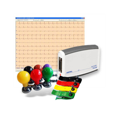
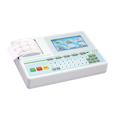
Click Here To Download Catalogue | AsCARD Green electrocardiograph is a 1- and 3-channel ECG unit which enables to make electrocardiogram in full 12 leads. Intended for ECG examinations of adult and paediatric patients aimed at identification of cardiological abnormalities, myocardial ischaemia or infarction. The device is intended for use in healthcare facilities by duly trained personnel. ECG examination may be recorded in manual or automatic mode with the ability to perform the analysis and interpretation.
Electrocardiograph is based on advanced microprocessor technology. It is equipped with a thermal printer with high-resolution head and graphical LCD display. A hightech membrane keyboard makes the AsCARD Green device operation intuitive, and its menu navigation exceptionally easy. This light-weight, small-footprint and battery powered cause that device can be easily transported to any location. With plastic casing and foil covered keyboard, the device is neat and easy to clean.
Technical Specifications:
Click Here To Download Catalogue | DETAILS
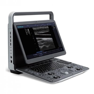
B + Compound
B + Compound utilizes several lines of sight for optimal contrast resolution, speckle reduction and border detection, with which P10 is ideal for superficial and abdominal imaging with better clarity and improved continuity of structures.
μ-Scan
The new generation μ-Scan imaging technology gives you better image quality by reducing noise, improving signal strength and improving visualization.
P10 offers a comprehensive selection of electronic probes to maximize its capabilities to meet a wide range of applications including abdomen, pediatric, OB/GYN, cardiovascular, musculoskeletal, etc. The advanced probe technologies also effectively enhance the image quality and confidence in reaching clinical diagnoses, even in difficult patients.
Convex Probe 3C-A
Ideal for an abundant of application such as abdomen, gynecology, obstetrics, urology and even abdomen biopsy.
Linear Probe L741
This linear probe is designed to satisfy vascular, breast, thyroid, and other small parts diagnosis, and its adjustable parameters could also present users a clear view of MSK and deep vessels.
Phase Array Probe 3P-A
For the purpose of adult and pediatric cardiology and emergency, the phase array probe provides elaborate presets for different exam modes, even for difficult patients.
Intracavitary Probe 6V1
Intracavitary probe could face application of gynecology, urology, prostate, and its temperature detection technology not only protects the patient but also extends the service life.
Click Here To Download Catalogue | ASPEL Ambulatory BP Machine - is a recorder of long-term records of non-invasive measurement of blood pressure intended for use in clinics, hospitals, outpatient centers and specialist surgeries. The recorder enables the assessment of blood pressure by the oscillometric method in adult patients, pregnant women, including preeclampsia and pediatric patients (from 3 years of age). Blood pressure is assessed by using an inflatable cuff, an accurate pressure transducer, and a deflation valve.
Features:
Save-2-Safe: Double security system
Thanks to the use of two independent measuring systems with an additional valve, it meets the highest standards and takes care of patient safety even better.
Start-Easy: Quick start in two moves
The quick launch function allows you to use the device instantly, easily allows you to start recording in holter mode.
Memo-Care: Cuff pressure memory
Recorder remembers the pressure in the cuff. Thanks to the use of Intelligent Solutions, it adapts individually to the patient.
Power-Usb: USB connection
The device can work without batteries: by connecting to a computer via a USB cable.
Technical Specification:
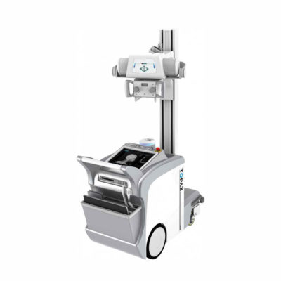
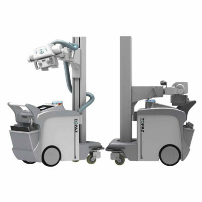
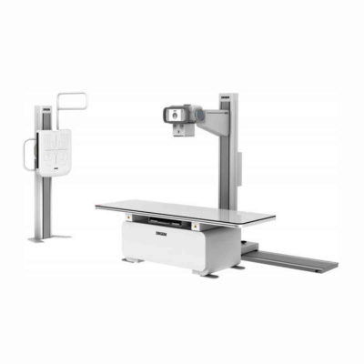
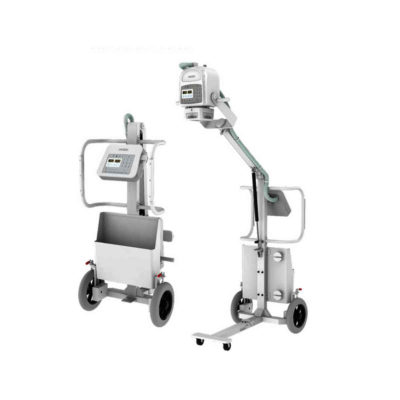
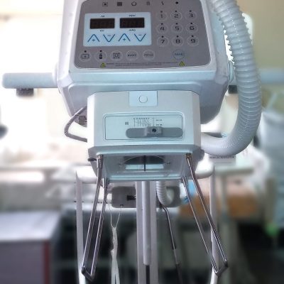
Click Here To Download Catalogue | DrGem Ceiling Analogue X-ray Machine is a diagnostic radiography system X-ray Machine that provides reliable high quality radiographic images with a reduced dose. The reliable high-frequency x-ray generators that are known worldwide for their excellent performance, lifetime and stability. Patient tables and wall stands are also offered.
Features of DrGem Ceiling Analogue X-ray Machine
Click Here To Download Catalogue | DETAILS
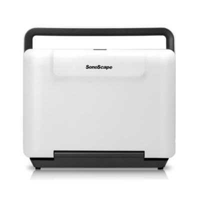
Super Wide-bandwidth Platform
Inheriting Wi-sono's ultra-wide system platform and with the advanced probe technology, high-resolution and deep penetration images are provided for precision medicine.
Spatial Compound Imaging
Spatial Compound Imaging utilizes several lines of sight for optimal contrast resolution, speckle reduction and border detection, with which P15 is ideal for superficial and abdominal imaging with better clarity and improved continuity of structures.
μ-Scan+
The new generation μ-Scan imaging technology gives you better image quality by reducing noise, improving signal strength and improving visualization.
Dynamic Color
Dynamic color improves upon already existing color Doppler technologies for a clearer capture of color flow and detailed visualization of even tiny veins with lower velocities.
Real-time Panoramic
With real-time panoramic, you can acquire an extended field of view for large organs or long vessels for easy measurement and diagnostic efficiency. Accomplished in real-time for the convenience of the sonographers, any mistake can also be easily back tracked and corrected without interrupting the scan.
3D/4D
Outstanding volume performance with speed and convenience makes P15 outshine others on volume imaging.
Tissue Doppler Imaging
Tissue Doppler Imaging allows clinical doctors to quantitatively evaluate local myocardial movements and functions, facilitating them with the ability to analyze and compare the motions of the different parts of the patient's heart.
Auto IMT
Quick measurement of intra-media vessel thickness ensures good reproducibility and high diagnostic efficiency.
Click Here To Download Catalogue | ||||||||||||||||||||||||||||||||||||||||||||||||||||||||||||||||||||||||||||||||||||||||||||||||||||||||||||||||||||||||||||||||||||||||||||||||||||||||||||||||||||||||||||||||||||||||||||||||||||||||||||||||||||||||||||||||||||||||||||||||||||||||||||||||||||||||||||||||||||||||||||||||||||||||||||
| Weight | N/A | N/A | N/A | N/A | N/A | N/A | ||||||||||||||||||||||||||||||||||||||||||||||||||||||||||||||||||||||||||||||||||||||||||||||||||||||||||||||||||||||||||||||||||||||||||||||||||||||||||||||||||||||||||||||||||||||||||||||||||||||||||||||||||||||||||||||||||||||||||||||||||||||||||||||||||||||||||||||||||||||||||||||||||||||||||||
| Dimensions | N/A | N/A | N/A | N/A | N/A | N/A | ||||||||||||||||||||||||||||||||||||||||||||||||||||||||||||||||||||||||||||||||||||||||||||||||||||||||||||||||||||||||||||||||||||||||||||||||||||||||||||||||||||||||||||||||||||||||||||||||||||||||||||||||||||||||||||||||||||||||||||||||||||||||||||||||||||||||||||||||||||||||||||||||||||||||||||
| Additional information |
|

Jorge Meléndez
Please send me price and more information
samson faluro
please send a request email to biz.development@halomedicals.com