
Deep Vein Thrombosis DVT-7700
$0.00
In Stock
DVT-7700 MAIN with HOSE and Accessories (Disposable sleeve thigh(M), Disposable sleeve calf(M), Disposable sleeve foot, and Tube (pair)) – Deep vein thrombosis (DVT) happens when a blood clot forms in a vein deep inside the body. Veins in the thigh, foot, and calf create a natural pumping system that helps flow blood back to the heart. But when an individual is immobile, blood circulation is decreased, and the risk of developing a blood clot goes up.
Delivery & Availability:
Typically 14-21 working days – excluding furniture and heavy/bulky equipment. Please contact us for further information.
Description
Deep vein thrombosis DVT-7700 MAIN is a DVT machine with HOSE and Accessories (Disposable sleeve thigh(M), Disposable sleeve calf(M), Disposable sleeve foot, and Tube (pair)) – Deep vein thrombosis (DVT) happens when a blood clot forms in a vein deep inside the body. Veins in the thigh, foot, and calf create a natural pumping system that helps flow blood back to the heart. But when an individual is immobile, blood circulation is decreased, and the risk of developing a blood clot goes up.
DVT machine Specifications:
Power requiremnet: 100V~240VAC, 50/60Hz
Power Consumption: Maximum 18w
Power Cord: Hospital Grade Plug (Detachable)
Operation Mode: A1,A2,A3,B1,B2,B3,B4,C1,D1,D2
Adjustable Timer: 0-99 Hours
Applied Pressure Leg sleeve: 30~60 mmHg (Caif, Thigh)
Food Cuffs: 130mmHg
Operation Mood: Continuous
Compression Type: Leg sleeve: Gradient, Sequential
Food Cuffs: Uniform
Dimension: 200mm (L) * 190mm (W) * 260 mm (H)
Weight: 2.3 kg (5.07 ib)
Battery: Capacity 10.8 Vdc 4,400 mAh Lithiumion (optional)
Run Time 6-8 Hours
Click Here To Download Catalogue
Reviews(2)
Quick Comparison
| Deep Vein Thrombosis DVT-7700 remove | Sonoscape S22 Ultrasound Machine remove | Sonoscape S8 Exp Portable Ultrasound remove | Sonoscape P10 Ultrasound Machine remove | Sonoscape P15 Ultrasound Machine With Four Probes remove | Bistos BT-770-12.1" Touchscreen Patient Monitor remove | |
|---|---|---|---|---|---|---|
| Name | Deep Vein Thrombosis DVT-7700 remove | Sonoscape S22 Ultrasound Machine remove | Sonoscape S8 Exp Portable Ultrasound remove | Sonoscape P10 Ultrasound Machine remove | Sonoscape P15 Ultrasound Machine With Four Probes remove | Bistos BT-770-12.1" Touchscreen Patient Monitor remove |
| Image | 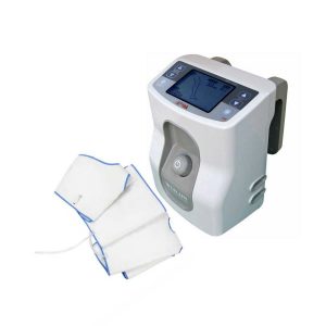 | 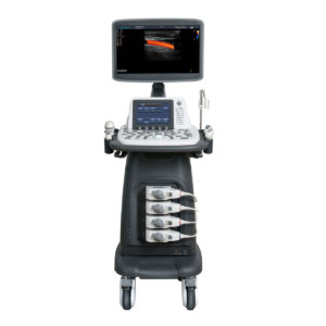 | 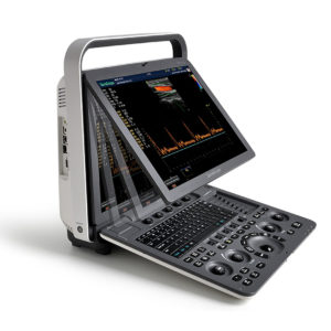 | 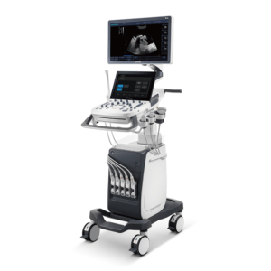 | 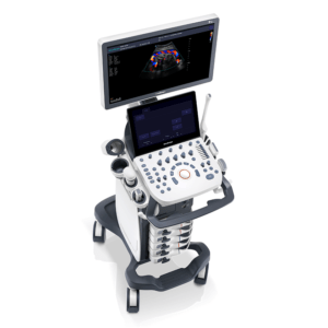 | 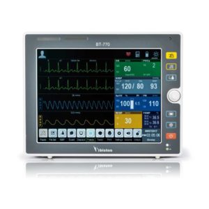 |
| SKU | SF1033560084-290 | SF1033560012-3 | SF1033560012-15 | SF1033560012-7 | SF1033560012-8 | SF1033560059-1 |
| Rating | ||||||
| Price |
|
|
|
|
|
|
| Stock | ||||||
| Availability | ||||||
| Add to cart | ||||||
| Description | In Stock DVT-7700 MAIN with HOSE and Accessories (Disposable sleeve thigh(M), Disposable sleeve calf(M), Disposable sleeve foot, and Tube (pair)) - Deep vein thrombosis (DVT) happens when a blood clot forms in a vein deep inside the body. Veins in the thigh, foot, and calf create a natural pumping system that helps flow blood back to the heart. But when an individual is immobile, blood circulation is decreased, and the risk of developing a blood clot goes up. Delivery & Availability: Typically 14-21 working days – excluding furniture and heavy/bulky equipment. Please contact us for further information. | Shipped from Abroad As SonoScape steps forward to add value and efficiency to ultrasound, the latest S22 was designed in a user-friendly platform to address current and future demanding needs. It represents an excellent mix in performance and price. Delivery & Availability: Typically 5-7 working days – excluding furniture and heavy/bulky equipment. Please contact us for further information. | Shipped from Abroad With ultra-modern innovative design and the clinically-proven technologies, S8 Exp is portable ultrasound scanner well equipped as a low-physical-effort and enhanced-image-quality ultrasound scanner, which not only provides optimized solutions for versatile applications, but does help to improve the user-experience for both routine and non-traditional challenges. Delivery & Availability: Typically 5-7 working days – excluding furniture and heavy/bulky equipment. Please contact us for further information. | Shipped from Abroad The P10 color Doppler ultrasound system is a new generation product from SonoScape. It is designed to give high quality images, rich probe configurations, various clinical tools and automatic analysis software to provide you with comprehensive solutions for your growing demand for clinical applications. Delivery & Availability: Typically 5-7 working days – excluding furniture and heavy/bulky equipment. Please contact us for further information. | In Stock A feature-rich system inheriting the Wi-Sono high-end platform, the P15 uses an array of advanced tools to help enhance the image quality. It's a cost-effective, simplified console with an intuitive user interface and multiple intelligent functions. Delivery & Availability: Typically 2 working days – excluding furniture and heavy/bulky equipment. Please contact us for further information. | Shipped from Abroad The Bistos BT-770 patient monitor is equipped with a 12.1" touchscreen display, which allows for an easy operation and readability with a powerful rechargeable battery guaranteeing a continuous operation of 5 hours to monitor ECG, SpO2, NIBP, temperature and respiration Delivery & Availability: Typically 14 working days – excluding furniture and heavy/bulky equipment. Please contact us for further information. |
| Content | Deep vein thrombosis DVT-7700 MAIN is a DVT machine with HOSE and Accessories (Disposable sleeve thigh(M), Disposable sleeve calf(M), Disposable sleeve foot, and Tube (pair)) - Deep vein thrombosis (DVT) happens when a blood clot forms in a vein deep inside the body. Veins in the thigh, foot, and calf create a natural pumping system that helps flow blood back to the heart. But when an individual is immobile, blood circulation is decreased, and the risk of developing a blood clot goes up.
DVT machine Specifications:
Power requiremnet: 100V~240VAC, 50/60Hz
Power Consumption: Maximum 18w
Power Cord: Hospital Grade Plug (Detachable)
Operation Mode: A1,A2,A3,B1,B2,B3,B4,C1,D1,D2
Adjustable Timer: 0-99 Hours
Applied Pressure Leg sleeve: 30~60 mmHg (Caif, Thigh)
Food Cuffs: 130mmHg
Operation Mood: Continuous
Compression Type: Leg sleeve: Gradient, Sequential
Food Cuffs: Uniform
Dimension: 200mm (L) * 190mm (W) * 260 mm (H)
Weight: 2.3 kg (5.07 ib)
Battery: Capacity 10.8 Vdc 4,400 mAh Lithiumion (optional)
Run Time 6-8 Hours
Click Here To Download Catalogue | DETAILS
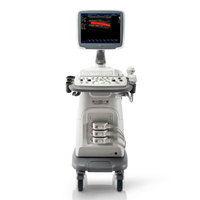
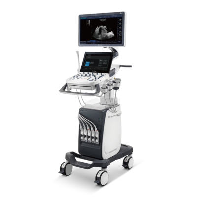
As SonoScape steps forward to add value and efficiency to ultrasound, the latest S22 was designed in a user-friendly platform to address current and future demanding needs. It represents an excellent mix in performance and price.
S22, is a shared service ultrasound system with a slim and elegant package that has combined mobility with utility to fit in specific clinical situations including emergency department, ICU, operating room and so on. Furthermore, its ergonomic design, easy operating and flexible data management will give you a memorable experience.
SPECIFICATION
• Large high-resolution widescreen LED
• Sensitive touch screen
• Four transducer sockets plus one socket for pencil probe
• A comprehensive selection of probes: linear, Convex, Micro-convex, Volumetric, Endocavity, Bi-plane, Phased Array, TEE, Intraoperative, Pencil
• Premium application technology: 4D, μ-scan speckle reduction, compound imaging, Pulse Inversion Harmonic Imaging, Color M-Mode, Steer M-Mode, PDI, TDI, Real-time Panoramic Imaging, Trapezoid Imaging, Auto-IMT…
• Full patient database and image management solutions: DICOM 3.0, AVI/JPG, USB 2.0, HDD, DVD, PDF report
• Multi-Language Input Keyboard
• Built-in battery
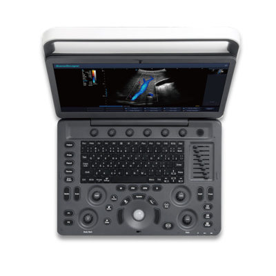
Click Here To Download Catalogue | Sonoscape S8 Exp Portable Ultrasound scannerDETAILS Agile and Versatile With ultra-modern innovative design and the clinically-proven technologies, S8 Exp Portable Ultrasound scanner is well equipped as a low-physical-effort and enhanced-image-quality ultrasound scanner, which not only provides optimized solutions for versatile applications but does help to improve the user experience for both routine and non-traditional challenges. Working with S8 Exp, it will trigger your unlimited reverie and endow you with endless charm. Carrying forward the classical design of SonoScape's portable ultrasound products, S8 Exp successfully combines the best ergonomics, attractive design and ease of use. This charismatic identity is also enhanced by a sophisticated color palette—with sedate grey as its interior paint color and pearl white as exterior cover, S8 Exp reveals a style of aristocrat and strong character among SonoScape's ultrasound systems. Workflow The S8 Exp is a portable ultrasound scanner that adapts to your workflow, whether you are in the consulting room, at the bedside, or at a remote location. With easy-to-use new platform designed for sonographers' needs and full connection interfaces for easy connectivity and data sharing, S8 Exp leads to improved user comfort and clinical outcome as well as patient throughput and working efficiency. Powerful Platform Embedded with SonoScape's core imaging technologies such as μ-scan, PHI and Spatial Compound, S8 Exp boasts exceptional 2D image, sensitive spectral, Color and Power Doppler, displaying well-defined anatomy and pathology and facilitating a highly optimized diagnostic user environment for conclusive diagnoses. Besides, S8 Exp offers a comprehensive selection of electronic probes to maximally extend its capabilities to meet a wide range of applications including the abdomen, pediatric, OB/GYN, cardiovascular, musculoskeletal, etc. The advanced probe technologies also effectively enhance the image quality and confidence in reaching clinical diagnoses even in difficult patients.Click Here To Download Catalogue | DETAILS
B + Compound
B + Compound utilizes several lines of sight for optimal contrast resolution, speckle reduction and border detection, with which P10 is ideal for superficial and abdominal imaging with better clarity and improved continuity of structures.
μ-Scan
The new generation μ-Scan imaging technology gives you better image quality by reducing noise, improving signal strength and improving visualization.
P10 offers a comprehensive selection of electronic probes to maximize its capabilities to meet a wide range of applications including abdomen, pediatric, OB/GYN, cardiovascular, musculoskeletal, etc. The advanced probe technologies also effectively enhance the image quality and confidence in reaching clinical diagnoses, even in difficult patients.
Convex Probe 3C-A
Ideal for an abundant of application such as abdomen, gynecology, obstetrics, urology and even abdomen biopsy.
Linear Probe L741
This linear probe is designed to satisfy vascular, breast, thyroid, and other small parts diagnosis, and its adjustable parameters could also present users a clear view of MSK and deep vessels.
Phase Array Probe 3P-A
For the purpose of adult and pediatric cardiology and emergency, the phase array probe provides elaborate presets for different exam modes, even for difficult patients.
Intracavitary Probe 6V1
Intracavitary probe could face application of gynecology, urology, prostate, and its temperature detection technology not only protects the patient but also extends the service life.
Click Here To Download Catalogue | DETAILS
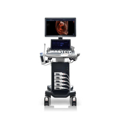
Super Wide-bandwidth Platform
Inheriting Wi-sono's ultra-wide system platform and with the advanced probe technology, high-resolution and deep penetration images are provided for precision medicine.
Spatial Compound Imaging
Spatial Compound Imaging utilizes several lines of sight for optimal contrast resolution, speckle reduction and border detection, with which P15 is ideal for superficial and abdominal imaging with better clarity and improved continuity of structures.
μ-Scan+
The new generation μ-Scan imaging technology gives you better image quality by reducing noise, improving signal strength and improving visualization.
Dynamic Color
Dynamic color improves upon already existing color Doppler technologies for a clearer capture of color flow and detailed visualization of even tiny veins with lower velocities.
Real-time Panoramic
With real-time panoramic, you can acquire an extended field of view for large organs or long vessels for easy measurement and diagnostic efficiency. Accomplished in real-time for the convenience of the sonographers, any mistake can also be easily back tracked and corrected without interrupting the scan.
3D/4D
Outstanding volume performance with speed and convenience makes P15 outshine others on volume imaging.
Tissue Doppler Imaging
Tissue Doppler Imaging allows clinical doctors to quantitatively evaluate local myocardial movements and functions, facilitating them with the ability to analyze and compare the motions of the different parts of the patient's heart.
Auto IMT
Quick measurement of intra-media vessel thickness ensures good reproducibility and high diagnostic efficiency.
Click Here To Download Catalogue |
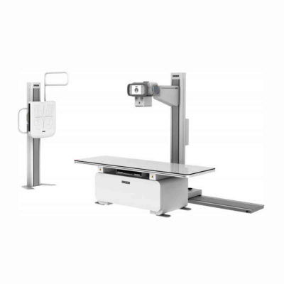
Bistos BT-770 is a 12.1" touchscreen patient monitor designed for easy operations.
SPECIFICATIONS
Click Here To Download Catalogue |
| Weight | N/A | N/A | N/A | N/A | N/A | N/A |
| Dimensions | N/A | N/A | N/A | N/A | N/A | N/A |
| Additional information |

Learn More
As I web-site possessor I believe the content matter here is rattling fantastic , appreciate it for your hard work. You should keep it up forever! Best of luck.
manhwaland
Really informative article.Much thanks again. Cool.