PACS System
$0.00
Shipped From Abroad
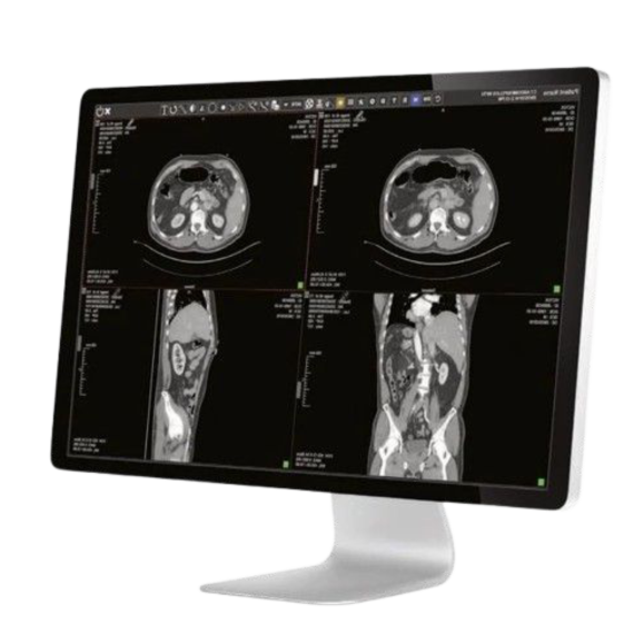
The Oryx Healthcare PACS Solution is a secure medical imaging platform that stores, manages, and distributes diagnostic images efficiently, enabling fast access, seamless workflow integration, and accurate clinical decision-making across hospitals, imaging centers, and healthcare networks.
Typically 10-21 working days – excluding furniture and heavy/bulky equipment. Please contact us for further information.
Description
The Oryx Healthcare PACS Solution provides a centralized digital system for acquiring, storing, viewing, and sharing medical images such as X-ray, CT, MRI, ultrasound, and fluoroscopy studies. Designed to enhance diagnostic efficiency, it supports DICOM standards and integrates smoothly with hospital information systems. The solution improves clinical workflows, reduces reliance on physical films, enables remote access, and ensures secure image archiving for faster diagnosis and improved patient care.
Key Features
-
Centralized digital storage for all diagnostic images
-
Full DICOM compatibility across imaging modalities
-
Fast image retrieval and advanced viewing tools
-
Integration with HIS, RIS, and EHR systems
-
Multi-user access across departments
-
Secure user authentication and data protection
-
Supports teleradiology and remote consultations
-
Scalable architecture for small clinics to large hospitals
Quick Comparison
| PACS System remove | Sonoscape E2 Ultrasound Machine remove | Topaz Digital X-ray Machine remove | Sonoscape S11 Ultrasound Machine remove | Anke MRI Openmark 5000 Permanent System remove | IBIS Neeo R9 Digital Surgical C-Arm remove | ||||||||||||||||||||||||||||||||||||||||||||||||||||||||||||||||||||||||||||||||||||||||||||||||||||||||||||||||||||||||||||||||||||||||||||||||||||||||||||||||||||||||||||||||||||||||||||||||||||||||||||||||||||||||||||||||||||||||||||||||||||||||||||||||||||||||||||||||||||||||||||||||||||||||||||||||
|---|---|---|---|---|---|---|---|---|---|---|---|---|---|---|---|---|---|---|---|---|---|---|---|---|---|---|---|---|---|---|---|---|---|---|---|---|---|---|---|---|---|---|---|---|---|---|---|---|---|---|---|---|---|---|---|---|---|---|---|---|---|---|---|---|---|---|---|---|---|---|---|---|---|---|---|---|---|---|---|---|---|---|---|---|---|---|---|---|---|---|---|---|---|---|---|---|---|---|---|---|---|---|---|---|---|---|---|---|---|---|---|---|---|---|---|---|---|---|---|---|---|---|---|---|---|---|---|---|---|---|---|---|---|---|---|---|---|---|---|---|---|---|---|---|---|---|---|---|---|---|---|---|---|---|---|---|---|---|---|---|---|---|---|---|---|---|---|---|---|---|---|---|---|---|---|---|---|---|---|---|---|---|---|---|---|---|---|---|---|---|---|---|---|---|---|---|---|---|---|---|---|---|---|---|---|---|---|---|---|---|---|---|---|---|---|---|---|---|---|---|---|---|---|---|---|---|---|---|---|---|---|---|---|---|---|---|---|---|---|---|---|---|---|---|---|---|---|---|---|---|---|---|---|---|---|---|---|---|---|---|---|---|---|---|---|---|---|---|---|---|---|---|---|---|---|---|---|---|---|---|---|---|---|---|---|---|---|---|---|---|---|---|---|---|---|---|---|---|---|---|---|---|---|---|---|---|---|---|---|
| Name | PACS System remove | Sonoscape E2 Ultrasound Machine remove | Topaz Digital X-ray Machine remove | Sonoscape S11 Ultrasound Machine remove | Anke MRI Openmark 5000 Permanent System remove | IBIS Neeo R9 Digital Surgical C-Arm remove | |||||||||||||||||||||||||||||||||||||||||||||||||||||||||||||||||||||||||||||||||||||||||||||||||||||||||||||||||||||||||||||||||||||||||||||||||||||||||||||||||||||||||||||||||||||||||||||||||||||||||||||||||||||||||||||||||||||||||||||||||||||||||||||||||||||||||||||||||||||||||||||||||||||||||||||||
| Image |  | 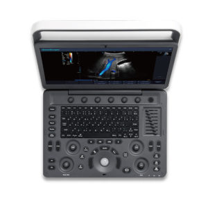 | 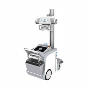 | 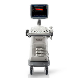 | 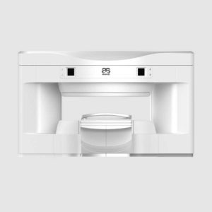 | 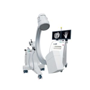 | |||||||||||||||||||||||||||||||||||||||||||||||||||||||||||||||||||||||||||||||||||||||||||||||||||||||||||||||||||||||||||||||||||||||||||||||||||||||||||||||||||||||||||||||||||||||||||||||||||||||||||||||||||||||||||||||||||||||||||||||||||||||||||||||||||||||||||||||||||||||||||||||||||||||||||||||
| SKU | SF1033560012-17 | SF1033560074-1 | SF1033560012-1 | SF1033560092-3 | SF1033560011-1 | ||||||||||||||||||||||||||||||||||||||||||||||||||||||||||||||||||||||||||||||||||||||||||||||||||||||||||||||||||||||||||||||||||||||||||||||||||||||||||||||||||||||||||||||||||||||||||||||||||||||||||||||||||||||||||||||||||||||||||||||||||||||||||||||||||||||||||||||||||||||||||||||||||||||||||||||||
| Rating | |||||||||||||||||||||||||||||||||||||||||||||||||||||||||||||||||||||||||||||||||||||||||||||||||||||||||||||||||||||||||||||||||||||||||||||||||||||||||||||||||||||||||||||||||||||||||||||||||||||||||||||||||||||||||||||||||||||||||||||||||||||||||||||||||||||||||||||||||||||||||||||||||||||||||||||||||||||
| Price |
|
|
|
|
|
| |||||||||||||||||||||||||||||||||||||||||||||||||||||||||||||||||||||||||||||||||||||||||||||||||||||||||||||||||||||||||||||||||||||||||||||||||||||||||||||||||||||||||||||||||||||||||||||||||||||||||||||||||||||||||||||||||||||||||||||||||||||||||||||||||||||||||||||||||||||||||||||||||||||||||||||||
| Stock | |||||||||||||||||||||||||||||||||||||||||||||||||||||||||||||||||||||||||||||||||||||||||||||||||||||||||||||||||||||||||||||||||||||||||||||||||||||||||||||||||||||||||||||||||||||||||||||||||||||||||||||||||||||||||||||||||||||||||||||||||||||||||||||||||||||||||||||||||||||||||||||||||||||||||||||||||||||
| Availability | |||||||||||||||||||||||||||||||||||||||||||||||||||||||||||||||||||||||||||||||||||||||||||||||||||||||||||||||||||||||||||||||||||||||||||||||||||||||||||||||||||||||||||||||||||||||||||||||||||||||||||||||||||||||||||||||||||||||||||||||||||||||||||||||||||||||||||||||||||||||||||||||||||||||||||||||||||||
| Add to cart | |||||||||||||||||||||||||||||||||||||||||||||||||||||||||||||||||||||||||||||||||||||||||||||||||||||||||||||||||||||||||||||||||||||||||||||||||||||||||||||||||||||||||||||||||||||||||||||||||||||||||||||||||||||||||||||||||||||||||||||||||||||||||||||||||||||||||||||||||||||||||||||||||||||||||||||||||||||
| Description | Shipped From Abroad
The Oryx Healthcare PACS Solution is a secure medical imaging platform that stores, manages, and distributes diagnostic images efficiently, enabling fast access, seamless workflow integration, and accurate clinical decision-making across hospitals, imaging centers, and healthcare networks.
Delivery & Availability:
Typically 10-21 working days – excluding furniture and heavy/bulky equipment. Please contact us for further information.
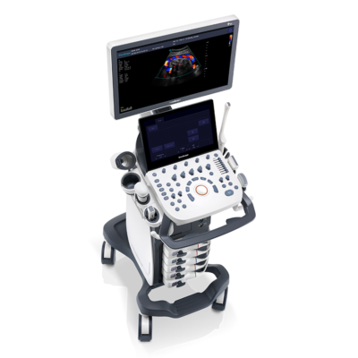
| Shipped from Abroad Sonoscape E2 portable ultrasound machine is a color Doppler ultrasound system that reaches beyond your expectations due to its compact and fashionable appearance. It fulfills GI, OB/GYN, Cardiac and POC applications to fit your routine scanning needs while its color mode will help you for more accurate and efficient diagnosis of lesions. E2 provides a wide range of applications to assist users with routine scanning. E2 provides automatic calculations to enhance your diagnostic confidence and save you time for patient communication. Delivery & Availability: Typically 14 working days – excluding furniture and heavy/bulky equipment. Please contact us for further information. | In Stock DRGEM’s TOPAZ X-ray machine is a state-of-the-art mobile digital radiography system, designed with maximum comfort for patients and users in mind. From its user-friendly software to smooth movements, TOPAZ is made to improve your workflow and provide you with high-quality images. Delivery & Availability: Typically 21 working days – excluding furniture and heavy/bulky equipment. Please contact us for further information. | In Stock A Value Choice beyond Your Expectation. SonoScape’s trolley color Doppler system S11 redefines price and performance with practical design. The S11 will go beyond your expectations but not your budget. Delivery & Availability: Typically 2 working days – excluding furniture and heavy/bulky equipment. Please contact us for further information. | Shipped from Abroad
OPENMARK 5000 is 0.51T MRI. It's approved by FDA and has CE mark. It adopts two-pillar magnet design with 280 degree openness and equipped with powerful
RF and gradient system, together with advanced imaging technology, making it as a high-end system which is comparable to high-field MRI.
Delivery & Availability: Typically 90 working days – excluding furniture and heavy/bulky equipment. Please contact us for further information. | Shipped from Abroad Our Neeo “C” arms are easy to place, use and are specifically designed to be used in orthopedics, traumatology, abdominal surgery, urology, cardiology and operating rooms. Delivery & Availability: Typically 21 working days – excluding furniture and heavy/bulky equipment. Please contact us for further information. | |||||||||||||||||||||||||||||||||||||||||||||||||||||||||||||||||||||||||||||||||||||||||||||||||||||||||||||||||||||||||||||||||||||||||||||||||||||||||||||||||||||||||||||||||||||||||||||||||||||||||||||||||||||||||||||||||||||||||||||||||||||||||||||||||||||||||||||||||||||||||||||||||||||||||||||||
| Content | The Oryx Healthcare PACS Solution provides a centralized digital system for acquiring, storing, viewing, and sharing medical images such as X-ray, CT, MRI, ultrasound, and fluoroscopy studies. Designed to enhance diagnostic efficiency, it supports DICOM standards and integrates smoothly with hospital information systems. The solution improves clinical workflows, reduces reliance on physical films, enables remote access, and ensures secure image archiving for faster diagnosis and improved patient care. Key Features
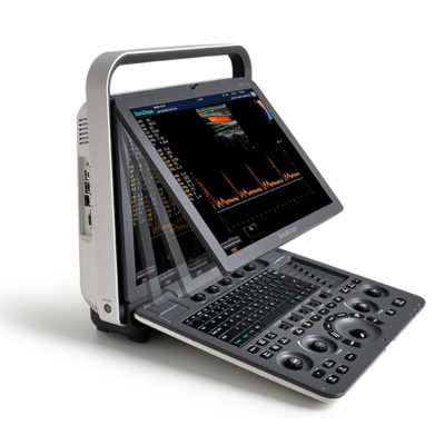
| SONOSCAPE E2 DETAILS
Auto Image Optimization
A portable ultrasound machine with the press of a button, the image is automatically adjusted and optimized, saving you time with parameter adjustments. Additionally, with Auto Focus on, the focus area follows the depth of the ROI box as it is moved in the scanning field, providing users with excellent image quality in the desired area of interest.
Automated Calculation
Auto IMT is used when determining the level of vascular sclerosis present in the patient by automatically tracing the thickness of the carotid vessels.
Auto trace provides users sensitive and accurate wave tracing, avoiding the error of manual trace and giving out calculation result in no time
In-Build Battery pack
This portable ultrasound machine was equipped with an in-build battery pack which enable the user to perform image scanning when AC power is not available.
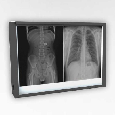
Click Here To Download Catalogue | TOPAZ X-ray machine is among the high end X-ray machine manufactured by DRGEM, a digital X-ray system that provides quality images with little or no effort.
It begins with Advanced Technology
Integrating high technology and over a decade of experience in conventional and digital radiography systems, DRGEM’s TOPAZ X-ray machine is a state-of-the-art mobile digital radiography system, designed with maximum comfort for patients and users. From its user-friendly software to smooth movements, TOPAZ X-ray machine is made to improve your workflow and provide you with high-quality images.
Full Featured Imaging Software & Excellent Digital Image Processing
With a high-performance, built-in touchscreen, TOPAZ X-ray machine offers a user-friendly interface and powerful software for easy operation and increased workflow. The anatomical view-based digital image processing, automatically optimizes and enhances the quality of the image. it also comes with automatic image storage and print with DICOM 3.0 networking capability. additionally, the system offers increasing exam throughput while decreasing examination time.
Click Here To Download Catalogue | DETAILS
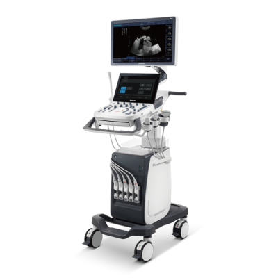
SonoScape’s trolley colour Doppler system S11 redefines price and performance with practical design. The S11 will go beyond your expectations but not your budget. As an easy-to-use ultrasound system, the S11 is integrated with a new software platform, especially optimized for a smooth workflow and convenient operation. The system speeds up the exam process and makes file management easier.
SPECIFICATION
- 15-inch high definition LCD monitor with articulating arm
- Compact and agile trolley design
- 3 active transducer sockets available for a wide range of applications
- Duplex, Color Doppler, DPI, PW Doppler, tissue harmonic imaging, μ-scan speckle reduction imaging, compound imaging, trapezoidal imaging
- Customized settings based on your own working style
- Full patient database and image management solutions
Click Here To Download Catalogue | OPENMARK 5000 is 0.51T MRI. It's approved by FDA and has CE mark. It adopts two-pillar magnet design with 280 degree openness and equipped with powerful
RF and gradient system, together with advanced imaging technology, making it as a high-end system which is comparable to high-field MRI.
Features:
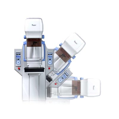
Click Here To Download Catalogue | Our Neeo “C” arms are easy to place, use and are specifically designed to be used in orthopedics, traumatology, abdominal surgery, urology, cardiology and operating rooms.
Using Neeo with the RTP (Real Time Processing) option it is possible to perform vascular, urological and cardiological diagnostics. One of the main functions, digital image subtraction, allows to see, as an example, the passage of contrast liquids in a tissue or in a venous or arterial duct; thanks to the possibility of looping, the acquired video can be reproduced several times to monitor more accurately the passage of the fluid within the area in question. Angiographic measurement is another useful function in the vascular field (QA Quantitative Angiography) that allows the measurement of stenoses. Finally, fluoroscopy allows the correct positioning of stents or expanders.
Neeo is used in various interventional and diagnostic procedures in traumatology and orthopedics wards and operating rooms as well. Thanks to low-dose fluoroscopy, it is possible to use the device for positioning bone or subcutaneous grafts, inserting K-wire (Kirschner wire) for stabilization of bone fragments or for the correct positioning of prostheses. The low dose emitted ensures safe use for both the patient and the surgeon or doctor on the operating field.
On the control panel there is a large touch screen display that allows to adjust the basic functions of the equipment. From this display it is possible to select and adjust the fluoroscopic data for the examination, activate or deactivate the laser pointer, select between pulsed, one shot or standard fluoroscopy, rotate the image and perform all operations on collimator. The four side buttons on the display offer the possibility to move the bow vertically thanks to an extremely silent motor.
Neeo has two 19 “medical grade monitors that can be positioned according to the needs of the medical practitioner. Work monitors and feedback monitors are separated to be managed independently. The possible movements are: rotation, revolution, tilting and possibility of height adjustment.
Features:
Click Here To Download Catalogue | |||||||||||||||||||||||||||||||||||||||||||||||||||||||||||||||||||||||||||||||||||||||||||||||||||||||||||||||||||||||||||||||||||||||||||||||||||||||||||||||||||||||||||||||||||||||||||||||||||||||||||||||||||||||||||||||||||||||||||||||||||||||||||||||||||||||||||||||||||||||||||||||||||||||||||||||
| Weight | N/A | N/A | N/A | N/A | N/A | N/A | |||||||||||||||||||||||||||||||||||||||||||||||||||||||||||||||||||||||||||||||||||||||||||||||||||||||||||||||||||||||||||||||||||||||||||||||||||||||||||||||||||||||||||||||||||||||||||||||||||||||||||||||||||||||||||||||||||||||||||||||||||||||||||||||||||||||||||||||||||||||||||||||||||||||||||||||
| Dimensions | N/A | N/A | N/A | N/A | N/A | N/A | |||||||||||||||||||||||||||||||||||||||||||||||||||||||||||||||||||||||||||||||||||||||||||||||||||||||||||||||||||||||||||||||||||||||||||||||||||||||||||||||||||||||||||||||||||||||||||||||||||||||||||||||||||||||||||||||||||||||||||||||||||||||||||||||||||||||||||||||||||||||||||||||||||||||||||||||
| Additional information |
|


Reviews
There are no reviews yet.