- Sorry, this product cannot be purchased.
Colonoscopy Training Model
$0.00
Shipped From Abroad
The Colonoscopy Training Model is a realistic medical simulation model for training lower gastrointestinal endoscopic procedures.
Typically 10-21 working days – excluding furniture and heavy/bulky equipment. Please contact us for further information.
Description
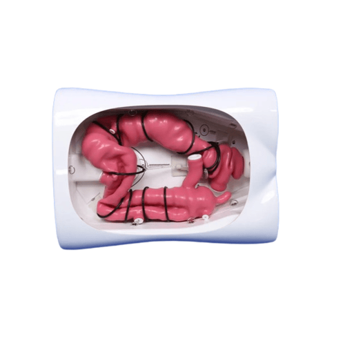
The Colonoscopy Training Model (Model: PLXH1001) by Preclinic is a realistic medical simulation model for training lower gastrointestinal endoscopic procedures. It’s designed from adult lower digestive tract CT data, using soft silica gel (hardness ~18A-30A), to mimic the colon anatomy including the ascending, transverse, and descending colon. Trainers can practice colonoscopy, biopsy, polypectomy, and other procedures using real endoscopic tools. The model includes fixed parts (colon, base, upper cover) and replaceable simulated polyp modules, providing realistic tactile feedback and versatility for different training scenarios.
Key Features & Specifications
| Feature | Details |
|---|---|
| Model | PLXH1001 Colonoscopy Training Model |
| Material | Highly soft silica gel, hardness ~ 18A-30A |
| Size | Length: ~68 cm (26.8 in); Width: ~23 cm (9.1 in); Height: ~25 cm (9.8 in) |
| Fixed Components | Colon model, base, upper cover |
| Replaceable Components | Simulated polyps; multiple lesion modules so you can simulate different pathologies (tumors, polyps, inflammation) |
| Procedures Supported | Colonoscopy, GI biopsy, polypectomy |
| Anatomical Accuracy | Includes anatomical parts: anus, rectum, sigmoid colon, descending/transverse/ascending colon, cecum, appendix and opening of small intestine. Based on adult CT data. |
| Compatibility | Works with most brands of colonoscopes and biopsy / polypectomy tools (forceps, snares, injection needles) |
| Lead Time | ~15 days |
| Port | Shanghai, China |
Quick Comparison
| Colonoscopy Training Model remove | 3B Scientific Human Skull with Facial Muscles - 3B Smart Anatomy remove | 3B Scientific Functional Human Shoulder Joint - 3B Smart Anatomy remove | 3B Scientific Hand Skeleton Model with Ligaments and Muscles - 3B Smart Anatomy remove | 3B Scientific Classic Unisex Human Torso Model, 12 part - 3B Smart Anatomy remove | 3B Scientific Functional Human Hip Joint Model - 3B Smart Anatomy remove | |||||||||||||||||||||||
|---|---|---|---|---|---|---|---|---|---|---|---|---|---|---|---|---|---|---|---|---|---|---|---|---|---|---|---|---|
| Name | Colonoscopy Training Model remove | 3B Scientific Human Skull with Facial Muscles - 3B Smart Anatomy remove | 3B Scientific Functional Human Shoulder Joint - 3B Smart Anatomy remove | 3B Scientific Hand Skeleton Model with Ligaments and Muscles - 3B Smart Anatomy remove | 3B Scientific Classic Unisex Human Torso Model, 12 part - 3B Smart Anatomy remove | 3B Scientific Functional Human Hip Joint Model - 3B Smart Anatomy remove | ||||||||||||||||||||||
| Image |  | 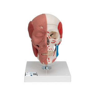 | 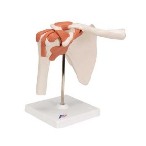 | 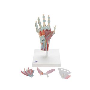 | 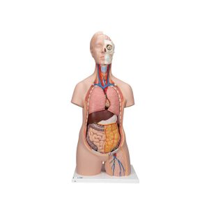 | 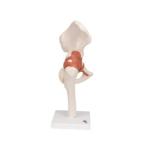 | ||||||||||||||||||||||
| SKU | SF1033560099-10 | SF1033560099-7 | SF1033560099-14 | SF1033560099-3 | SF1033560099-5 | |||||||||||||||||||||||
| Rating | ||||||||||||||||||||||||||||
| Price |
|
|
|
|
|
| ||||||||||||||||||||||
| Stock | ||||||||||||||||||||||||||||
| Availability | ||||||||||||||||||||||||||||
| Add to cart | ||||||||||||||||||||||||||||
| Description | Shipped From Abroad
The Colonoscopy Training Model is a realistic medical simulation model for training lower gastrointestinal endoscopic procedures.
Delivery & Availability:
Typically 10-21 working days – excluding furniture and heavy/bulky equipment. Please contact us for further information.
| Ship from abroad
This model of the face musculature by 3B Scientific is used to easily demonstrate the causes of temporomandibular disorders and other dysfunctional disturbances of the TMJ and masticatory muscles.
| Ship from abroad
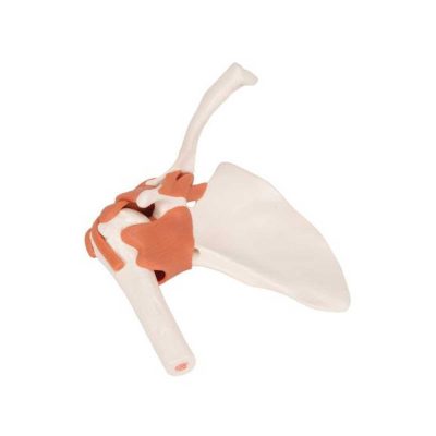
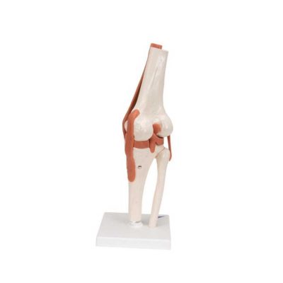
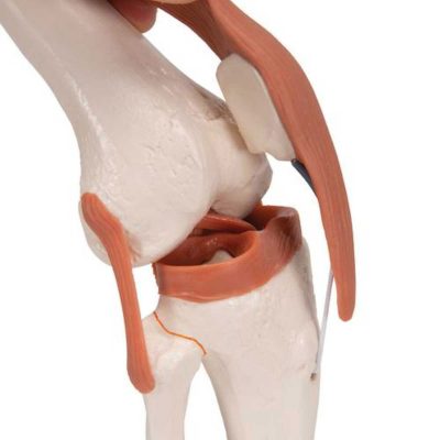
This high quality life-size functional shoulder joint model shows the anatomy and mechanics of the shoulder joint. Consisting of the scapula, clavical, portion of humerus and joint ligaments, this fully flexible shoulder joint model clearly demonstrates abduction, anteversion, retroversion and internal/external rotation. The Functional shoulder joint model comes on a stand for easy study and display.
| Ship from abroad
The bones, muscles, tendons, ligaments, nerves, arteries, and veins are all featured in this high quality 4 part model of the hand and lower forearm. The dorsal side of the hand shows the extensor muscles as well as portions of the tendons at the wrist as they pass under the extensor retunaculum. The palmar face of the hand is represented in three layers, the first two are removable to allow detailed study of the deeper anatomical layer of the hand. In addition clinically important structures such as the median nerve and superficial palmar arterial arch can be explored in detail in the hand model. The deepest anatomical layer allows for study of the intrinsic muscles and deep palmar arterial arch in addition to other details of the anatomy of the hand. This high quality anatomically correct hand model with ligaments and muscles is great for detailed study.
| Ship from abroad
This 12 part anatomically correct human torso is an educational tool of true quality. The unisex torso is hand-painted true to detail and made of high-quality plastic.
| Ship from abroad
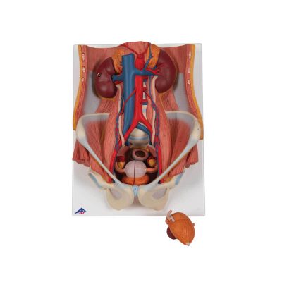
This life size functional hip joint model clearly shows the anatomy and mechanics of the human hip joint. This fully flexible hip joint demonstrates abduction, anteversion, retroversion and internal/external rotation. This high quality functional joint consists of a portion of femur, hip bone and joint ligaments. Comes on a stand for easy display in the classroom or doctor's office.
| ||||||||||||||||||||||
| Content | The Colonoscopy Training Model (Model: PLXH1001) by Preclinic is a realistic medical simulation model for training lower gastrointestinal endoscopic procedures. It’s designed from adult lower digestive tract CT data, using soft silica gel (hardness ~18A-30A), to mimic the colon anatomy including the ascending, transverse, and descending colon. Trainers can practice colonoscopy, biopsy, polypectomy, and other procedures using real endoscopic tools. The model includes fixed parts (colon, base, upper cover) and replaceable simulated polyp modules, providing realistic tactile feedback and versatility for different training scenarios.
Key Features & Specifications
|
| This high quality life-size functional shoulder joint model shows the anatomy and mechanics of the shoulder joint. Consisting of the scapula, clavical, portion of humerus and joint ligaments, this fully flexible shoulder joint model clearly demonstrates abduction, anteversion, retroversion and internal/external rotation. The Functional shoulder joint model comes on a stand for easy study and display. | The bones, muscles, tendons, ligaments, nerves, arteries, and veins are all featured in this high quality 4 part model of the hand and lower forearm. The dorsal side of the hand shows the extensor muscles as well as portions of the tendons at the wrist as they pass under the extensor retunaculum. The palmar face of the hand is represented in three layers, the first two are removable to allow detailed study of the deeper anatomical layer of the hand. In addition clinically important structures such as the median nerve and superficial palmar arterial arch can be explored in detail in the hand model. The deepest anatomical layer allows for study of the intrinsic muscles and deep palmar arterial arch in addition to other details of the anatomy of the hand. This high quality anatomically correct hand model with ligaments and muscles is great for detailed study. |
This 12 part anatomically correct human torso is an educational tool of true quality. The unisex torso is hand-painted true to detail and made of high-quality plastic. This classic human torso was developed and modeled in Germany. Whether you are a student studying human anatomy in a biology classroom or a doctor explaining something to a patient, this human torso model is a valuable tool.
The following components of this unisex torso are removable:
All the organs in this human torso are hand painted for a quality product. This great human anatomy educational tool and makes learning the location of the human organs easy. It is also supplied with the 3B Torso Guide. | This life size functional hip joint model clearly shows the anatomy and mechanics of the human hip joint. This fully flexible hip joint demonstrates abduction, anteversion, retroversion and internal/external rotation. This high quality functional joint consists of a portion of femur, hip bone and joint ligaments. Comes on a stand for easy display in the classroom or doctor's office. | ||||||||||||||||||||||
| Weight | N/A | N/A | N/A | N/A | N/A | N/A | ||||||||||||||||||||||
| Dimensions | N/A | N/A | N/A | N/A | N/A | N/A | ||||||||||||||||||||||
| Additional information |


Reviews
There are no reviews yet.