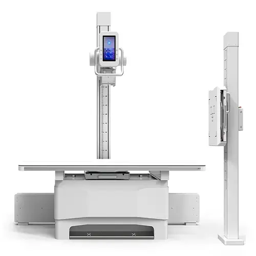
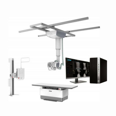
| Description | Built as a floor-mounted digital X-ray machine, it features a coordinated X-ray tube and flat panel detector that maintain precise alignment throughout imaging.
Shipped From China
Delivery & Availability:
Typically 10-21 working days – excluding furniture and heavy/bulky equipment. Please contact us for further information.
| Shipped from Abroad
SuperMark 1.5T is a new generation superconducting MRI system based on years of experience in production and research. It's applicable to whole body scan, such as, nervous system, spine, joint soft tissue, pelvic and abdominal cavity, etc
Delivery & Availability:
Typically 90 working days – excluding furniture and heavy/bulky equipment. Please contact us for further information. | In Stock
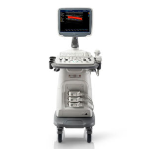
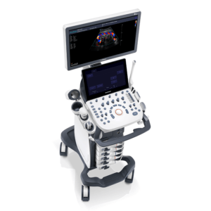
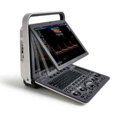
A Value Choice beyond Your Expectation. SonoScape’s trolley color Doppler system S11 redefines price and performance with practical design. The S11 will go beyond your expectations but not your budget.
Delivery & Availability:
Typically 2 working days – excluding furniture and heavy/bulky equipment. Please contact us for further information. | In Stock
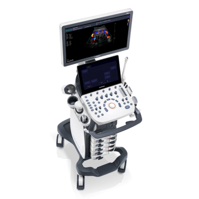
A feature-rich system inheriting the Wi-Sono high-end platform, the P15 uses an array of advanced tools to help enhance the image quality. It's a cost-effective, simplified console with an intuitive user interface and multiple intelligent functions.
Delivery & Availability:
Typically 2 working days – excluding furniture and heavy/bulky equipment. Please contact us for further information. | In stock
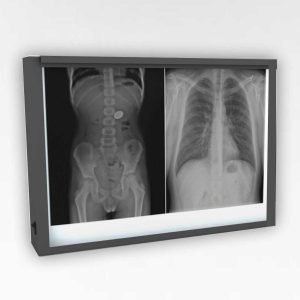
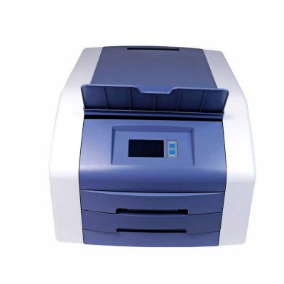
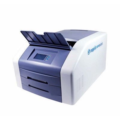
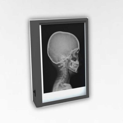
Double x-ray film viewer, Compact, Solid with Backlight of LED’s based panel, Long Life Approximate LED’s life 50, 000 Hrs., Uniform Light at the total surface area, No Heat Emission, Wall Mounted, Can be used for tracing on X-Ray, Auto-sensor, Screen. Size: 430 mm x 710 mm.
Delivery & Availability:
Typically 5-7 working days – excluding furniture and heavy/bulky equipment. Please contact us for further information. | Shipped from Abroad
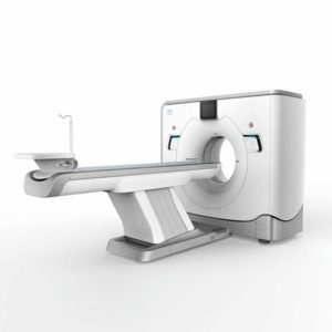
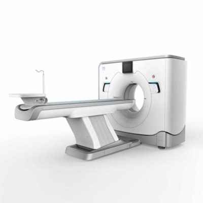
The ANATOM 64 CT scanner is the latest innovation for cardiac imaging based on Precision Platform system. The excellent design of Ahart technology which innovatively combined single spiral scan + gated imaging + mA modulation for easy heart imaging at extremely low radiation dose. We provide you ANATOM 64 Clarity/Precision of two models which are low/high configurations for preferences. It also offers you conventional clinical applications of low dose, better image quality and faster exams.
Delivery & Availability:
Typically 90 working days – excluding furniture and heavy/bulky equipment. Please contact us for further information. |
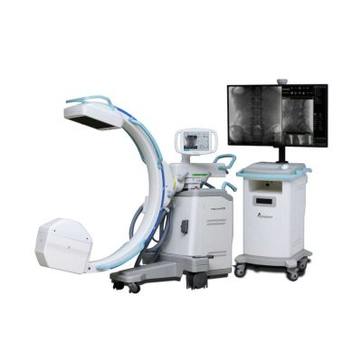
| Content | Built as a floor-mounted digital X-ray machine, it features a coordinated X-ray tube and flat panel detector that maintain precise alignment throughout imaging. Optional AEC and DAP functions can be added to strengthen exposure control and ensure consistent diagnostic quality. Facilities needing faster patient positioning can upgrade the radiographic table to a six-way floating tabletop, improving comfort and overall workflow.
The machine integrates a floor-mounted table with a wall-mounted chest stand, creating a clean and efficient room layout. Its smooth mechanical movement and intuitive operating controls give radiographers greater flexibility when adjusting patient positions and selecting projection angles.
As a versatile high-end DR platform, the SHO-DDX01 supports a wide range of routine clinical examinations, including head, chest, abdominal, lumbar spine, and extremity imaging. It accommodates PA, lateral, oblique, and angled views, making it suitable for hospitals and clinics of all sizes.
Features
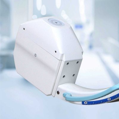
- The X-ray tube operates at a high rotational speed and is supported by an efficient heat-dissipation system, allowing the equipment to perform reliably during long clinical sessions while maintaining an extended service life.
- The high-frequency generator provides strong output performance with stable, high-quality X-ray emission, ensuring dependable imaging results across routine and demanding examinations.
- A smart high-voltage control system incorporates multiple built-in protection functions to maintain safe and stable operation throughout daily clinical use.
- The advanced a-Si CsI flat panel detector delivers high conversion efficiency, enabling clear, detailed digital images with reduced radiation dose.
- A robust digital imaging platform offers comprehensive post-processing capabilities, giving clinicians the tools needed to support a wide variety of diagnostic requirements.
Specifications
| Ambient Conditions |
| Ambient temperature |
10℃–40℃ |
| Relative humidity |
30%–75% |
| Atmospheric pressure |
70–106 kPa |
| Main head |
10.4-inch touchscreen control panel |
| X-Ray Generator |
| Model |
SHO-DDX01 Series |
| Output power (kW) |
40 |
50 |
65 |
| Current (mA) |
10-500 |
10-630 |
10-800 |
| Voltage (kV) |
40-125 |
40-150 |
40-150 |
| mAs |
0.1-400 |
0.1-630 |
0.1-630 |
| Exposure time |
0.002~6.3S |
| APR program |
Equipped |
| Intelligent protection system |
Includes a self-protection system with automatic alarms and fault code diagnostics. |
| X-ray Tube |
| Output power (kW) |
40 |
50 |
65 |
| Anode type |
Rotating anode |
| Revolving speed of anode (rpm) |
50Hz: 2800 60Hz: 3000 |
| Focus size |
1.0/2.0 |
0.6/1.2 |
0.6/1.2 |
| Anode heat storage capacity (kHU) |
150 |
300 |
400 |
| Maximum Anode Heat Dissipation (W) |
475 |
750 |
1000 |
| Housing heat storage capacity (kJ) |
900 |
900 |
1000 |
| X-ray tube Rotation angel (°) |
±180 |
±180 |
±180 |
| X-ray Detector (Wired Flat Panel Detector) |
| Type |
Amorphous Silicon |
| Scintillator |
Cesium Iodide |
| Active area |
17x17 Inch (43 cm*43 cm) |
| Active pixel |
3072*3072 |
| Pixel pitch |
139 um |
| A/D Conversion |
16 bits |
| DQE |
≧70% |
| Spatial resolution |
36 Lp/cm |
| Acquisition time |
≤2 s |
| Collimator |
| Type |
Manual collimator |
| Max. Window |
440 mm × 440 mm (SID = 100 cm) |
| Lamp |
AC/DC 24 V, 5 W |
| Lamp timer |
Automatic illumination with timer for lamp (30S) |
| Inherent filtration |
1.0 mmAl |
| Illuminant |
LED |
| Grid |
| Densities |
40 lp/cm |
| Ratios |
10:1 |
| Size |
18" × 18" |
| Focal distance |
100 cm (Radiography table), 180 cm (Bucky stand) |
| Photography Table |
| Type |
Radiography table |
| Tabletop size |
2300 mm × 800 mm x 60 mm |
| Load-bearing |
300 kg |
| Floating front and back |
740 mm ±3% |
| Floating left and right |
210 mm ±3% |
| FPD tray movement range |
650 mm ±3% |
| Column stand movement |
| Tube stand |
| Up-down movement (to photography table) |
1500 mm ±3% |
| Stand left-right movement |
1900 mm ±3% |
| Rotational angles of stand |
+180°~-180° |
| Bucky stand |
| Detector vertical movement range (distance between detector center and ground) |
1500 mm ±3% |
| The lowest point of the detector center relative to the ground |
≤ 46 cm |
| Automatic tracking function |
Yes, electric correspondence between the X-ray tube and flat panel detector |
| Workstation |
| Monitor |
23" LCD Dell brand |
| CPU |
≥ 2.8 GHz |
| Memory |
≥ 4 GB |
| HDD |
≥ 1 TB |
| DICOM3.0 |
Query for integration with any PACS |
| Functions |
Import/export function |
| Image info |
| Management of patient info |
| Post processing |
| Measurement etc. |
| Console |
| Control room console |
10.4 inch LCD color touch screen console for exposure parameter setting and exposure |
| Controller |
Wired controller for electric movement |
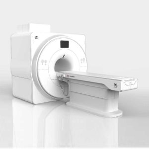
| SuperMark 1.5T is a new generation superconducting MRI system based on years of experience in production and research. It's applicable to whole body scan, such as, nervous system, spine, joint soft tissue, pelvic and abdominal cavity, etc. SuperMark 1.5T provides not only conventional pulse sequences and clinical diagnosis functions, but also provides advanced functional applications, for instance, 3D angiography and water imaging. It adopts brand new ANKE APEX operating system which ensures easy operation and fast diagnosis.
Technical Advantages:
- Reliable short cavity superconducting magnet system with zero liquid helium
consumption
- New generation fully digitalized and extensible multichannel spectrometer
- Powerful high efficiency and high fidelity gradient system; Multi-channel PA RF
receiving coil with intelligent identification
- English operating system and high extensible computer system
- High resolution conventional clinical images; Practical advanced functional
imaging
Superconducting MRI System:
- Highly open and humanization design -> Streamlined design
- Rich sequences and technology satisfy clinical needs -> Efficient service
Low Investment:
- High cost performance superconducting MRI system
- Zero liquid helium consumption, low running and maintenance cost
- Core technology by independent R & D supports full upgrade
- Low electric consumption
- Compact magnet design, minimum installation space: 35 square meters
High Return:
- High resolution thin slice images improve diagnosis
- Short cavity magnet design makes patients comfortable
- Fast scan speed improves work efficiency
Technical Specifications:
| No. |
Technique Description |
Parameter |
| 1 |
Magnet System |
|
| 1.1 |
Magnet Type |
Permanent Magnet
Automatic constant temperature
system |
| 1.2 |
Field Strength |
0.51T |
| 1.3 |
Magnet Shape |
Dual-pillar shape |
| 1.4 |
Homogeneity(40cm,DSV,VRMS) |
≤1.6ppm |
| 1.5 |
Shim Method |
Active/Passive |
| 1.6 |
Magnet Vertical Gap (Cover) |
40cm |
| 1.7 |
Magnetic Pole Dia. (Exclude Cover) |
145cm |
| 1.8 |
Accessibility(Horizontal Opening Angle, |
280° |
| 1.9 |
5 Gauss fringe field |
X-axis:horizontal ≤2.5m
Y-axis:Vertical ≤2.5m
Z-axis:horizontal ≤2.5m |
| 2 |
Patient Couch and Communication |
|
| 2.1 |
Patient Couch Driven mode |
Motor-driven |
| 2.2 |
Max. Patient Weight |
≥200kg(440lbs) |
| 2.3 |
Patient Positioning Tools |
Laser Light Localizer for positioning of center slice Motor-driven transfer to center of imaging volume |
| 2.4 |
Position accuracy |
±1mm |
| 2.5 |
Emergency Call Key |
Yes |
| 2.6 |
Intercom System |
Yes |
| 3 |
Gradient System |
|
| 3.1 |
Gradient Field Strength(Single Axis) |
≥30mT/m |
| 3.2 |
Gradient Slew Rate (Single Axis) |
≥100mT/m/ms |
| 3.3 |
Rise Time |
≤0.3ms |
| 3.4 |
Gradient Cooling System ( Gradient coils
and Power electronics) |
Air Cooling |
| 4 |
RF System |
|
| 4.1 |
RF System Type |
Digital Transmit and
Receive signal |
| 4.2 |
Number of RF Channels |
4 |
| 4.3 |
Transmitter Amplifier Peak Power |
6kW |
| 4.4 |
RF Bandwidth of Receiver |
≥1.25MHz |
| 4.5 |
Head Coil |
Yes |
| 4.6 |
Neck Coil |
Yes |
| 4.7 |
Body/Spine Coil (17 inch) |
Yes |
| 4.8 |
Body/Spine Coil (21 inch) |
Yes |
| 4.9 |
Knee Coil |
Yes |
| 4.10 |
Shoulder Coil |
Yes |
| 4.11 |
Flexible Coil |
Optional |
| 4.12 |
Breast Coil |
Optional |
| 5 |
Computer System |
|
| 5.1 |
Host Computer |
DELL Computer (for MR) |
| 5.2 |
System Software |
Windows XP |
| 5.3 |
Operation Software |
APEX |
| 5.4 |
CPU Clock rate |
3.0GHz |
| 5.5 |
Main Memory |
4GB |
| 5.6 |
Color LCD Monitor |
19” |
| 5.7 |
Keyboard and Mouse |
Standard |
| 5.8 |
Image Reconstruction Speed(256 x 256
Matrix) |
200 frame/Sec. |
| 5.9 |
Hard Disk |
500GB |
| 5.10 |
Image Storage Capacity(256 x 256
Matrix) |
500,000 |
| 5.11 |
Media Driver |
DVD RW |
| 5.12 |
DICOM 3.0 |
Yes |
| 5.13 |
Ethernet |
Yes |
| 5.14 |
Operation Console |
Yes |
| 5.15 |
Operation Chair |
Yes |
| 6 |
Scanning Parameter |
|
| 6.1 |
Max. FOV |
410mm |
| 6.2 |
Min. FOV |
5mm |
| 6.3 |
Min. TE(SE) |
5ms |
| 6.4 |
Min. TR(SE) |
11ms |
| 6.5 |
Min. TE(GR) |
1ms |
| 6.6 |
Min. TR(GR) |
3ms |
| 6.7 |
Min. 2D Thickness |
1.0mm |
| 6.8 |
Min. 3D Thickness |
0.1mm |
| 6.9 |
Max. Image Matrix |
512x512 |
| 7 |
Scanning Sequence & Imaging Technique |
|
| 7.1 |
Spin Echo 2D/3D (SE 2D/3D) |
Yes |
| 7.2 |
DE/QE |
Yes |
| 7.3 |
Fast Spin Echo 2D/3D(FSE 2D/3D) |
Yes |
| 7.4 |
Single Shot FSE 2D/3D |
Yes |
| 7.5 |
Inversion Recovery(IR) |
Yes |
| 7.6 |
Fast Inversion Recovery(FIR) |
Yes |
| 7.7 |
Gradient Echo 2D/3D(GR 2D/3D) |
Yes |
| 7.8 |
Fast GR 2D/3D |
Yes |
| 7.9 |
SPGR |
Yes |
| 7.10 |
FLAIR |
Yes |
| 7.11 |
Fat Imaging |
Yes |
| 7.12 |
Fat Suppression imaging |
Yes |
| 7.13 |
Water-Fat Separation imaging |
Yes |
| 7.14 |
TOF MRA(2D/3D) |
Yes |
| 7.15 |
MRCP(2D/3D) |
Yes |
| 7.16 |
MRU (2D/3D) |
Yes |
| 7.17 |
MRM |
Yes |
| 7.18 |
Fast Hydrograph Imaging |
Yes |
| 7.19 |
Diffusion Weighted Imaging(DWI) |
Yes |
| 7.20 |
Max. b Value |
1000s/mm2 |
| 7.21 |
Breath Hold Technology |
Yes |
| 7.22 |
Magnetization Transfer Contrast(MTC) |
Yes |
| 7.23 |
Multi-slice and Angle-free Presaturation |
Yes |
| 7.24 |
Saturation Tracking |
Yes |
| 7.25 |
Maximum Intensity Projection(MIP) |
Yes |
| 7.26 |
Multi-Angle Projection(MAP) |
Yes |
| 7.27 |
3D Reconstruction |
Yes |
| 7.28 |
Multi-planar Reconstruction(MPR) |
Yes |
| 7.29 |
Multi-Artifacts Eliminating technology |
Yes |
| 7.30 |
Checking with Part Metal Implant |
Yes |
| 7.31 |
Online Image Filtration |
Yes |
| 7.32 |
Online Post Procession |
Yes |
| 7.33 |
3D Scout |
Yes |
| 7.34 |
Scanning Protocol Preset |
Yes |
| 7.35 |
Scanning Protocol Queue Waiting |
Yes |
| 7.36 |
Advanced Image Post Processing |
Yes |
| 7.37 |
Image Fusion Technology of Vascular |
Yes |
| 7.38 |
Image Fusion Technology of Spine |
Yes |
| DETAILS
SonoScape’s trolley colour Doppler system S11 redefines price and performance with practical design. The S11 will go beyond your expectations but not your budget. As an easy-to-use ultrasound system, the S11 is integrated with a new software platform, especially optimized for a smooth workflow and convenient operation. The system speeds up the exam process and makes file management easier.
SPECIFICATION
- 15-inch high definition LCD monitor with articulating arm
- Compact and agile trolley design
- 3 active transducer sockets available for a wide range of applications
- Duplex, Color Doppler, DPI, PW Doppler, tissue harmonic imaging, μ-scan speckle reduction imaging, compound imaging, trapezoidal imaging
- Customized settings based on your own working style
- Full patient database and image management solutions
| DETAILS
Super Wide-bandwidth Platform
Inheriting Wi-sono's ultra-wide system platform and with the advanced probe technology, high-resolution and deep penetration images are provided for precision medicine.
Spatial Compound Imaging
Spatial Compound Imaging utilizes several lines of sight for optimal contrast resolution, speckle reduction and border detection, with which P15 is ideal for superficial and abdominal imaging with better clarity and improved continuity of structures.
μ-Scan+
The new generation μ-Scan imaging technology gives you better image quality by reducing noise, improving signal strength and improving visualization.
Dynamic Color
Dynamic color improves upon already existing color Doppler technologies for a clearer capture of color flow and detailed visualization of even tiny veins with lower velocities.
Real-time Panoramic
With real-time panoramic, you can acquire an extended field of view for large organs or long vessels for easy measurement and diagnostic efficiency. Accomplished in real-time for the convenience of the sonographers, any mistake can also be easily back tracked and corrected without interrupting the scan.
3D/4D
Outstanding volume performance with speed and convenience makes P15 outshine others on volume imaging.
Tissue Doppler Imaging
Tissue Doppler Imaging allows clinical doctors to quantitatively evaluate local myocardial movements and functions, facilitating them with the ability to analyze and compare the motions of the different parts of the patient's heart.
Auto IMT
Quick measurement of intra-media vessel thickness ensures good reproducibility and high diagnostic efficiency.
| Double x-ray film viewer, Compact, Solid with Backlight of LED’s based panel, Long Life Approximate LED’s life 50, 000 Hrs., Uniform Light at the total surface area, No Heat Emission, Wall Mounted, Can be used for tracing on X-Ray, Auto-sensor, Screen. Size: 430 mm x 710 mm. | The ANATOM 64 CT scanner is the latest innovation for cardiac imaging based on Precision Platform system. The excellent design of Ahart technology which innovatively combined single spiral scan + gated imaging + mA modulation for easy heart imaging at extremely low radiation dose. We provide you ANATOM 64 Clarity/Precision of two models which are low/high configurations for preferences. It also offers you conventional clinical applications of low dose, better image quality and faster exams.
Features:
- Modularized OptiWave HD detector features low-cost & easy maintenance, high spatial resolution and long lifetime
- Admir3D iterative technology delivers optimal dose efficiency and noise reduction without compromising image quality
- High configurations of main components ensure the best results and maximum patient throughput
- Uniquely and creatively uses 140kV and 80kV dual energy scan mode for brain imaging on 16-slice CT to offers you extraordinary image quality both in low and high density resolutions
- AdoseTM mA modulation ensures you low dose imaging without compromising image quality particularly useful in pediatric applications
- Equipped with dedicated Abast and Amast for bone and metal artifacts
- The brilliant Ahart technology enables you to experience so easy and low-dose cardiac imaging applications
Technical Specifications:
| Model |
ANATOM 64 Precision |
ANATOM 64 Fit |
| Rack type |
Low pressure slip ring |
Low pressure slip ring |
| Scan aperture |
70cm |
70cm |
| Rack Physical inclination |
± 30 ° |
N.A |
| Rack digital inclination |
± 50 ° |
± 50 ° |
| cooling method |
Air-cooled |
Air-cooled |
| Focus to the center distance |
56 cm |
53 cm |
| |
|
|
| Maximum power (non-equivalent) |
80kW |
42kW |
| Votage (kV) |
80kV / 100kV / 120kV / 140kV |
70kV / 80kV / 100kV / 120kV /
140kV |
| |
|
|
| Heat capacity |
8MHU |
5.0MHU |
| Heat dissipation rate |
931 kHU / min |
748kHU / min |
| cooling method |
Oil cool |
Oil cool |
| Large focus size |
1.1mm × 1.2mm |
1.2mm × 1.4mm |
| Small focus size |
0.6mm × 1.2mm |
0.7mm × 0.8mm |
| Tube current range |
10-670mA |
10-350mA |
| |
|
|
| Detector type |
Optiwave detectors |
Optiwave detectors |
| Number of Z-axis |
32 |
32 |
| The width of the Z-axis |
20mm |
20mm |
| The number of elements per row |
912 |
848 |
| Total number of detectors |
29184 |
27136 |
| Acquisition mode |
64x0.625, 32x0.625,
16x0.625 |
64x0.625, 32x0.625,
16x0.625 |
| |
|
|
| Scanning range |
1800mm |
1800mm |
| Horizontal positioning accuracy |
± 0.25mm |
± 0.25mm |
| weight capacity |
205kg |
205kg |
| Minimum height |
43cm |
43cm |
| Anti - collision protection device |
Yes |
Yes |
| Foot control switch |
Yes |
Yes |
| IV rack |
Yes |
Yes |
| |
|
|
| CPU |
3.5GHz |
3.5GHz |
| RAM |
16 GB × 4 |
16 GB × 4 |
| Hard drive capacity |
1T × 2 |
1T × 2 |
| Display size |
24 inch LCD monitor |
24 inch LCD monitor |
| Display resolution |
1920 × 1200 |
1920 × 1200 |
| Computer operating system |
Windows 7 |
Windows 7 |
| Image reconstruction speed |
65 frames/ second |
65 frames/ second |
| Number of image store |
1000000 |
1000000 |
| Data external storage mode |
CD / DVD / USB |
CD / DVD / USB |
| |
|
|
| Minimum Scan Time of 360 degree |
0.39sec |
0.75sec |
| Sub-millimeter acquisition layers |
64 |
64 |
| Double sub-millimeter acquisition
layers |
64 |
64 |
| Thinnest acquisition thickness |
0.625mm |
0.625mm |
| The thinnest reconstruction
thickness |
0.3125mm |
0.625mm |
| Conventional reconstruction
thickness (mm) |
0.3125 mm, 0.625 mm, 1.25 mm, 2.5
mm, 5.0 mm, 7.5 mm, 10 mm |
0.625 mm, 1.25 mm, 2.5 mm, 5.0
mm, 7.5 mm, 10 mm |
| The reconstruction matrix |
512 x 512, 1024 x 1024 |
512 x 512, 1024 x 1024 |
| Display matrix |
1024 × 1024 |
1024 × 1024 |
| Max FOV |
52cm |
50cm |
| The maximum display field of view |
70cm |
50cm |
| Maximum scan length |
1800mm |
1800mm |
| Maximum continuous helix scan
time |
120s |
120s |
| Pitch range |
0.5-1.5 |
0.5, 1.0, 1.5 |
| |
|
|
| High contrast resolution |
21 Lp / cm @ 0% MTF |
21 Lp / cm @ 0% MTF |
| Low contrast resolution |
2mm @ 0.3% |
2mm @ 0.3% |
| Image noise |
≤ 0.25 |
≤ 0.29 |
| |
|
|
| MPR |
Yes |
Yes |
| CPR |
Yes |
Yes |
| SSD |
Yes |
Yes |
| VR |
Yes |
Yes |
| MIP |
Yes |
Yes |
| MinIP |
Yes |
Yes |
| Virtual endoscopy |
Yes |
Yes |
| CT angiography |
Yes |
Yes |
| Tissue segmentation |
Yes |
Yes |
| One-key bone removal |
Yes |
Yes |
| Automatically patient table removal |
Yes |
Yes |
| Contrast Agent Automatic Tracking
Technology- bolus tracking |
Yes |
Yes |
| Automatic linkage trigger
technology |
Yes |
Yes |
| Cine mode display |
Yes |
Yes |
| Bone artifact suppression technique |
AbastTM |
AbastTM |
| Metal artifact suppression technique |
AbastTM |
AbastTM |
| Iterative reconstruction technique |
Admir3D global iteration |
Admir3D full-domain iteration |
| Low - dose children 's scanning
technology |
Yes |
Yes |
| Low - dose lung scan |
Yes |
Yes |
| Gray matter enhancement
technology |
AccuHead |
AccuHead |
| High resolution imaging of the lung |
AccuLung |
AccuLung |
| Inner ear high resolution imaging |
AccuOtica |
AccuOtica |
| Body high resolution imaging |
AccuBody |
AccuBody |
| Bone high resolution imaging |
AccuBone |
AccuBone |
| Head dual-energy imaging |
Ahead |
Ahead |
| CT perfusion imaging |
Optional |
Optional |
| Quantitative analysis of blood
vessels |
Optional |
Optional |
| Heart coronary artery imaging |
Aheart |
N.A |
| ECG gated |
Yes |
N.A |
| Low dose cardiac scan |
Yes |
N.A |
| |
|
|
| Green energy saving technology |
AccuSaving |
AccuSaving |
| Dual-energy scan technology |
Optional |
Optional |
|

Reviews
There are no reviews yet.