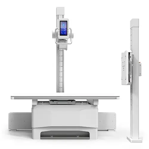
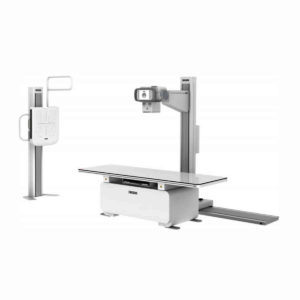
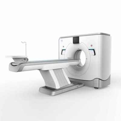
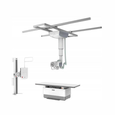
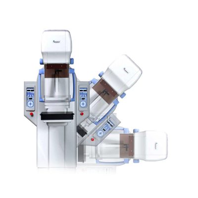
| Description | Built as a floor-mounted digital X-ray machine, it features a coordinated X-ray tube and flat panel detector that maintain precise alignment throughout imaging.
Shipped From China
Delivery & Availability:
Typically 10-21 working days – excluding furniture and heavy/bulky equipment. Please contact us for further information.
| In Stock
The GXR-SD is a diagnostic digital radiography system that provides reliable high quality digital radiographic images with a reduced dose. The GXR-SD DR systems offer comprehensive digital solutions to all radiography needs, featuring ACQUIDR digital imaging system with stationary or portable digital flat-panel detectors as well as reliable high-frequency x-ray generators that are known worldwide for their excellent performance, lifetime and stability. Patient tables and wall stands are also offered.
Delivery & Availability:
Typically 21 working days – excluding furniture and heavy/bulky equipment. Please contact us for further information. | Shipped from Abroad
SonoScape has developed a new probe and function for the E1 Exp. With these additions the E1 Exp will bring users a more efficient examination experience with satisfying image quality and a smooth workflow.
Delivery & Availability:
Typically 5-7 working days – excluding furniture and heavy/bulky equipment. Please contact us for further information. | In Stock
GXR Analogue X-ray system matches with a radiographic room which perfectly fits your workow and can be easily upgraded to DR system with the help of DR interface and PC interface in GXR generator as well as Bucky suitable to Flat Panel Detector. GXR X-ray system is equipped with a high frequency X-ray generator which consistently produces high quality radiograph in favor of high quality X-ray output with a very small kV ripple and accurate mA and mAs. GXR X-ray system is designed to provide convenience to operator and comfort to patient.
Delivery & Availability:
Typically 21 working days – excluding furniture and heavy/bulky equipment. Please contact us for further information. | Shipped from Abroad
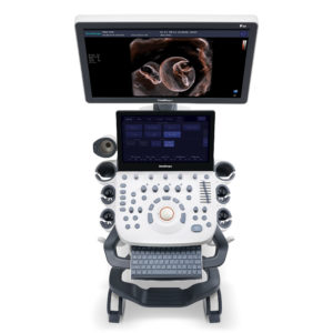
Incorporating innovative technologies, P20’s user-friendly design with a simple operation panel, intuitive user interface and a variety of intelligent auxiliary scanning tools, will significantly improve your daily examination experience. Besides general imaging applications, P20 has entitled with diagnostic 4D technology which has an extraordinary performance in obstetrics and gynecology applications.
Delivery & Availability:
Typically 5-7 working days – excluding furniture and heavy/bulky equipment. Please contact us for further information. | In Stock
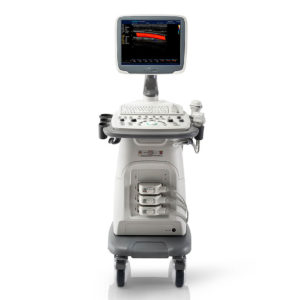
A Value Choice beyond Your Expectation. SonoScape’s trolley color Doppler system S11 redefines price and performance with practical design. The S11 will go beyond your expectations but not your budget.
Delivery & Availability:
Typically 2 working days – excluding furniture and heavy/bulky equipment. Please contact us for further information. |
| Content | Built as a floor-mounted digital X-ray machine, it features a coordinated X-ray tube and flat panel detector that maintain precise alignment throughout imaging. Optional AEC and DAP functions can be added to strengthen exposure control and ensure consistent diagnostic quality. Facilities needing faster patient positioning can upgrade the radiographic table to a six-way floating tabletop, improving comfort and overall workflow.
The machine integrates a floor-mounted table with a wall-mounted chest stand, creating a clean and efficient room layout. Its smooth mechanical movement and intuitive operating controls give radiographers greater flexibility when adjusting patient positions and selecting projection angles.
As a versatile high-end DR platform, the SHO-DDX01 supports a wide range of routine clinical examinations, including head, chest, abdominal, lumbar spine, and extremity imaging. It accommodates PA, lateral, oblique, and angled views, making it suitable for hospitals and clinics of all sizes.
Features
- The X-ray tube operates at a high rotational speed and is supported by an efficient heat-dissipation system, allowing the equipment to perform reliably during long clinical sessions while maintaining an extended service life.
- The high-frequency generator provides strong output performance with stable, high-quality X-ray emission, ensuring dependable imaging results across routine and demanding examinations.
- A smart high-voltage control system incorporates multiple built-in protection functions to maintain safe and stable operation throughout daily clinical use.
- The advanced a-Si CsI flat panel detector delivers high conversion efficiency, enabling clear, detailed digital images with reduced radiation dose.
- A robust digital imaging platform offers comprehensive post-processing capabilities, giving clinicians the tools needed to support a wide variety of diagnostic requirements.
Specifications
| Ambient Conditions |
| Ambient temperature |
10℃–40℃ |
| Relative humidity |
30%–75% |
| Atmospheric pressure |
70–106 kPa |
| Main head |
10.4-inch touchscreen control panel |
| X-Ray Generator |
| Model |
SHO-DDX01 Series |
| Output power (kW) |
40 |
50 |
65 |
| Current (mA) |
10-500 |
10-630 |
10-800 |
| Voltage (kV) |
40-125 |
40-150 |
40-150 |
| mAs |
0.1-400 |
0.1-630 |
0.1-630 |
| Exposure time |
0.002~6.3S |
| APR program |
Equipped |
| Intelligent protection system |
Includes a self-protection system with automatic alarms and fault code diagnostics. |
| X-ray Tube |
| Output power (kW) |
40 |
50 |
65 |
| Anode type |
Rotating anode |
| Revolving speed of anode (rpm) |
50Hz: 2800 60Hz: 3000 |
| Focus size |
1.0/2.0 |
0.6/1.2 |
0.6/1.2 |
| Anode heat storage capacity (kHU) |
150 |
300 |
400 |
| Maximum Anode Heat Dissipation (W) |
475 |
750 |
1000 |
| Housing heat storage capacity (kJ) |
900 |
900 |
1000 |
| X-ray tube Rotation angel (°) |
±180 |
±180 |
±180 |
| X-ray Detector (Wired Flat Panel Detector) |
| Type |
Amorphous Silicon |
| Scintillator |
Cesium Iodide |
| Active area |
17x17 Inch (43 cm*43 cm) |
| Active pixel |
3072*3072 |
| Pixel pitch |
139 um |
| A/D Conversion |
16 bits |
| DQE |
≧70% |
| Spatial resolution |
36 Lp/cm |
| Acquisition time |
≤2 s |
| Collimator |
| Type |
Manual collimator |
| Max. Window |
440 mm × 440 mm (SID = 100 cm) |
| Lamp |
AC/DC 24 V, 5 W |
| Lamp timer |
Automatic illumination with timer for lamp (30S) |
| Inherent filtration |
1.0 mmAl |
| Illuminant |
LED |
| Grid |
| Densities |
40 lp/cm |
| Ratios |
10:1 |
| Size |
18" × 18" |
| Focal distance |
100 cm (Radiography table), 180 cm (Bucky stand) |
| Photography Table |
| Type |
Radiography table |
| Tabletop size |
2300 mm × 800 mm x 60 mm |
| Load-bearing |
300 kg |
| Floating front and back |
740 mm ±3% |
| Floating left and right |
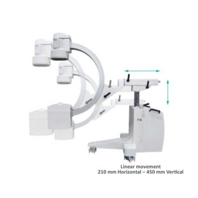
210 mm ±3% |
| FPD tray movement range |
650 mm ±3% |
| Column stand movement |
| Tube stand |
| Up-down movement (to photography table) |
1500 mm ±3% |
| Stand left-right movement |
1900 mm ±3% |
| Rotational angles of stand |
+180°~-180° |
| Bucky stand |
| Detector vertical movement range (distance between detector center and ground) |
1500 mm ±3% |
| The lowest point of the detector center relative to the ground |
≤ 46 cm |
| Automatic tracking function |
Yes, electric correspondence between the X-ray tube and flat panel detector |
| Workstation |
| Monitor |
23" LCD Dell brand |
| CPU |
≥ 2.8 GHz |
| Memory |
≥ 4 GB |
| HDD |
≥ 1 TB |
| DICOM3.0 |
Query for integration with any PACS |
| Functions |
Import/export function |
| Image info |
| Management of patient info |
| Post processing |
| Measurement etc. |
| Console |
| Control room console |
10.4 inch LCD color touch screen console for exposure parameter setting and exposure |
| Controller |
Wired controller for electric movement |
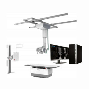
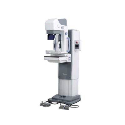
| DrGem Ceiling Mounted Digital X-ray is a diagnostic digital radiography system that provides reliable high quality digital radiographic images with a reduced dose. The GXR-SD DR systems offer comprehensive digital solutions to all radiography needs, featuring ACQUIDR digital imaging system with stationary or portable digital flat-panel detectors as well as reliable high-frequency x-ray generators that are known worldwide for their excellent performance, lifetime and stability. Patient tables and wall stands are also offered.
Features:
- TS-CSA-A (Vertical movement, 1.6m stroke, rail length 3x4meter) including HV cable 15m
- WBS-TA: Vertical movement
- V Stroke:1,450mm in Uprigh Bucky Position,
- 1,526mm in Horizontal Bucky position.
- PBT-4 is a 4 way Floating Tabletop. A large tabletop with extended travel enables all radiography studies with minimal patient movement. Fully fat tabletop without a frame on the edge makes cleanliness and odors free
- Digital Flat Panel Detector (FPD) – Wireless 17X14 (Csl, 4336W) with Auto Exposure Detection (AED) function, there is no DR trigger cable between detector and generator.
- Full Featured Imaging Software & Excellent Digital Image Processing:
- Provides convenient user interface and easy operation
- Anatomical view-based digital image processing automatically optimizes and enhances the quality of the captured image for the pictured anatomy.
- Radiographic stand & automatic collimator control function
- DICOM 3.0 networking interface includes Worklist, Print, Store, Query for integration with any PACS or RIS
- Included – Software, HP Laptop Computer
- CPU≥3.2GHz
- Memory capacity:≥4GB
- Hard drive capacity :≥500 GB
- Resolution: 1280 x 1024
- Display size: 21 inch color LCD screen
- 64 bit Windows 10 operation system
- Core: i5
Technical Specification:
- Power Rating - 32KW
- Generator - GXR-32S
- Rotor - Dual Speed Starter(DSS)
- Input Power - 400/480VAC, Three phase
- Line Frequency - 50/60Hz
- X-ray tube - DXT-12M, (0.6/1.2mm, 300kHU)
- Tube Voltage - 40 to 150kV, 1kV Step
- Tube Current – 10 to 640mA
- Output - 640mA@81kV, 500mA@104kV, 400mA@130kV, 320mA@150kV
- Time Range - 1ms to 10s
- mAs Range - 0.1 to 800mAs
- Reproducibility - Coecient of Variation : kV < 0.005, Time < 0.005,mAs < 0.01
- Accuracy - kV < ±(1%+1kV), mA < ±(3%+1mA), Time <±(1%+0.5ms), mAs < ±(3%+0.1mAs)
- Linearity - Coecient of Linearity < 0.01 : CL = (X1-X2)/(X1+X2), where X is mR/mAs
- Mechanical Parts:
-TS-CSA-A (Vertical movement, 1.6m, stroke rail length 3x4meter) including HV cable 15m
- PBT-4: 4 way Floating Tabletop.
- WBS-TA: a. Vertical movement
- V Stroke:1,450mm in Upright Bucky
- Position, 1,526mm in Horizontal Bucky position.
- HVC-15: 15M HV cable
- Auto Collimator
| DETAILS
Efficient Diagnosis
μ-Scan, Speckle Reduction & Edge Enhancement
Spatial Compound Imaging
PIH - Pure Inversion Harmonic
Wide Scan - Enlarged Image Area
Tissue-Specific Imaging
SR Flow
Ergonomic Designs
Up to 2 Transducer Ports
Light Weight and Compact
15.6 inch Anti-flickering HD LED Screen
Tilting Monitor Angle Adjustment
Backlit Keyboard and Intelligent Panel
Long-lasting Battery for 90 mins
Ease of Use
Quick Boot Up
Auto-Brightness Adjustment
Auto Image Optimization
Auto IMT
Auto Trace
Equipped Accessories
Wi-Fi and Bluetooth Available
DICOM
500GB Hard Disk
Height Adjustable Trolley
Durable, Carry-on Site Suitcase
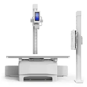
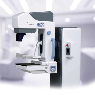
| DrGem GXR Floor Mounted Analogue X-ray system matches with a radiographic room which perfectly fits your workflow and can be easily upgraded to DR system with the help of DR interface and PC interface in GXR generator as well as Bucky suitable to Flat Panel Detector. GXR (Analogue X-ray)system is equipped with a high frequency X-ray generator which consistently produces high quality radiograph in favor of high quality X-ray output with a very small kV ripple and accurate mA and mAs. GXR (Analogue X-ray) system is designed to provide convenience to operator and comfort to patient.
Features of DrGem GXR Floor Mounted Analogue X-ray:
- 4 way Tabletop Patient Table (PBT-4)
A large tabletop with extended travel enables all radiography studies with minimal patient movement, and supporting patient weight up to 300kg. Fully at tabletop without a frame on the edge makes cleanliness and odors free
- Floor Mount Tube Stand (TS-FM6)
Floor Rail type tube stand provides all radiography studies with smooth movement on the rail.
- Wall Bucky Stand (WBS)
Elegant design, durable and easy-to-use Wall Bucky Stand provides full satisfaction.
Technical Specifications of DrGem GXR Floor Mounted Analogue X-ray:
- Power Rating - 32KW
- Generator - GXR-32S
- Rotor - Dual Speed Starter(DSS)
- Input Power - 400/480VAC, Three phase
- Line Frequency - 50/60Hz
- X-ray tube - DXT-12M, (0.6/1.2mm, 300kHU)
- Tube Voltage - 40 to 150kV, 1kV Step
- Tube Current – 10 to 640mA
- Output - 640mA@81kV, 500mA@104kV, 400mA@130kV, 320mA@150kV
- Time Range - 1ms to 10s
- mAs Range - 0.1 to 800mAs
- Reproducibility - Coecient of Variation : kV < 0.005, Time < 0.005,mAs < 0.01
- Accuracy - kV < ±(1%+1kV), mA < ±(3%+1mA), Time <±(1%+0.5ms), mAs < ±(3%+0.1mAs)
- Linearity - Coecient of Linearity < 0.01 : CL = (X1-X2)/(X1+X2), where X is mR/mAs
| DETAILS
Upgraded Images with More Clarity
SonoScape never stops making progress in improving the image quality of its ultrasound products to enhance the confidence of diagnosis for doctors. With extraordinary images provided by P20, the anatomy structures are clearer than ever.
C-Xlasto Imaging
With C-xlasto Imaging, P20 enables comprehensive quantitative elastic analysis. Meanwhile, C-xlasto on P20 is supported by linear, convex and transvaginal probes, to ensure good reproducibility and highly consistent quantitative elastic results.
S-Live
S-Live allows for detailed visualization of subtle anatomical features, thereby enabling intuitive diagnosis with real-time 3D images and enriching patient communication.
Pelvic Floor 4D
Transperineal 4D pelvic floor ultrasound can provide useful clinical values in assessing the vaginal delivery impact on the female anterior compartment, judging whether the pelvic organs are prolapsed or not and the extent, determining if the pelvic muscles were torn accurately.
Anatomic M Mode
Anatomic M Mode helps you observe the myocardial motion at different phases by freely placing sample lines. It accurately measures the myocardial thickness and the heart size of even difficult patients and supports the myocardial function and LV wall-motion assessment.
Tissue Doppler Imaging
P20 is endowed with Tissue Doppler Imaging which provides velocities and other clinical information on myocardial functions, facilitating clinical doctors with the ability to analyze and compare the motions of different parts of the patient's heart.
| DETAILS
SonoScape’s trolley colour Doppler system S11 redefines price and performance with practical design. The S11 will go beyond your expectations but not your budget. As an easy-to-use ultrasound system, the S11 is integrated with a new software platform, especially optimized for a smooth workflow and convenient operation. The system speeds up the exam process and makes file management easier.
SPECIFICATION
- 15-inch high definition LCD monitor with articulating arm
- Compact and agile trolley design
- 3 active transducer sockets available for a wide range of applications
- Duplex, Color Doppler, DPI, PW Doppler, tissue harmonic imaging, μ-scan speckle reduction imaging, compound imaging, trapezoidal imaging
- Customized settings based on your own working style
- Full patient database and image management solutions
|

Reviews
There are no reviews yet.