Anke Supermark 1.5T MRI Machine
$0.00
Shipped from Abroad
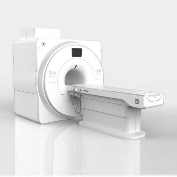
SuperMark 1.5T is a new generation superconducting MRI system based on years of experience in production and research. It’s applicable to whole body scan, such as, nervous system, spine, joint soft tissue, pelvic and abdominal cavity, etc
Delivery & Availability:
Typically 90 working days – excluding furniture and heavy/bulky equipment. Please contact us for further information.
Description
SuperMark 1.5T is a new generation superconducting MRI system based on years of experience in production and research. It’s applicable to whole body scan, such as, nervous system, spine, joint soft tissue, pelvic and abdominal cavity, etc. SuperMark 1.5T provides not only conventional pulse sequences and clinical diagnosis functions, but also provides advanced functional applications, for instance, 3D angiography and water imaging. It adopts brand new ANKE APEX operating system which ensures easy operation and fast diagnosis.
Technical Advantages:
- Reliable short cavity superconducting magnet system with zero liquid helium
consumption - New generation fully digitalized and extensible multichannel spectrometer
- Powerful high efficiency and high fidelity gradient system; Multi-channel PA RF
receiving coil with intelligent identification - English operating system and high extensible computer system
- High resolution conventional clinical images; Practical advanced functional
imaging
Superconducting MRI System:
- Highly open and humanization design -> Streamlined design
- Rich sequences and technology satisfy clinical needs -> Efficient service
Low Investment:
- High cost performance superconducting MRI system
- Zero liquid helium consumption, low running and maintenance cost
- Core technology by independent R & D supports full upgrade
- Low electric consumption
- Compact magnet design, minimum installation space: 35 square meters
High Return:
- High resolution thin slice images improve diagnosis
- Short cavity magnet design makes patients comfortable
- Fast scan speed improves work efficiency
Technical Specifications:
| No. | Technique Description | Parameter |
| 1 | Magnet System | |
| 1.1 | Magnet Type | Permanent Magnet
Automatic constant temperature system |
| 1.2 | Field Strength | 0.51T |
| 1.3 | Magnet Shape | Dual-pillar shape |
| 1.4 | Homogeneity(40cm,DSV,VRMS) | ≤1.6ppm |
| 1.5 | Shim Method | Active/Passive |
| 1.6 | Magnet Vertical Gap (Cover) | 40cm |
| 1.7 | Magnetic Pole Dia. (Exclude Cover) | 145cm |
| 1.8 | Accessibility(Horizontal Opening Angle, | 280° |
| 1.9 | 5 Gauss fringe field | X-axis:horizontal ≤2.5m
Y-axis:Vertical ≤2.5m Z-axis:horizontal ≤2.5m |
| 2 | Patient Couch and Communication | |
| 2.1 | Patient Couch Driven mode | Motor-driven |
| 2.2 | Max. Patient Weight | ≥200kg(440lbs) |
| 2.3 | Patient Positioning Tools | Laser Light Localizer for positioning of center slice Motor-driven transfer to center of imaging volume |
| 2.4 | Position accuracy | ±1mm |
| 2.5 | Emergency Call Key | Yes |
| 2.6 | Intercom System | Yes |
| 3 | Gradient System | |
| 3.1 | Gradient Field Strength(Single Axis) | ≥30mT/m |
| 3.2 | Gradient Slew Rate (Single Axis) | ≥100mT/m/ms |
| 3.3 | Rise Time | ≤0.3ms |
| 3.4 | Gradient Cooling System ( Gradient coils
and Power electronics) |
Air Cooling |
| 4 | RF System | |
| 4.1 | RF System Type | Digital Transmit and
Receive signal |
| 4.2 | Number of RF Channels | 4 |
| 4.3 | Transmitter Amplifier Peak Power | 6kW |
| 4.4 | RF Bandwidth of Receiver | ≥1.25MHz |
| 4.5 | Head Coil | Yes |
| 4.6 | Neck Coil | Yes |
| 4.7 | Body/Spine Coil (17 inch) | Yes |
| 4.8 | Body/Spine Coil (21 inch) | Yes |
| 4.9 | Knee Coil | Yes |
| 4.10 | Shoulder Coil | Yes |
| 4.11 | Flexible Coil | Optional |
| 4.12 | Breast Coil | Optional |
| 5 | Computer System | |
| 5.1 | Host Computer | DELL Computer (for MR) |
| 5.2 | System Software | Windows XP |
| 5.3 | Operation Software | APEX |
| 5.4 | CPU Clock rate | 3.0GHz |
| 5.5 | Main Memory | 4GB |
| 5.6 | Color LCD Monitor | 19” |
| 5.7 | Keyboard and Mouse | Standard |
| 5.8 | Image Reconstruction Speed(256 x 256
Matrix) |
200 frame/Sec. |
| 5.9 | Hard Disk | 500GB |
| 5.10 | Image Storage Capacity(256 x 256
Matrix) |
500,000 |
| 5.11 | Media Driver | DVD RW |
| 5.12 | DICOM 3.0 | Yes |
| 5.13 | Ethernet | Yes |
| 5.14 | Operation Console | Yes |
| 5.15 | Operation Chair | Yes |
| 6 | Scanning Parameter | |
| 6.1 | Max. FOV | 410mm |
| 6.2 | Min. FOV | 5mm |
| 6.3 | Min. TE(SE) | 5ms |
| 6.4 | Min. TR(SE) | 11ms |
| 6.5 | Min. TE(GR) | 1ms |
| 6.6 | Min. TR(GR) | 3ms |
| 6.7 | Min. 2D Thickness | 1.0mm |
| 6.8 | Min. 3D Thickness | 0.1mm |
| 6.9 | Max. Image Matrix | 512×512 |
| 7 | Scanning Sequence & Imaging Technique | |
| 7.1 | Spin Echo 2D/3D (SE 2D/3D) | Yes |
| 7.2 | DE/QE | Yes |
| 7.3 | Fast Spin Echo 2D/3D(FSE 2D/3D) | Yes |
| 7.4 | Single Shot FSE 2D/3D | Yes |
| 7.5 | Inversion Recovery(IR) | Yes |
| 7.6 | Fast Inversion Recovery(FIR) | Yes |
| 7.7 | Gradient Echo 2D/3D(GR 2D/3D) | Yes |
| 7.8 | Fast GR 2D/3D | Yes |
| 7.9 | SPGR | Yes |
| 7.10 | FLAIR | Yes |
| 7.11 | Fat Imaging | Yes |
| 7.12 | Fat Suppression imaging | Yes |
| 7.13 | Water-Fat Separation imaging | Yes |
| 7.14 | TOF MRA(2D/3D) | Yes |
| 7.15 | MRCP(2D/3D) | Yes |
| 7.16 | MRU (2D/3D) | Yes |
| 7.17 | MRM | Yes |
| 7.18 | Fast Hydrograph Imaging | Yes |
| 7.19 | Diffusion Weighted Imaging(DWI) | Yes |
| 7.20 | Max. b Value | 1000s/mm2 |
| 7.21 | Breath Hold Technology | Yes |
| 7.22 | Magnetization Transfer Contrast(MTC) | Yes |
| 7.23 | Multi-slice and Angle-free Presaturation | Yes |
| 7.24 | Saturation Tracking | Yes |
| 7.25 | Maximum Intensity Projection(MIP) | Yes |
| 7.26 | Multi-Angle Projection(MAP) | Yes |
| 7.27 | 3D Reconstruction | Yes |
| 7.28 | Multi-planar Reconstruction(MPR) | Yes |
| 7.29 | Multi-Artifacts Eliminating technology | Yes |
| 7.30 | Checking with Part Metal Implant | Yes |
| 7.31 | Online Image Filtration | Yes |
| 7.32 | Online Post Procession | Yes |
| 7.33 | 3D Scout | Yes |
| 7.34 | Scanning Protocol Preset | Yes |
| 7.35 | Scanning Protocol Queue Waiting | Yes |
| 7.36 | Advanced Image Post Processing | Yes |
| 7.37 | Image Fusion Technology of Vascular | Yes |
| 7.38 | Image Fusion Technology of Spine | Yes |
Click Here To Download Catalogue
Additional information
| Model | Advanced, Advanced Plus, Basic, Smart |
|---|
Quick Comparison
| Anke Supermark 1.5T MRI Machine remove | DrGem Diamond All-In-One Digital X-ray Machine remove | Jade Mobile X-ray machine (Analogue) remove | ASPEL AsPEKT 712 Holter Monitor and Software remove | Sonoscape P50 Ultrasound Machine remove | Sonoscape P10 Ultrasound Machine remove | ||||||||||||||||||||||||||||||||||||||||||||||||||||||||||||||||||||||||||||||||||||||||||||||||||||||||||||||||||||||||||||||||||||||||||||||||||||||||||||||||||||||||||||||||||||||||||||||||||||||||||||||||||||||||||||||||||||||||||||||||||||||||||||||||||||||||||||||||||||||||||||||||||||||||||||||||
|---|---|---|---|---|---|---|---|---|---|---|---|---|---|---|---|---|---|---|---|---|---|---|---|---|---|---|---|---|---|---|---|---|---|---|---|---|---|---|---|---|---|---|---|---|---|---|---|---|---|---|---|---|---|---|---|---|---|---|---|---|---|---|---|---|---|---|---|---|---|---|---|---|---|---|---|---|---|---|---|---|---|---|---|---|---|---|---|---|---|---|---|---|---|---|---|---|---|---|---|---|---|---|---|---|---|---|---|---|---|---|---|---|---|---|---|---|---|---|---|---|---|---|---|---|---|---|---|---|---|---|---|---|---|---|---|---|---|---|---|---|---|---|---|---|---|---|---|---|---|---|---|---|---|---|---|---|---|---|---|---|---|---|---|---|---|---|---|---|---|---|---|---|---|---|---|---|---|---|---|---|---|---|---|---|---|---|---|---|---|---|---|---|---|---|---|---|---|---|---|---|---|---|---|---|---|---|---|---|---|---|---|---|---|---|---|---|---|---|---|---|---|---|---|---|---|---|---|---|---|---|---|---|---|---|---|---|---|---|---|---|---|---|---|---|---|---|---|---|---|---|---|---|---|---|---|---|---|---|---|---|---|---|---|---|---|---|---|---|---|---|---|---|---|---|---|---|---|---|---|---|---|---|---|---|---|---|---|---|---|---|---|---|---|---|---|---|---|---|---|---|---|---|---|---|---|---|---|---|---|
| Name | Anke Supermark 1.5T MRI Machine remove | DrGem Diamond All-In-One Digital X-ray Machine remove | Jade Mobile X-ray machine (Analogue) remove | ASPEL AsPEKT 712 Holter Monitor and Software remove | Sonoscape P50 Ultrasound Machine remove | Sonoscape P10 Ultrasound Machine remove | |||||||||||||||||||||||||||||||||||||||||||||||||||||||||||||||||||||||||||||||||||||||||||||||||||||||||||||||||||||||||||||||||||||||||||||||||||||||||||||||||||||||||||||||||||||||||||||||||||||||||||||||||||||||||||||||||||||||||||||||||||||||||||||||||||||||||||||||||||||||||||||||||||||||||||||||
| Image |  | 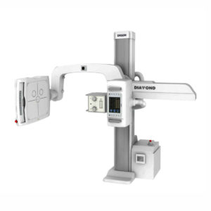 | 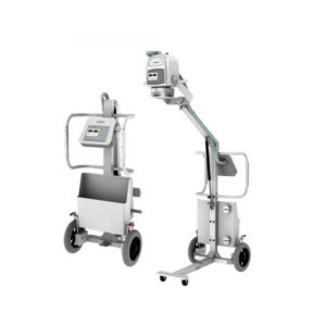 | 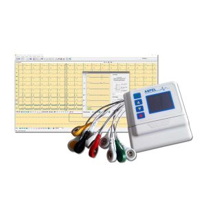 | 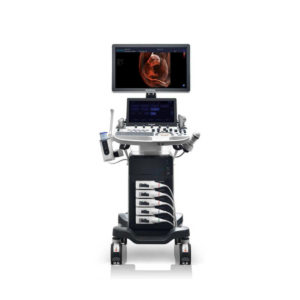 | 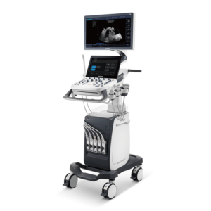 | |||||||||||||||||||||||||||||||||||||||||||||||||||||||||||||||||||||||||||||||||||||||||||||||||||||||||||||||||||||||||||||||||||||||||||||||||||||||||||||||||||||||||||||||||||||||||||||||||||||||||||||||||||||||||||||||||||||||||||||||||||||||||||||||||||||||||||||||||||||||||||||||||||||||||||||||
| SKU | SF1033560092-4 | SF1033560074-3 | SF1033560074-2 | SF1033560075-4 | SF1033560012-11 | SF1033560012-7 | |||||||||||||||||||||||||||||||||||||||||||||||||||||||||||||||||||||||||||||||||||||||||||||||||||||||||||||||||||||||||||||||||||||||||||||||||||||||||||||||||||||||||||||||||||||||||||||||||||||||||||||||||||||||||||||||||||||||||||||||||||||||||||||||||||||||||||||||||||||||||||||||||||||||||||||||
| Rating | |||||||||||||||||||||||||||||||||||||||||||||||||||||||||||||||||||||||||||||||||||||||||||||||||||||||||||||||||||||||||||||||||||||||||||||||||||||||||||||||||||||||||||||||||||||||||||||||||||||||||||||||||||||||||||||||||||||||||||||||||||||||||||||||||||||||||||||||||||||||||||||||||||||||||||||||||||||
| Price |
|
|
|
|
|
| |||||||||||||||||||||||||||||||||||||||||||||||||||||||||||||||||||||||||||||||||||||||||||||||||||||||||||||||||||||||||||||||||||||||||||||||||||||||||||||||||||||||||||||||||||||||||||||||||||||||||||||||||||||||||||||||||||||||||||||||||||||||||||||||||||||||||||||||||||||||||||||||||||||||||||||||
| Stock | |||||||||||||||||||||||||||||||||||||||||||||||||||||||||||||||||||||||||||||||||||||||||||||||||||||||||||||||||||||||||||||||||||||||||||||||||||||||||||||||||||||||||||||||||||||||||||||||||||||||||||||||||||||||||||||||||||||||||||||||||||||||||||||||||||||||||||||||||||||||||||||||||||||||||||||||||||||
| Availability | |||||||||||||||||||||||||||||||||||||||||||||||||||||||||||||||||||||||||||||||||||||||||||||||||||||||||||||||||||||||||||||||||||||||||||||||||||||||||||||||||||||||||||||||||||||||||||||||||||||||||||||||||||||||||||||||||||||||||||||||||||||||||||||||||||||||||||||||||||||||||||||||||||||||||||||||||||||
| Add to cart | |||||||||||||||||||||||||||||||||||||||||||||||||||||||||||||||||||||||||||||||||||||||||||||||||||||||||||||||||||||||||||||||||||||||||||||||||||||||||||||||||||||||||||||||||||||||||||||||||||||||||||||||||||||||||||||||||||||||||||||||||||||||||||||||||||||||||||||||||||||||||||||||||||||||||||||||||||||
| Description | Shipped from Abroad
SuperMark 1.5T is a new generation superconducting MRI system based on years of experience in production and research. It's applicable to whole body scan, such as, nervous system, spine, joint soft tissue, pelvic and abdominal cavity, etc
Delivery & Availability: Typically 90 working days – excluding furniture and heavy/bulky equipment. Please contact us for further information. | Shipped from Abroad DrGem Diamond All-In-One Digital X-ray Machine is a fully automatic digital radiography system providing state-of-the-art image quality, image processing and user interface. With a wide selection of anatomical studies on the imaging software, DIAMOND automatically sets up the x-ray generator’s preprogrammed exposure technique settings, motorized radiographic stand positioning, x-ray collimation and post-image processing for the selected study. Specifically designed to increase workflow, this fully digital system offers convenient auto-positioning and advanced image processing to achieve big performance with little effort. Delivery & Availability: Typically 21 working days – excluding furniture and heavy/bulky equipment. Please contact us for further information. | In Stock JADE is one of the lightest portable X-ray systems on the market, allowing it to be used in any imaginable way including bedside, operating rooms, intensive care units and in veterinary fields. With a simple, easy-to-use operator console, three-way control, two-step foldable stand and auto lock system, JADE is a user-friendly portable X-ray system. Delivery & Availability: Typically 21 working days – excluding furniture and heavy/bulky equipment. Please contact us for further information. | Shipped from Abroad The Holta Monitor allows quick analysis of ECG examination and detection, reviewing and editing capability in the qualitative assessment of VE, VT, Single SVE, PSVT, Pauses, Irregular Rhythm, VT, IVR, Brady - and Tachycardia, Couplets, ST-segment elevation and depression, Maximum, Minimum and averaged Heart Rates, artifacts Delivery & Availability: Typically 10 working days – excluding furniture and heavy/bulky equipment. Please contact us for further information. | Shipped from Abroad Easily accomplish more with SonoScape’s new P50 ultrasound system. Incorporating single crystal clarity, automatic corrections and calculation, and user defined flexibility promises a confident diagnostic experience as well as opening new doors of opportunity for ultrasound use. Delivery & Availability: Typically 7-14 working days – excluding furniture and heavy/bulky equipment. Please contact us for further information. | Shipped from Abroad The P10 color Doppler ultrasound system is a new generation product from SonoScape. It is designed to give high quality images, rich probe configurations, various clinical tools and automatic analysis software to provide you with comprehensive solutions for your growing demand for clinical applications. Delivery & Availability: Typically 5-7 working days – excluding furniture and heavy/bulky equipment. Please contact us for further information. | |||||||||||||||||||||||||||||||||||||||||||||||||||||||||||||||||||||||||||||||||||||||||||||||||||||||||||||||||||||||||||||||||||||||||||||||||||||||||||||||||||||||||||||||||||||||||||||||||||||||||||||||||||||||||||||||||||||||||||||||||||||||||||||||||||||||||||||||||||||||||||||||||||||||||||||||
| Content | SuperMark 1.5T is a new generation superconducting MRI system based on years of experience in production and research. It's applicable to whole body scan, such as, nervous system, spine, joint soft tissue, pelvic and abdominal cavity, etc. SuperMark 1.5T provides not only conventional pulse sequences and clinical diagnosis functions, but also provides advanced functional applications, for instance, 3D angiography and water imaging. It adopts brand new ANKE APEX operating system which ensures easy operation and fast diagnosis.
Technical Advantages:
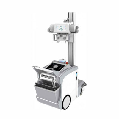
Click Here To Download Catalogue | DrGem Diamond All-In-One Digital X-ray Machine is a fully automatic digital radiography system providing state-of-the-art image quality, image processing and user interface. With a wide selection of anatomical studies on the imaging software, DIAMOND automatically sets up the x-ray generator’s pre-programmed exposure technique settings, motorized radiographic stand positioning, x-ray collimation and post-image processing for the selected study. Specifically designed to increase workflow, this fully digital system offers convenient auto-positioning and advanced image processing to achieve big performance with little effort.
Features of DrGem Diamond All-In-One Digital X-ray Machine:
Outstanding Image Quality -
Digital radiography via at panel detector improves your workflow, exam speed and comfort with efficiency. Digital at panel detector with Csl screen provides excellent spatial resolution, MTF, DQE and stability based on ne pixel pitch. A 3-field ion-chamber is provided for AEC function.
Automatic Collimation –
Automatic x-ray eld size control of the motorized collimator corresponds to dierent SIDs. Includes user adjustable lamp timer with on/oswitch.
Automatic Positioning –
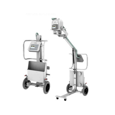
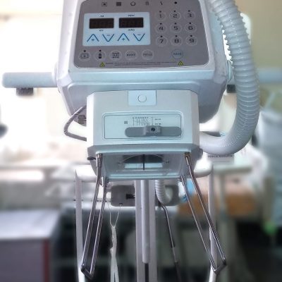
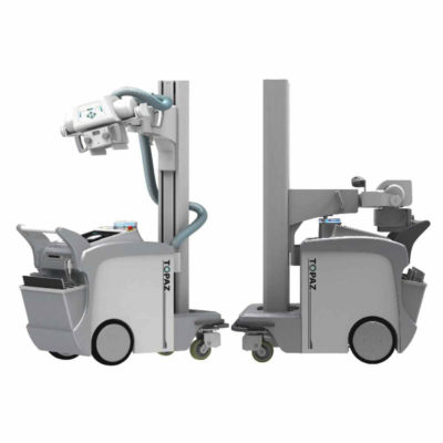
Click Here To Download Catalogue | JADE Mobile X-ray machine is one of the lightest portable X-ray systems on the market, allowing it to be used in any imaginable way including bedside, operating rooms, intensive care units and veterinary fields. With a simple, easy-to-use operator console, three-way control, two-step foldable stand and auto-lock system, the JADE Mobile X-ray machine is a user-friendly portable X-ray system.
Convenient & Intuitive Operation:
JADE is one of the lightest portable X-ray systems on the market, allowing it to be used in any imaginable way including bedside, operating rooms, intensive care units and in veterinary fields. With a simple, easy-to-use operator console, three-way control, two-step foldable stand and auto-lock system, JADE is a user-friendly portable X-ray system.
Compact & Powerful Design:
JADE Mobile X-ray machine is an innovative, highly versatile portable X-ray system suitable for a variety of clinical uses. Utilizing the unique technology used in DRGEM’s universally recognized X-ray generators, JADE is a compact but powerful unit with a 4kW output and thoughtfully designed components to increase efficiency and maximize workflow. The core part of X-ray source adopts high-quality tube assembly, X-ray collimator and high frequency X-ray generator with excellent performance, lifetime and stability.
Features:
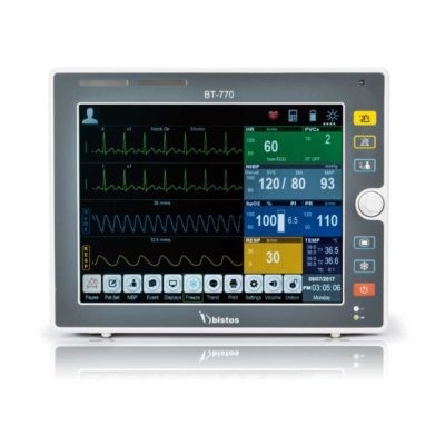
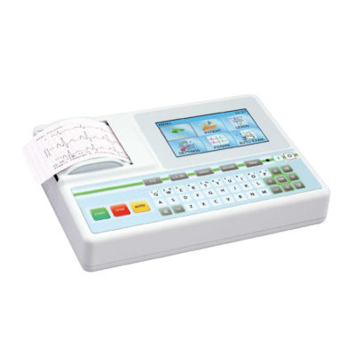
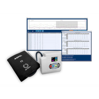
Click Here To Download Catalogue | The Holter Monitor allows quick analysis of ECG examination (arrhythmias and ST segment).
Technical specifications:
HolCARD 24W Software:
Click Here To Download Catalogue | DETAILS
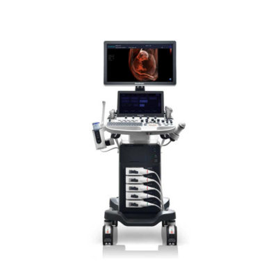
Powerful Compact Precision
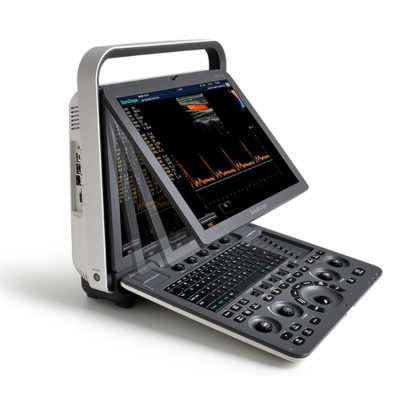
Taking into consideration the evolving expectations and needs for ultrasound, the P50 is a slim and unobtrusive trolley system that is comfortable in tight, congested spaces with little room to work in. Providing everything you need for a comfortable examination in a small space for both you and your patient.
Single Crystal Transducer
Wideband single crystal probes greatly improve the signal ratio, acquire stunning images and provide superior sensitivity and resolution for both the near and far-fields.
μ-Scan+
The new generation μ-Scan imaging technologies give you better image quality by reducing noise, improving signal strength and improving visualization.
Dynamic Color
Dynamic colour improves upon already existing colour Doppler technologies for clear capture of colour flow and detail visualization of even tiny veins with lower velocities.
Solution for Radiology
P50, is a leading-edge ultrasound system that can meet the demands of any clinical setting. You can experience a superior performance in multi-dimensional imaging for a full range of clinical applications – abdominal, breast and cardiovascular.
C-xlasto Imaging
By understanding that tissue stiffness varies depending on the type of tissue, we can use C-xlasto Imaging to easily find abnormalities and tumours within soft tissue. The differences in tissue responses are detected and visualized in real-time by the elastography algorithms through different representations, which can be particularly helpful in analyzing breast, thyroid and musculoskeletal structures. Predominately used only in linear probes, SonoScape’s new transvaginal and bi-plane probe for gynaecology and urology are breaking the mould and expanding elastography applications.
Real-time Color Panoramic
With the combination of colour flow and real-time panoramic, visualizing the blood flow of an entire vein or artery is now an easy task. Accomplished in real-time for the convenience of the sonographers, any mistakes can also be easily backtracked and corrected without interrupting the scan.
Contrast Imaging
Contrast Imaging on P50 makes full use of the infra harmonic signal and second harmonic signal to improve the image resolution and deep penetration. What’s more, the Dynamic Acoustic Control technology effectively controls the acoustic pressure for the contrast agent, decreasing the required agent dose and assures uniform image quality, guaranteeing longer contrast agent duration and better lesion perfusion of delayed phase observation.
Solution for OB/GYN
P50 has superior image quality, automated measurement tools, and a variety of volume technologies to provide ideal solutions for clinical examinations such as pregnancy examinations, and gynecologic disease diagnosis. With a new 4D transvaginal probe, P50 helps you to see and detect fetal abnormalities and significantly improves your diagnostic confidence during your examinations.
S-Live Silhouette
A unique transparent 3D anatomical image of the fetus for improved initial anatomical review. By using this new application, the system can create completely different fetal images from conventional ultrasound images, which can depict the fetal's intracorporeal anatomical structure.
Pelvic Floor 4D
Working in conjunction with SonoScape’s latest transvaginal probes, trans-perineal 4D pelvic floor ultrasound provides a useful clinical assessment of the impact of vaginal delivery on the female anterior compartment. Allowing doctors to judge whether the pelvic organs prolapsed or not, the extent of prolapse, and determining whether the pelvic muscles tore correctly.
S-Guide
S-Guide gives the user an extensive list of example obstetric ultrasound images as reference guides and a convenient checklist system to keep track of their progress during their obstetrics examination.
Auto Face
Automatically removes masking layers in front of the fetus’s face for a clearer vision of the fetus’s face.
AVC Follicle
AVC Follicle automatically identifies how many follicles are present and calculates their individual volumes.
Solution for Cardiology
P50 provides clear 2D clinical images and Doppler sensitivity to assess critical cardiac performance. Compatible with SonoScape’s single crystal probes, the P50 can provide images with better resolution and penetration in Cardiac diagnosis.
Tissue Doppler Imaging
Tissue Doppler Imaging allows clinical doctors to quantitatively evaluate local myocardial movements and functions, facilitating them with the ability to analyze and compare the motions of the different parts of the patient’s heart.
Stress Echo
Stress echocardiography is the combination of 2D echocardiography with physical, pharmacological or electrical stress of the patient. It also then provides users with report management tools such as configurable template editor, multiple loops to select one for storage, wall motion scoring, stress echo report, etc
Auto IMT
Auto IMT is used when determining the level of vascular sclerosis present in the patient by automatically tracing and calculating the thickness of the carotid vessels. What distinguishes the P50 is that it provides an instant and accurate Mean and Max index at the touch of a single button.
Auto EF
Automated 2D Cardiac Quantification is a fully intelligent trace function for endocardium with 19 easily-adjustable points providing rapid access to proven 2D EF and volumes.
Click Here To Download Catalogue | DETAILS
B + Compound
B + Compound utilizes several lines of sight for optimal contrast resolution, speckle reduction and border detection, with which P10 is ideal for superficial and abdominal imaging with better clarity and improved continuity of structures.
μ-Scan
The new generation μ-Scan imaging technology gives you better image quality by reducing noise, improving signal strength and improving visualization.
P10 offers a comprehensive selection of electronic probes to maximize its capabilities to meet a wide range of applications including abdomen, pediatric, OB/GYN, cardiovascular, musculoskeletal, etc. The advanced probe technologies also effectively enhance the image quality and confidence in reaching clinical diagnoses, even in difficult patients.
Convex Probe 3C-A
Ideal for an abundant of application such as abdomen, gynecology, obstetrics, urology and even abdomen biopsy.
Linear Probe L741
This linear probe is designed to satisfy vascular, breast, thyroid, and other small parts diagnosis, and its adjustable parameters could also present users a clear view of MSK and deep vessels.
Phase Array Probe 3P-A
For the purpose of adult and pediatric cardiology and emergency, the phase array probe provides elaborate presets for different exam modes, even for difficult patients.
Intracavitary Probe 6V1
Intracavitary probe could face application of gynecology, urology, prostate, and its temperature detection technology not only protects the patient but also extends the service life.
Click Here To Download Catalogue | |||||||||||||||||||||||||||||||||||||||||||||||||||||||||||||||||||||||||||||||||||||||||||||||||||||||||||||||||||||||||||||||||||||||||||||||||||||||||||||||||||||||||||||||||||||||||||||||||||||||||||||||||||||||||||||||||||||||||||||||||||||||||||||||||||||||||||||||||||||||||||||||||||||||||||||||
| Weight | N/A | N/A | N/A | N/A | N/A | N/A | |||||||||||||||||||||||||||||||||||||||||||||||||||||||||||||||||||||||||||||||||||||||||||||||||||||||||||||||||||||||||||||||||||||||||||||||||||||||||||||||||||||||||||||||||||||||||||||||||||||||||||||||||||||||||||||||||||||||||||||||||||||||||||||||||||||||||||||||||||||||||||||||||||||||||||||||
| Dimensions | N/A | N/A | N/A | N/A | N/A | N/A | |||||||||||||||||||||||||||||||||||||||||||||||||||||||||||||||||||||||||||||||||||||||||||||||||||||||||||||||||||||||||||||||||||||||||||||||||||||||||||||||||||||||||||||||||||||||||||||||||||||||||||||||||||||||||||||||||||||||||||||||||||||||||||||||||||||||||||||||||||||||||||||||||||||||||||||||
| Additional information |
|


Reviews
There are no reviews yet.