Atlas Venous Stent
$0.00
Shipped From Abroad
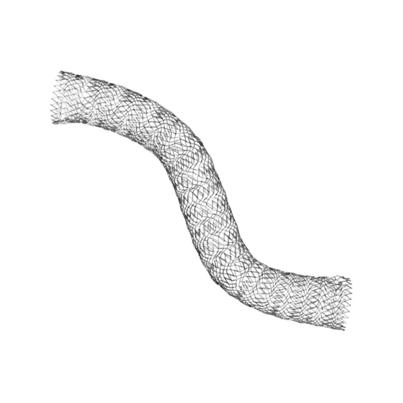
The Atlas™ Venous Stent is a self-expanding nitinol implant designed to restore venous outflow by maintaining vessel patency, improving blood flow, and reducing symptoms of chronic venous insufficiency in large veins such as the iliac and femoral segments.
Typically 10-21 working days – excluding furniture and heavy/bulky equipment. Please contact us for further information.
Description
The Atlas™ Venous Stent, introduced by Invamed—a global medical device manufacturer—is meticulously crafted for venous outflow restoration and chronic venous insufficiency management. Leveraging a self-expanding nitinol design, Atlas™ offers strong radial force, adaptive conformability, and minimal recoil, supporting durable patency in challenging venous anatomies.
Self-Expanding Nitinol Framework
Delivers consistent radial force to maintain lumen patency, especially in large, compressible venous segments.
High Conformability
Adapts to tortuous or dynamic venous structures (e.g., iliac vein, femoral vein), minimizing stress at stent edges and promoting stable apposition.
Minimal Recoil & Migration
Engineered for durable scaffolding, preserving vessel diameter and reducing the risk of stent migration under variable venous pressures.
Optimized for Venous Hemodynamics
Large cell design fosters robust flow, reducing intraluminal pressure gradients and supporting potential reduction in venous hypertension symptoms.
Wide Clinical Utility
Indicated for iliofemoral venous outflow obstructions, post-thrombotic syndrome, or venous stenoses from extrinsic compression or scar tissue.
Contraindications
Severe vessel tortuosity or diameter mismatch preventing stent passage, active local infection in the venous target, or contraindications to endovascular procedures.
Delivers consistent radial force to maintain lumen patency, especially in large, compressible venous segments.
Adapts to tortuous or dynamic venous structures (e.g., iliac vein, femoral vein), minimizing stress at stent edges and promoting stable apposition.
Engineered for durable scaffolding, preserving vessel diameter and reducing the risk of stent migration under variable venous pressures.
Large cell design fosters robust flow, reducing intraluminal pressure gradients and supporting potential reduction in venous hypertension symptoms.
Indicated for iliofemoral venous outflow obstructions, post-thrombotic syndrome, or venous stenoses from extrinsic compression or scar tissue.
Severe vessel tortuosity or diameter mismatch preventing stent passage, active local infection in the venous target, or contraindications to endovascular procedures.
Technical Specifications
| Specification | Detail / Value |
| Product Name | Atlas™ Venous Stent |
| Stent Material | Nitinol (nickel-titanium) self-expanding alloy |
| Deployment Mechanism | Over-the-wire via introducer sheath (typically 6F–9F, model-specific) |
| Radial Force | Moderate to high, designed to resist extrinsic compression |
| Stent Diameter Range | ~10–20 mm nominal (expanded), ensuring coverage of varied venous lumen sizes |
| Stent Length Range | ~40–120 mm, accommodating short or long-segment lesions |
| Radiopaque Markers | Proximal and distal markers for fluoroscopic visualization |
| Sterility | Sterile, single-use |
| Shelf Life | ~3–5 years if stored at 15–25 °C |
| Clinical Indications | Iliofemoral venous stenosis, post-thrombotic syndrome, extrinsic compression (May-Thurner), chronic venous insufficiency |
Size & Ordering Matrix7
| Stent Diameter (mm) | Stent Length (mm) | Sheath Compatibility (F) | Product Code | Notes |
| 10 | 40 | 6F | AVS-10×40-6F | Smaller diameter for narrower veins or moderate lesions |
| 10 | 60 | 6F | AVS-10×60-6F | Slightly longer coverage in the same diameter |
| 14 | 60 | 7F | AVS-14×60-7F | Popular mid-range diameter for common iliofemoral lesions |
| 14 | 80 | 7F | AVS-14×80-7F | Extended length for multi-segment stenoses |
| 16 | 80 | 8F | AVS-16×80-8F | Enhanced diameter for advanced venous outflow compromise |
| 16 | 100 | 8F | AVS-16×100-8F | Longer coverage in larger veins |
| 20 | 120 | 9F | AVS-20×120-9F | Maximum diameter & length for extensive venous pathology |
Quick Comparison
| Atlas Venous Stent remove | Sonoscape P10 Ultrasound Machine remove | ASPEL AsPEKT 712 Holter Monitor and Software remove | FlowMir Disposable Turbine with Cardboard Mouthpiece remove | Bistos BT- 720 Patient Monitor remove | ASPEL AsCARD Coral PC Based ECG Machine remove | |||||||||||||||||||||||||||||||||||||||||||||||||||||||||||||||||||||||||||||||||||||||||||||||||||||||||||||||||||||||||||||||||||||||||||||||
|---|---|---|---|---|---|---|---|---|---|---|---|---|---|---|---|---|---|---|---|---|---|---|---|---|---|---|---|---|---|---|---|---|---|---|---|---|---|---|---|---|---|---|---|---|---|---|---|---|---|---|---|---|---|---|---|---|---|---|---|---|---|---|---|---|---|---|---|---|---|---|---|---|---|---|---|---|---|---|---|---|---|---|---|---|---|---|---|---|---|---|---|---|---|---|---|---|---|---|---|---|---|---|---|---|---|---|---|---|---|---|---|---|---|---|---|---|---|---|---|---|---|---|---|---|---|---|---|---|---|---|---|---|---|---|---|---|---|---|---|---|---|---|---|---|---|---|---|---|
| Name | Atlas Venous Stent remove | Sonoscape P10 Ultrasound Machine remove | ASPEL AsPEKT 712 Holter Monitor and Software remove | FlowMir Disposable Turbine with Cardboard Mouthpiece remove | Bistos BT- 720 Patient Monitor remove | ASPEL AsCARD Coral PC Based ECG Machine remove | ||||||||||||||||||||||||||||||||||||||||||||||||||||||||||||||||||||||||||||||||||||||||||||||||||||||||||||||||||||||||||||||||||||||||||||||
| Image |  | 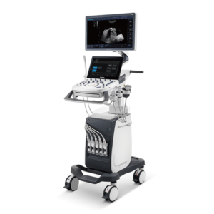 | 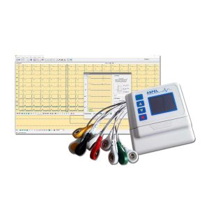 | 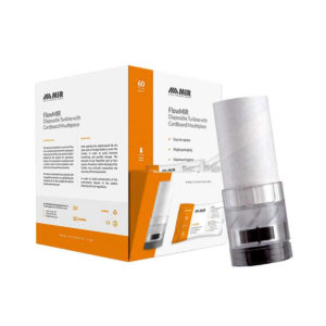 |  |  | ||||||||||||||||||||||||||||||||||||||||||||||||||||||||||||||||||||||||||||||||||||||||||||||||||||||||||||||||||||||||||||||||||||||||||||||
| SKU | SF1033560012-7 | SF1033560075-4 | SF1033560084-22 | SF1033560059-8 | SF1033560075-11 | |||||||||||||||||||||||||||||||||||||||||||||||||||||||||||||||||||||||||||||||||||||||||||||||||||||||||||||||||||||||||||||||||||||||||||||||
| Rating | ||||||||||||||||||||||||||||||||||||||||||||||||||||||||||||||||||||||||||||||||||||||||||||||||||||||||||||||||||||||||||||||||||||||||||||||||||||
| Price |
|
|
|
|
|
| ||||||||||||||||||||||||||||||||||||||||||||||||||||||||||||||||||||||||||||||||||||||||||||||||||||||||||||||||||||||||||||||||||||||||||||||
| Stock | ||||||||||||||||||||||||||||||||||||||||||||||||||||||||||||||||||||||||||||||||||||||||||||||||||||||||||||||||||||||||||||||||||||||||||||||||||||
| Availability | ||||||||||||||||||||||||||||||||||||||||||||||||||||||||||||||||||||||||||||||||||||||||||||||||||||||||||||||||||||||||||||||||||||||||||||||||||||
| Add to cart | ||||||||||||||||||||||||||||||||||||||||||||||||||||||||||||||||||||||||||||||||||||||||||||||||||||||||||||||||||||||||||||||||||||||||||||||||||||
| Description | Shipped From Abroad
The Atlas™ Venous Stent is a self-expanding nitinol implant designed to restore venous outflow by maintaining vessel patency, improving blood flow, and reducing symptoms of chronic venous insufficiency in large veins such as the iliac and femoral segments. Delivery & Availability:
Typically 10-21 working days – excluding furniture and heavy/bulky equipment. Please contact us for further information.
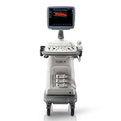
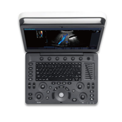
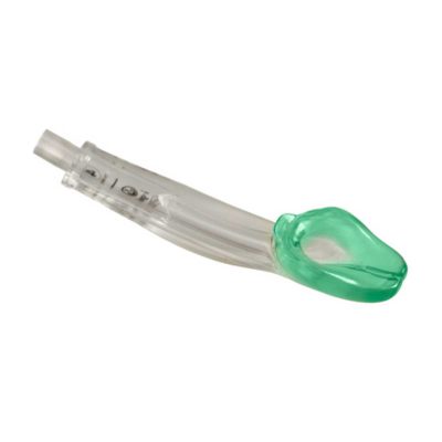
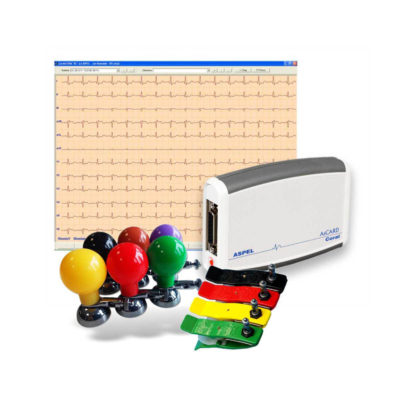
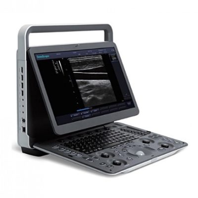
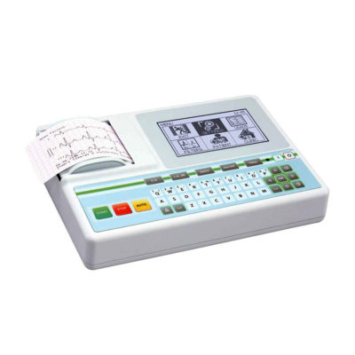
| Shipped from Abroad The P10 color Doppler ultrasound system is a new generation product from SonoScape. It is designed to give high quality images, rich probe configurations, various clinical tools and automatic analysis software to provide you with comprehensive solutions for your growing demand for clinical applications. Delivery & Availability: Typically 5-7 working days – excluding furniture and heavy/bulky equipment. Please contact us for further information. | Shipped from Abroad The Holta Monitor allows quick analysis of ECG examination and detection, reviewing and editing capability in the qualitative assessment of VE, VT, Single SVE, PSVT, Pauses, Irregular Rhythm, VT, IVR, Brady - and Tachycardia, Couplets, ST-segment elevation and depression, Maximum, Minimum and averaged Heart Rates, artifacts Delivery & Availability: Typically 10 working days – excluding furniture and heavy/bulky equipment. Please contact us for further information. | In stock Spirometry testing requires maximum accuracy and hygiene. Each Flowmir disposable turbine, which includes a cardboard mouthpiece, has been individually factory calibrated with a computerized system and it is packaged individually. FlowMIR® is an inexpensive alternative to a costly reusable flowmeter and replaces the need for an antibacterial filter Typically 2 working days – excluding furniture and heavy/bulky equipment. Please contact us for further information. | Shipped from abroad Bistos BT - 720 Patient Monitor: Bistos Portable Patient Vital Signs Monitor, SpO2, Pulse, *NIBP, BT-720. The Bistos BT-720 is a compact size vital signs monitor with the standard parameters of SpO2 and Pulse. You can also add the NIBP and/or Masimo SpO2 at an additional cost. The BT-720 has a 4.3" color touch screen The PC software allows the vital signs information to be transferred and analyzed on a computer. Delivery & Availability: Typically 7 working days – excluding furniture and heavy/bulky equipment. Please contact us for further information. | Shipped from Abroad AsCARD Coral electrocardiograph is a 3-, 6-, 12-channel ECG equipped with CardioTEKA software allows transmission of full 12 ECG leads to the user PC through USB interface. It is intended for carrying out ECG examinations in adults and pediatric patients in all types of health care centres. ECG procedures can be performed by qualified personnel only. AsCARD Coral can cooperate also with CardioTEST system as 12-channel ECG device allows transmission of full 12 ECG leads to the user PC through USB interface. Delivery & Availability: Typically 10 working days – excluding furniture and heavy/bulky equipment. Please contact us for further information. | ||||||||||||||||||||||||||||||||||||||||||||||||||||||||||||||||||||||||||||||||||||||||||||||||||||||||||||||||||||||||||||||||||||||||||||||
| Content | The Atlas™ Venous Stent, introduced by Invamed—a global medical device manufacturer—is meticulously crafted for venous outflow restoration and chronic venous insufficiency management. Leveraging a self-expanding nitinol design, Atlas™ offers strong radial force, adaptive conformability, and minimal recoil, supporting durable patency in challenging venous anatomies.
Technical Specifications
Size & Ordering Matrix7
| DETAILS
B + Compound
B + Compound utilizes several lines of sight for optimal contrast resolution, speckle reduction and border detection, with which P10 is ideal for superficial and abdominal imaging with better clarity and improved continuity of structures.
μ-Scan
The new generation μ-Scan imaging technology gives you better image quality by reducing noise, improving signal strength and improving visualization.
P10 offers a comprehensive selection of electronic probes to maximize its capabilities to meet a wide range of applications including abdomen, pediatric, OB/GYN, cardiovascular, musculoskeletal, etc. The advanced probe technologies also effectively enhance the image quality and confidence in reaching clinical diagnoses, even in difficult patients.
Convex Probe 3C-A
Ideal for an abundant of application such as abdomen, gynecology, obstetrics, urology and even abdomen biopsy.
Linear Probe L741
This linear probe is designed to satisfy vascular, breast, thyroid, and other small parts diagnosis, and its adjustable parameters could also present users a clear view of MSK and deep vessels.
Phase Array Probe 3P-A
For the purpose of adult and pediatric cardiology and emergency, the phase array probe provides elaborate presets for different exam modes, even for difficult patients.
Intracavitary Probe 6V1
Intracavitary probe could face application of gynecology, urology, prostate, and its temperature detection technology not only protects the patient but also extends the service life.
Click Here To Download Catalogue | The Holter Monitor allows quick analysis of ECG examination (arrhythmias and ST segment).
Technical specifications:
HolCARD 24W Software:
Click Here To Download Catalogue |
| Bistos BT - 720 Patient Monitor: Bistos Portable Patient Vital Signs Monitor, SpO2, Pulse, *NIBP, BT-720. The Bistos BT-720 is a compact size vital signs monitor with the standard parameters of SpO2 and Pulse. You can also add the NIBP and/or Masimo SpO2 at an additional cost. The BT-720 has a 4.3" color touch screen The PC software allows the vital signs information to be transferred and analyzed on a computer.
Features:
Click Here To Download Catalogue |
AsCARD Coral electrocardiograph is a 3-, 6-, 12-channel ECG equipped with CardioTEKA software allows transmission of full 12 ECG leads to the user PC through USB interface. It is intended for carrying out ECG examinations in adults and pediatric patients in all types of health care centres. ECG procedures can be performed by qualified personnel only. AsCARD Coral can cooperate also with CardioTEST system as 12-channel ECG device allows transmission of full 12 ECG leads to the user PC through USB interface.
Technical Specification:
Click Here To Download Catalogue | ||||||||||||||||||||||||||||||||||||||||||||||||||||||||||||||||||||||||||||||||||||||||||||||||||||||||||||||||||||||||||||||||||||||||||||||
| Weight | N/A | N/A | N/A | N/A | N/A | N/A | ||||||||||||||||||||||||||||||||||||||||||||||||||||||||||||||||||||||||||||||||||||||||||||||||||||||||||||||||||||||||||||||||||||||||||||||
| Dimensions | N/A | N/A | N/A | N/A | N/A | N/A | ||||||||||||||||||||||||||||||||||||||||||||||||||||||||||||||||||||||||||||||||||||||||||||||||||||||||||||||||||||||||||||||||||||||||||||||
| Additional information | ||||||||||||||||||||||||||||||||||||||||||||||||||||||||||||||||||||||||||||||||||||||||||||||||||||||||||||||||||||||||||||||||||||||||||||||||||||

Reviews
There are no reviews yet.