
Coronis Fusion 4MP (MDCC‑4430)
$0.00
Shipped From Abroad
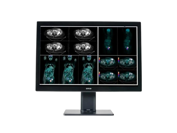
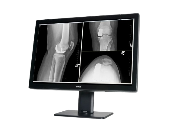
The Barco Coronis Fusion 4MP (MDCC-4430) is a high-fidelity medical display designed for diagnostic radiology. With a 4-megapixel resolution, wide color gamut, and image-enhancement features, it supports mammography, CT, MRI, and multi-modality imaging workflows.
Typically 10-21 working days – excluding furniture and heavy/bulky equipment. Please contact us for further information.
Description
The Barco Coronis Fusion 4MP (MDCC-4430) delivers optimized diagnostic visualization for radiologists by combining high-quality optics with advanced processing. Its native resolution of 2560 × 1600 pixels and large active area (655 × 410 mm) provide generous screen real estate for image review. The display includes uniformity correction and Barco’s image-enhancement technologies to maintain consistency across the display. It supports multi-modality imaging in breast, vascular, and general radiology settings. With efficient design and compatibility for clinical workflows, it balances performance and ergonomics in diagnostic environments.
Key Features
-
Native 4MP resolution (2560 × 1600)
-
Active screen area: 655 × 410 mm
-
Uniformity correction to ensure brightness consistency
-
Image-enhancement tools for clarity in clinical reading
-
Wide gamut/color support (multi-modality capability)
-
Optimized for breast imaging, mammography, CT, and MRI
-
Efficient, lightweight design for diagnostic reading rooms
A brighter diagnosis
Coronis Fusion, Barco’s renowned multi-modality display for radiologists, now comes in a new, energy-efficient, and lightweight design. Bright on so many levels, Coronis Fusion has been designed to help radiologists provide care with confidence.
See more details quickly thanks to the high brightness, high contrast ratio, and best-in-class image quality. What’s more, the wide color gamut in combination with SteadyColor™ calibration technology helps you see even more colors and more details on the 30-inch screen.
Smart workflow
Designed for your comfort and to boost productivity, Coronis Fusion comes with smart image-enhancing features and workflow tools. The display’s wide viewing angle, combined with the SoftGlow™ task and wall light, helps reduce eye strain. Thanks to SpotView, radiologists can further improve detection accuracy as well as reading productivity.
Effortless quality & compliance
Like all of Barco’s medical display systems, Coronis Fusion comes with QAWeb Enterprise, a cloud-based technology for automated calibration, Quality Assurance, and compliance to ensure maximum uptime of the display with no need for human intervention.
A one-stop-shop solution
Barco medical display systems include exclusive display controllers that are validated with the latest workstations and with all major PACS applications. All components are under the full 5-year warranty (including the display backlight) for complete peace of mind.
Ensuring diagnostic confidence with MDR Class IIa
Our radiology displays are MDR-certified as Class IIa. Their product information has been reviewed and cleared by independent medical and technical experts, and is audited yearly. In other words, we ensure diagnostic confidence and peace of mind for our users.
Specifications
| Category | Specification |
|---|---|
| Screen Technology | IPS |
| Active Screen Size (Diagonal) | 772 mm (30.4″) |
| Active Screen Size (H × V) | 655 × 410 mm (25.8″ × 16.1″) |
| Aspect Ratio | 16:10 |
| Resolution | Native 4MP (2560 × 1600 pixels); Configurable to 2 × 2MP+ (1280 × 1600 pixels); 2 × 2MP (1200 × 1600 pixels) |
| Pixel Pitch | 0.256 mm |
| Color Imaging / Gray Imaging | Yes / Yes |
| Bit Depth | 30-bit |
| Viewing Angle (H/V) | 178° |
| Uniformity Correction | Color PPU |
| SteadyColor | Yes (with Barco Display Controller and QAWeb Enterprise 2.2+) |
| Ambient Light Presets / Sensor | Yes / Yes |
| Front Sensor | Yes (I-Guard, Coronis) |
| Maximum Luminance (Typical) | 1050 cd/m² |
| DICOM Calibrated Luminance | 600 cd/m² |
| Contrast Ratio | 2000:1 |
| Response Time | 18 ms |
| Housing Color | Black / White |
| Video Input Signals | 2 × DisplayPort 1.2 |
| Video Output Signals | 1 × DisplayPort (MST) |
| USB Ports | 1 × USB 2.0 upstream; 2 × USB 2.0 downstream; 1 × USB 2.0 downstream with high-power charging |
| Power Rating | 100–240 Vac, 50/60 Hz, 3.6–1.6 A |
| Power Consumption | 75 W (nominal @ 600 cd/m²); <0.5 W (hibernate/standby) |
| Dimensions with Stand (W × H × D) | 714 × 524~624 × 240 mm |
| Dimensions without Stand (W × H × D) | 714 × 478 × 74 mm |
| Dimensions Packaged (W × H × D) | 800 × 650 × 295 mm |
| Net Weight with Stand | MDCC-4430: 17.7 kg; MDCC-4430 NC: 16.3 kg |
| Net Weight without Stand | MDCC-4430: 13.1 kg; MDCC-4430 NC: 11.7 kg |
| Net Weight Packaged | MDCC-4430: 22.3 kg; MDCC-4430 NC: 20.9 kg |
| Tilt / Swivel / Pivot | -5° to +25° / ±30° / N/A |
| Height Adjustment Range | 100 mm |
| Mounting Standard | VESA (100 mm) |
| Screen Protection | MDCC-4430: Protective, anti-reflective glass; MDCC-4430 NC: No glass cover |
| Recommended Modalities | All digital images except digital mammography |
| Certifications | FDA 510(k) K191845, CE0123, CCC, INMETRO, KC, BIS, EAC |
| Safety Standards | IEC/EN/UL/CSA 60950-1, 62368-1, 60601-1, AAMI ES 60601-1 |
| EMI Standards | IEC/EN 60601-1-2, FCC Part 15 Class B, ICES-001 Level B, VCCI |
| Environmental Compliance | China Energy Label, EU RoHS, China RoHS, REACH, Canada Health, WEEE, Packaging Directive |
| Supplied Accessories | User guide, Documentation disc, System sheet, Video cables, Mains cables, USB cable |
| Optional Accessories | Graphics board, Touch pad, QA software (QAWeb) |
| Warranty | 5 years (includes 40,000 hrs backlight warranty) |
| Operating Temperature | 0–35°C (specs: 10–30°C) |
| Storage Temperature | -20–60°C |
| Operating Humidity | 20–85% RH (non-condensing) |
| Storage Humidity | 20–85% RH (non-condensing) |
| Operating Pressure | ≥70 kPa |
| Storage Pressure | 50–106 kPa |
Quick Comparison
| Coronis Fusion 4MP (MDCC‑4430) remove | LED Double X-ray Viewing Box remove | SIGNERS SUPiA Dry Thermal X-ray Film Printer remove | Anke Anatom 64 Clarity Multi-Slice Spiral CT Scan remove | Sonoscape S22 Ultrasound Machine remove | Sonoscape P20 Ultrasound Machine remove | |||||||||||||||||||||||||||||||||||||||||||||||||||||||||||||||||||||||||||||||||||||||||||||||||||||||||||||||||||||||||||||||||||||||||||||||||||||||||||||||||||||||||||||||||||||||||||||||||||||||||||||||||||||||||||||||||||||||||||||||||||||||||||||||||||||||||||||||||||||||||||||||||||||||||||||||||||||||||||||||||||||||||||||||||||||||||||||||||||||||||||||||||||||||||||
|---|---|---|---|---|---|---|---|---|---|---|---|---|---|---|---|---|---|---|---|---|---|---|---|---|---|---|---|---|---|---|---|---|---|---|---|---|---|---|---|---|---|---|---|---|---|---|---|---|---|---|---|---|---|---|---|---|---|---|---|---|---|---|---|---|---|---|---|---|---|---|---|---|---|---|---|---|---|---|---|---|---|---|---|---|---|---|---|---|---|---|---|---|---|---|---|---|---|---|---|---|---|---|---|---|---|---|---|---|---|---|---|---|---|---|---|---|---|---|---|---|---|---|---|---|---|---|---|---|---|---|---|---|---|---|---|---|---|---|---|---|---|---|---|---|---|---|---|---|---|---|---|---|---|---|---|---|---|---|---|---|---|---|---|---|---|---|---|---|---|---|---|---|---|---|---|---|---|---|---|---|---|---|---|---|---|---|---|---|---|---|---|---|---|---|---|---|---|---|---|---|---|---|---|---|---|---|---|---|---|---|---|---|---|---|---|---|---|---|---|---|---|---|---|---|---|---|---|---|---|---|---|---|---|---|---|---|---|---|---|---|---|---|---|---|---|---|---|---|---|---|---|---|---|---|---|---|---|---|---|---|---|---|---|---|---|---|---|---|---|---|---|---|---|---|---|---|---|---|---|---|---|---|---|---|---|---|---|---|---|---|---|---|---|---|---|---|---|---|---|---|---|---|---|---|---|---|---|---|---|---|---|---|---|---|---|---|---|---|---|---|---|---|---|---|---|---|---|---|---|---|---|---|---|---|---|---|---|---|---|---|---|---|---|---|---|---|---|---|---|---|---|---|---|---|---|---|---|---|---|---|---|---|---|---|---|---|---|---|---|---|---|---|---|---|---|---|---|---|---|---|---|---|---|---|
| Name | Coronis Fusion 4MP (MDCC‑4430) remove | LED Double X-ray Viewing Box remove | SIGNERS SUPiA Dry Thermal X-ray Film Printer remove | Anke Anatom 64 Clarity Multi-Slice Spiral CT Scan remove | Sonoscape S22 Ultrasound Machine remove | Sonoscape P20 Ultrasound Machine remove | ||||||||||||||||||||||||||||||||||||||||||||||||||||||||||||||||||||||||||||||||||||||||||||||||||||||||||||||||||||||||||||||||||||||||||||||||||||||||||||||||||||||||||||||||||||||||||||||||||||||||||||||||||||||||||||||||||||||||||||||||||||||||||||||||||||||||||||||||||||||||||||||||||||||||||||||||||||||||||||||||||||||||||||||||||||||||||||||||||||||||||||||||||||||||||
| Image | 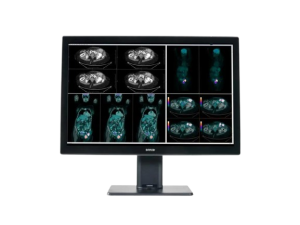 | 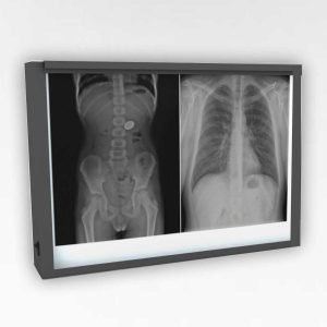 | 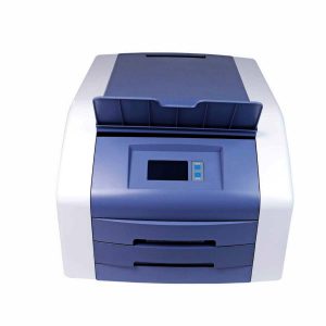 | 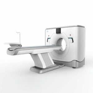 | 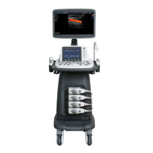 | 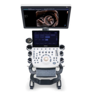 | ||||||||||||||||||||||||||||||||||||||||||||||||||||||||||||||||||||||||||||||||||||||||||||||||||||||||||||||||||||||||||||||||||||||||||||||||||||||||||||||||||||||||||||||||||||||||||||||||||||||||||||||||||||||||||||||||||||||||||||||||||||||||||||||||||||||||||||||||||||||||||||||||||||||||||||||||||||||||||||||||||||||||||||||||||||||||||||||||||||||||||||||||||||||||||
| SKU | SF1033560084-193 | SF1033560050-02 | SF1033560092-2 | SF1033560012-3 | SF1033560012-9 | |||||||||||||||||||||||||||||||||||||||||||||||||||||||||||||||||||||||||||||||||||||||||||||||||||||||||||||||||||||||||||||||||||||||||||||||||||||||||||||||||||||||||||||||||||||||||||||||||||||||||||||||||||||||||||||||||||||||||||||||||||||||||||||||||||||||||||||||||||||||||||||||||||||||||||||||||||||||||||||||||||||||||||||||||||||||||||||||||||||||||||||||||||||||||||
| Rating | ||||||||||||||||||||||||||||||||||||||||||||||||||||||||||||||||||||||||||||||||||||||||||||||||||||||||||||||||||||||||||||||||||||||||||||||||||||||||||||||||||||||||||||||||||||||||||||||||||||||||||||||||||||||||||||||||||||||||||||||||||||||||||||||||||||||||||||||||||||||||||||||||||||||||||||||||||||||||||||||||||||||||||||||||||||||||||||||||||||||||||||||||||||||||||||||||
| Price |
|
|
|
|
|
| ||||||||||||||||||||||||||||||||||||||||||||||||||||||||||||||||||||||||||||||||||||||||||||||||||||||||||||||||||||||||||||||||||||||||||||||||||||||||||||||||||||||||||||||||||||||||||||||||||||||||||||||||||||||||||||||||||||||||||||||||||||||||||||||||||||||||||||||||||||||||||||||||||||||||||||||||||||||||||||||||||||||||||||||||||||||||||||||||||||||||||||||||||||||||||
| Stock | ||||||||||||||||||||||||||||||||||||||||||||||||||||||||||||||||||||||||||||||||||||||||||||||||||||||||||||||||||||||||||||||||||||||||||||||||||||||||||||||||||||||||||||||||||||||||||||||||||||||||||||||||||||||||||||||||||||||||||||||||||||||||||||||||||||||||||||||||||||||||||||||||||||||||||||||||||||||||||||||||||||||||||||||||||||||||||||||||||||||||||||||||||||||||||||||||
| Availability | ||||||||||||||||||||||||||||||||||||||||||||||||||||||||||||||||||||||||||||||||||||||||||||||||||||||||||||||||||||||||||||||||||||||||||||||||||||||||||||||||||||||||||||||||||||||||||||||||||||||||||||||||||||||||||||||||||||||||||||||||||||||||||||||||||||||||||||||||||||||||||||||||||||||||||||||||||||||||||||||||||||||||||||||||||||||||||||||||||||||||||||||||||||||||||||||||
| Add to cart | ||||||||||||||||||||||||||||||||||||||||||||||||||||||||||||||||||||||||||||||||||||||||||||||||||||||||||||||||||||||||||||||||||||||||||||||||||||||||||||||||||||||||||||||||||||||||||||||||||||||||||||||||||||||||||||||||||||||||||||||||||||||||||||||||||||||||||||||||||||||||||||||||||||||||||||||||||||||||||||||||||||||||||||||||||||||||||||||||||||||||||||||||||||||||||||||||
| Description | Shipped From Abroad
The Barco Coronis Fusion 4MP (MDCC-4430) is a high-fidelity medical display designed for diagnostic radiology. With a 4-megapixel resolution, wide color gamut, and image-enhancement features, it supports mammography, CT, MRI, and multi-modality imaging workflows.
Delivery & Availability:
Typically 10-21 working days – excluding furniture and heavy/bulky equipment. Please contact us for further information.
| In stock
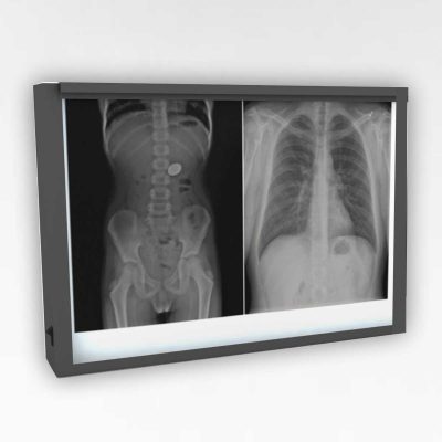
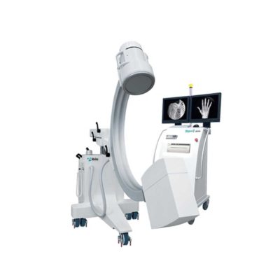
Double x-ray film viewer, Compact, Solid with Backlight of LED’s based panel, Long Life Approximate LED’s life 50, 000 Hrs., Uniform Light at the total surface area, No Heat Emission, Wall Mounted, Can be used for tracing on X-Ray, Auto-sensor, Screen. Size: 430 mm x 710 mm.
| Shipped from Abroad
Signers Digital Dry Thermal X-ray Film Printer - Is a dry imager designed to output information processed through DICOM network protocol, supports 4 films sizes. 8”x10”, 10”x12”, 11”x14” and 14”x17” Achieve the convenience of multiple sizes.
Delivery & Availability: Typically 14 working days – excluding furniture and heavy/bulky equipment. Please contact us for further information. | Shipped from Abroad
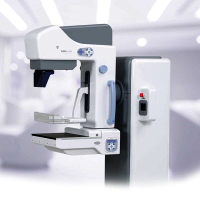
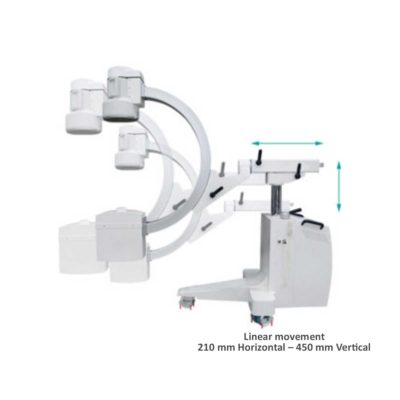
The ANATOM 64 CT scanner is the latest innovation for cardiac imaging based on Precision Platform system. The excellent design of Ahart technology which innovatively combined single spiral scan + gated imaging + mA modulation for easy heart imaging at extremely low radiation dose. We provide you ANATOM 64 Clarity/Precision of two models which are low/high configurations for preferences. It also offers you conventional clinical applications of low dose, better image quality and faster exams.
Delivery & Availability: Typically 90 working days – excluding furniture and heavy/bulky equipment. Please contact us for further information. | Shipped from Abroad As SonoScape steps forward to add value and efficiency to ultrasound, the latest S22 was designed in a user-friendly platform to address current and future demanding needs. It represents an excellent mix in performance and price. Delivery & Availability: Typically 5-7 working days – excluding furniture and heavy/bulky equipment. Please contact us for further information. | Shipped from Abroad Incorporating innovative technologies, P20’s user-friendly design with a simple operation panel, intuitive user interface and a variety of intelligent auxiliary scanning tools, will significantly improve your daily examination experience. Besides general imaging applications, P20 has entitled with diagnostic 4D technology which has an extraordinary performance in obstetrics and gynecology applications. Delivery & Availability: Typically 5-7 working days – excluding furniture and heavy/bulky equipment. Please contact us for further information. | ||||||||||||||||||||||||||||||||||||||||||||||||||||||||||||||||||||||||||||||||||||||||||||||||||||||||||||||||||||||||||||||||||||||||||||||||||||||||||||||||||||||||||||||||||||||||||||||||||||||||||||||||||||||||||||||||||||||||||||||||||||||||||||||||||||||||||||||||||||||||||||||||||||||||||||||||||||||||||||||||||||||||||||||||||||||||||||||||||||||||||||||||||||||||||
| Content | The Barco Coronis Fusion 4MP (MDCC-4430) delivers optimized diagnostic visualization for radiologists by combining high-quality optics with advanced processing. Its native resolution of 2560 × 1600 pixels and large active area (655 × 410 mm) provide generous screen real estate for image review. The display includes uniformity correction and Barco’s image-enhancement technologies to maintain consistency across the display. It supports multi-modality imaging in breast, vascular, and general radiology settings. With efficient design and compatibility for clinical workflows, it balances performance and ergonomics in diagnostic environments. Key Features
A brighter diagnosisCoronis Fusion, Barco's renowned multi-modality display for radiologists, now comes in a new, energy-efficient, and lightweight design. Bright on so many levels, Coronis Fusion has been designed to help radiologists provide care with confidence. See more details quickly thanks to the high brightness, high contrast ratio, and best-in-class image quality. What's more, the wide color gamut in combination with SteadyColor™ calibration technology helps you see even more colors and more details on the 30-inch screen.Smart workflowDesigned for your comfort and to boost productivity, Coronis Fusion comes with smart image-enhancing features and workflow tools. The display's wide viewing angle, combined with the SoftGlow™ task and wall light, helps reduce eye strain. Thanks to SpotView, radiologists can further improve detection accuracy as well as reading productivity.Effortless quality & complianceLike all of Barco's medical display systems, Coronis Fusion comes with QAWeb Enterprise, a cloud-based technology for automated calibration, Quality Assurance, and compliance to ensure maximum uptime of the display with no need for human intervention.A one-stop-shop solutionBarco medical display systems include exclusive display controllers that are validated with the latest workstations and with all major PACS applications. All components are under the full 5-year warranty (including the display backlight) for complete peace of mind.Ensuring diagnostic confidence with MDR Class IIaOur radiology displays are MDR-certified as Class IIa. Their product information has been reviewed and cleared by independent medical and technical experts, and is audited yearly. In other words, we ensure diagnostic confidence and peace of mind for our users.Specifications
| Double x-ray film viewer, Compact, Solid with Backlight of LED’s based panel, Long Life Approximate LED’s life 50, 000 Hrs., Uniform Light at the total surface area, No Heat Emission, Wall Mounted, Can be used for tracing on X-Ray, Auto-sensor, Screen. Size: 430 mm x 710 mm. | Signers Digital Dry Thermal X-ray Film Printer - Is a dry imager designed to output information processed through DICOM network protocol, supports 4 films sizes. 8”x10”, 10”x12”, 11”x14” and 14”x17” Achieve the convenience of multiple sizes.
Technical Specifications:
Click Here To Download Catalogue | The ANATOM 64 CT scanner is the latest innovation for cardiac imaging based on Precision Platform system. The excellent design of Ahart technology which innovatively combined single spiral scan + gated imaging + mA modulation for easy heart imaging at extremely low radiation dose. We provide you ANATOM 64 Clarity/Precision of two models which are low/high configurations for preferences. It also offers you conventional clinical applications of low dose, better image quality and faster exams.
Features:
Click Here To Download Catalogue | DETAILS
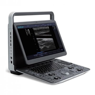
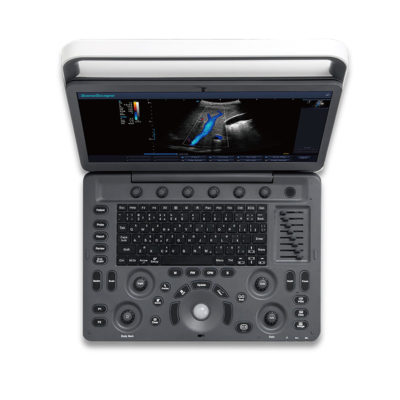
As SonoScape steps forward to add value and efficiency to ultrasound, the latest S22 was designed in a user-friendly platform to address current and future demanding needs. It represents an excellent mix in performance and price.
S22, is a shared service ultrasound system with a slim and elegant package that has combined mobility with utility to fit in specific clinical situations including emergency department, ICU, operating room and so on. Furthermore, its ergonomic design, easy operating and flexible data management will give you a memorable experience.
SPECIFICATION
• Large high-resolution widescreen LED
• Sensitive touch screen
• Four transducer sockets plus one socket for pencil probe
• A comprehensive selection of probes: linear, Convex, Micro-convex, Volumetric, Endocavity, Bi-plane, Phased Array, TEE, Intraoperative, Pencil
• Premium application technology: 4D, μ-scan speckle reduction, compound imaging, Pulse Inversion Harmonic Imaging, Color M-Mode, Steer M-Mode, PDI, TDI, Real-time Panoramic Imaging, Trapezoid Imaging, Auto-IMT…
• Full patient database and image management solutions: DICOM 3.0, AVI/JPG, USB 2.0, HDD, DVD, PDF report
• Multi-Language Input Keyboard
• Built-in battery
Click Here To Download Catalogue | DETAILS
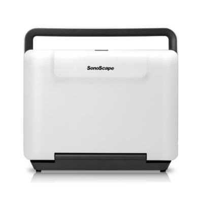
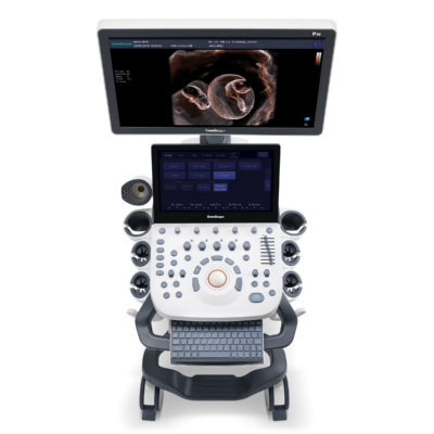
Upgraded Images with More Clarity
SonoScape never stops making progress in improving the image quality of its ultrasound products to enhance the confidence of diagnosis for doctors. With extraordinary images provided by P20, the anatomy structures are clearer than ever.
C-Xlasto Imaging
With C-xlasto Imaging, P20 enables comprehensive quantitative elastic analysis. Meanwhile, C-xlasto on P20 is supported by linear, convex and transvaginal probes, to ensure good reproducibility and highly consistent quantitative elastic results.
S-Live
S-Live allows for detailed visualization of subtle anatomical features, thereby enabling intuitive diagnosis with real-time 3D images and enriching patient communication.
Pelvic Floor 4D
Transperineal 4D pelvic floor ultrasound can provide useful clinical values in assessing the vaginal delivery impact on the female anterior compartment, judging whether the pelvic organs are prolapsed or not and the extent, determining if the pelvic muscles were torn accurately.
Anatomic M Mode
Anatomic M Mode helps you observe the myocardial motion at different phases by freely placing sample lines. It accurately measures the myocardial thickness and the heart size of even difficult patients and supports the myocardial function and LV wall-motion assessment.
Tissue Doppler Imaging
P20 is endowed with Tissue Doppler Imaging which provides velocities and other clinical information on myocardial functions, facilitating clinical doctors with the ability to analyze and compare the motions of different parts of the patient's heart.
Click Here To Download Catalogue | ||||||||||||||||||||||||||||||||||||||||||||||||||||||||||||||||||||||||||||||||||||||||||||||||||||||||||||||||||||||||||||||||||||||||||||||||||||||||||||||||||||||||||||||||||||||||||||||||||||||||||||||||||||||||||||||||||||||||||||||||||||||||||||||||||||||||||||||||||||||||||||||||||||||||||||||||||||||||||||||||||||||||||||||||||||||||||||||||||||||||||||||||||||||||||
| Weight | N/A | N/A | N/A | N/A | N/A | N/A | ||||||||||||||||||||||||||||||||||||||||||||||||||||||||||||||||||||||||||||||||||||||||||||||||||||||||||||||||||||||||||||||||||||||||||||||||||||||||||||||||||||||||||||||||||||||||||||||||||||||||||||||||||||||||||||||||||||||||||||||||||||||||||||||||||||||||||||||||||||||||||||||||||||||||||||||||||||||||||||||||||||||||||||||||||||||||||||||||||||||||||||||||||||||||||
| Dimensions | N/A | N/A | N/A | N/A | N/A | N/A | ||||||||||||||||||||||||||||||||||||||||||||||||||||||||||||||||||||||||||||||||||||||||||||||||||||||||||||||||||||||||||||||||||||||||||||||||||||||||||||||||||||||||||||||||||||||||||||||||||||||||||||||||||||||||||||||||||||||||||||||||||||||||||||||||||||||||||||||||||||||||||||||||||||||||||||||||||||||||||||||||||||||||||||||||||||||||||||||||||||||||||||||||||||||||||
| Additional information |
|

Reviews
There are no reviews yet.