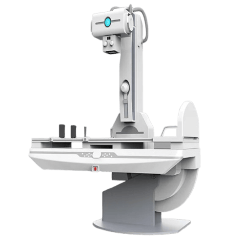
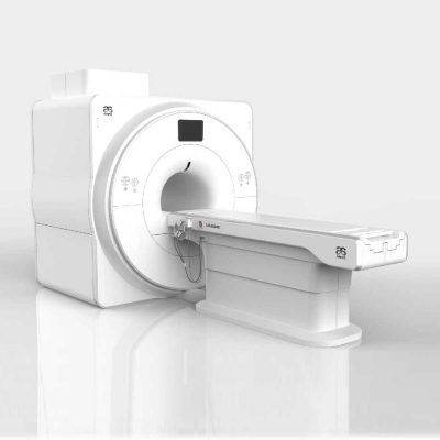
Digital Gastrointestinal Machine
$0.00
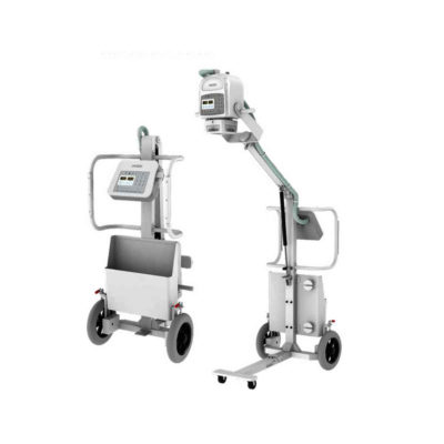
The digital gastrointestinal machine is designed for high-performance, long-duration diagnostics, making it ideal for gastrointestinal examinations and fluoroscopy procedures.
Shipped From China
Typically 10-21 working days – excluding furniture and heavy/bulky equipment. Please contact us for further information.
Description
The digital gastrointestinal machine is designed for high-performance, long-duration diagnostics, making it ideal for gastrointestinal examinations and fluoroscopy procedures. With its high-frequency inverter and high-voltage generator, you get a constant and stable DC output, ensuring clear and consistent imaging throughout every procedure. Equipped with a dynamic flat panel detector, this system allows for direct digital radiography and fluoroscopy in multiple positions. The high-density grid further enhances X-ray quality, while its low-dose fluoroscopy design provides safe and reliable imaging for gastrointestinal disease diagnosis.
This compact and versatile digital gastrointestinal machine supports radiography, fluoroscopy, stomach exams, and gastrointestinal imaging, giving you a complete diagnostic solution. Its wide-range movement and automatic DR functionality reduce patient repositioning, making your workflow smoother and more efficient.
Specifications
Ambient Conditions
- Ambient temperature: 10℃ to 40℃
- Relative humidity: 30-75%
- Atmospheric: 70-106 kPa
X-Ray Generator
- Model: SHO-DDR01 Series
- Output power (kW): 50, 65, 80
- Current (mA): 10-630, 10-800, 0-1000
- Voltage (kV): 40-150, 40-150, 40-150
- mAs (mAs): 0.1-630, 0.1-800, 0.1-1000
- Exposure time (ms): 1ms-6300
- Focus size (mm): 0.6/1.2
X-Ray Tube
- Anode type: Rotating Anode
- Focal Spot value: 0.6 mm / 1.2 mm
- Maximum voltage: 150 kV
- Revolving speed of anode: 2800 rpm
- Inherent filtration: 1.0 mmAl
- Anode heat storage capacity: 300 kHU
- Maximum cooling rate of anode: 870 W
- Housing heat storage capacity: 900 kJ
X-Ray Detector (Iray Brand Flat Panel Detector)
- Type: Amorphous Silicon
- Scintillator: Cesium Iodide
- Active Area: 17×17 inch (43×43 cm)
- Active Pixel: 3072×3072
- Pixel Pitch: 139 um
- Limiting resolution: 36 Lp/cm
- Data acquisition time: ≤2s
Collimator
- Type: Electric and multileaf collimator
- Power: 150 W; 24 VAC 1
- Lamp timer: Automatic illumination with timer for lamp (30 s)
- Filtration: 1.2 mmAL
Mobile Patient Table
- Type: Table for gastrointestinal imaging
- Tabletop lateral movement: 250 mm
- Rotating range: -30° to +30°
- Height of bed surface: 950 mm
- Bed bearing weight: 135 kg
- Bed rotation angle range and deviation: +90° to 0° to -20°
Mechanical movement
- SID: 1000-1800 mm
- Column lateral movement range: 0-1000 mm
Workstation
- Monitor: 23’’ LCD Dell brand
- CPU: ≥2.8 GHz
- Memory: ≥4 GB
- HDD: ≥1 TB
- DICOM3.0: Query for integration with any PACS
- Functions: Import/export function
Quick Comparison
| Digital Gastrointestinal Machine remove | DrGem Diamond All-In-One Digital X-ray Machine remove | Anke Anatom 32 Fit Multi-Slice Spiral CT Scan remove | Jade Mobile X-ray machine (Analogue) remove | SIGNERS SUPiA Dry Thermal X-ray Film Printer remove | DrGem Ceiling Analogue X-ray Machine remove | |||||||||||||||||||||||||||||||||||||||||||||||||||||||||||||||||||||||||||||||||||||||||||||||||||||||||||||||||||||||||||||||||||||||||||||||||||||||||||||||||||||||||||||||||||||||||||||||||||||||||||||||||||||||||||||||||||||||||||||||||||||||||||||||||||||||||||||||||||||||||||||||||||||||||||||||||||||||||
|---|---|---|---|---|---|---|---|---|---|---|---|---|---|---|---|---|---|---|---|---|---|---|---|---|---|---|---|---|---|---|---|---|---|---|---|---|---|---|---|---|---|---|---|---|---|---|---|---|---|---|---|---|---|---|---|---|---|---|---|---|---|---|---|---|---|---|---|---|---|---|---|---|---|---|---|---|---|---|---|---|---|---|---|---|---|---|---|---|---|---|---|---|---|---|---|---|---|---|---|---|---|---|---|---|---|---|---|---|---|---|---|---|---|---|---|---|---|---|---|---|---|---|---|---|---|---|---|---|---|---|---|---|---|---|---|---|---|---|---|---|---|---|---|---|---|---|---|---|---|---|---|---|---|---|---|---|---|---|---|---|---|---|---|---|---|---|---|---|---|---|---|---|---|---|---|---|---|---|---|---|---|---|---|---|---|---|---|---|---|---|---|---|---|---|---|---|---|---|---|---|---|---|---|---|---|---|---|---|---|---|---|---|---|---|---|---|---|---|---|---|---|---|---|---|---|---|---|---|---|---|---|---|---|---|---|---|---|---|---|---|---|---|---|---|---|---|---|---|---|---|---|---|---|---|---|---|---|---|---|---|---|---|---|---|---|---|---|---|---|---|---|---|---|---|---|---|---|---|---|---|---|---|---|---|---|---|---|---|---|---|---|---|---|---|---|---|---|---|---|---|---|---|---|---|---|---|---|---|---|---|---|---|---|---|---|---|---|---|
| Name | Digital Gastrointestinal Machine remove | DrGem Diamond All-In-One Digital X-ray Machine remove | Anke Anatom 32 Fit Multi-Slice Spiral CT Scan remove | Jade Mobile X-ray machine (Analogue) remove | SIGNERS SUPiA Dry Thermal X-ray Film Printer remove | DrGem Ceiling Analogue X-ray Machine remove | ||||||||||||||||||||||||||||||||||||||||||||||||||||||||||||||||||||||||||||||||||||||||||||||||||||||||||||||||||||||||||||||||||||||||||||||||||||||||||||||||||||||||||||||||||||||||||||||||||||||||||||||||||||||||||||||||||||||||||||||||||||||||||||||||||||||||||||||||||||||||||||||||||||||||||||||||||||||||
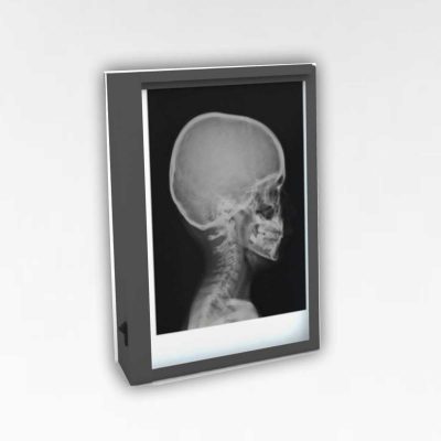
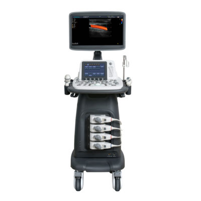
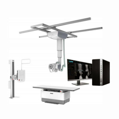
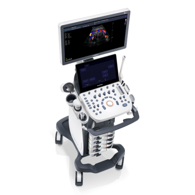
| Image |  | 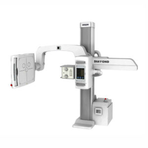 | 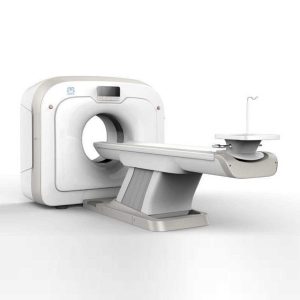 | 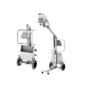 | 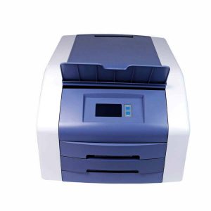 | 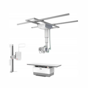 | ||||||||||||||||||||||||||||||||||||||||||||||||||||||||||||||||||||||||||||||||||||||||||||||||||||||||||||||||||||||||||||||||||||||||||||||||||||||||||||||||||||||||||||||||||||||||||||||||||||||||||||||||||||||||||||||||||||||||||||||||||||||||||||||||||||||||||||||||||||||||||||||||||||||||||||||||||||||||
| SKU | SF1033560074-3 | SF1033560092-1 | SF1033560074-2 | SF1033560050-02 | SF1033560074-7 | |||||||||||||||||||||||||||||||||||||||||||||||||||||||||||||||||||||||||||||||||||||||||||||||||||||||||||||||||||||||||||||||||||||||||||||||||||||||||||||||||||||||||||||||||||||||||||||||||||||||||||||||||||||||||||||||||||||||||||||||||||||||||||||||||||||||||||||||||||||||||||||||||||||||||||||||||||||||||
| Rating | ||||||||||||||||||||||||||||||||||||||||||||||||||||||||||||||||||||||||||||||||||||||||||||||||||||||||||||||||||||||||||||||||||||||||||||||||||||||||||||||||||||||||||||||||||||||||||||||||||||||||||||||||||||||||||||||||||||||||||||||||||||||||||||||||||||||||||||||||||||||||||||||||||||||||||||||||||||||||||||||
| Price |
|
|
|
|
|
| ||||||||||||||||||||||||||||||||||||||||||||||||||||||||||||||||||||||||||||||||||||||||||||||||||||||||||||||||||||||||||||||||||||||||||||||||||||||||||||||||||||||||||||||||||||||||||||||||||||||||||||||||||||||||||||||||||||||||||||||||||||||||||||||||||||||||||||||||||||||||||||||||||||||||||||||||||||||||
| Stock | ||||||||||||||||||||||||||||||||||||||||||||||||||||||||||||||||||||||||||||||||||||||||||||||||||||||||||||||||||||||||||||||||||||||||||||||||||||||||||||||||||||||||||||||||||||||||||||||||||||||||||||||||||||||||||||||||||||||||||||||||||||||||||||||||||||||||||||||||||||||||||||||||||||||||||||||||||||||||||||||
| Availability | ||||||||||||||||||||||||||||||||||||||||||||||||||||||||||||||||||||||||||||||||||||||||||||||||||||||||||||||||||||||||||||||||||||||||||||||||||||||||||||||||||||||||||||||||||||||||||||||||||||||||||||||||||||||||||||||||||||||||||||||||||||||||||||||||||||||||||||||||||||||||||||||||||||||||||||||||||||||||||||||
| Add to cart | ||||||||||||||||||||||||||||||||||||||||||||||||||||||||||||||||||||||||||||||||||||||||||||||||||||||||||||||||||||||||||||||||||||||||||||||||||||||||||||||||||||||||||||||||||||||||||||||||||||||||||||||||||||||||||||||||||||||||||||||||||||||||||||||||||||||||||||||||||||||||||||||||||||||||||||||||||||||||||||||
| Description | The digital gastrointestinal machine is designed for high-performance, long-duration diagnostics, making it ideal for gastrointestinal examinations and fluoroscopy procedures.
Shipped From China
Delivery & Availability:
Typically 10-21 working days – excluding furniture and heavy/bulky equipment. Please contact us for further information.
| Shipped from Abroad DrGem Diamond All-In-One Digital X-ray Machine is a fully automatic digital radiography system providing state-of-the-art image quality, image processing and user interface. With a wide selection of anatomical studies on the imaging software, DIAMOND automatically sets up the x-ray generator’s preprogrammed exposure technique settings, motorized radiographic stand positioning, x-ray collimation and post-image processing for the selected study. Specifically designed to increase workflow, this fully digital system offers convenient auto-positioning and advanced image processing to achieve big performance with little effort. Delivery & Availability: Typically 21 working days – excluding furniture and heavy/bulky equipment. Please contact us for further information. | Shipped from Abroad
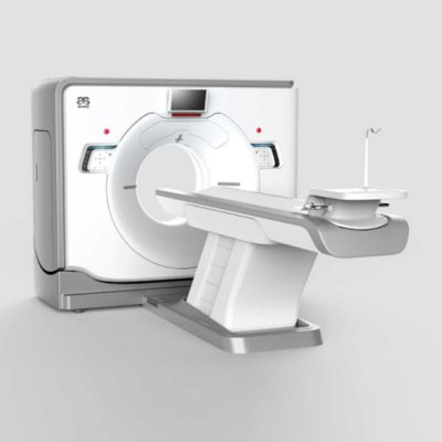
This Machine gives a possibility to perform computed tomography without any problems and on high quality level. This device is used to conduct exams of internal organs and their functioning. With its help, a physician has a possibility to assess the condition of the human body as a whole.
Delivery & Availability: Typically 90 working days – excluding furniture and heavy/bulky equipment. Please contact us for further information. | In Stock JADE is one of the lightest portable X-ray systems on the market, allowing it to be used in any imaginable way including bedside, operating rooms, intensive care units and in veterinary fields. With a simple, easy-to-use operator console, three-way control, two-step foldable stand and auto lock system, JADE is a user-friendly portable X-ray system. Delivery & Availability: Typically 21 working days – excluding furniture and heavy/bulky equipment. Please contact us for further information. | Shipped from Abroad
Signers Digital Dry Thermal X-ray Film Printer - Is a dry imager designed to output information processed through DICOM network protocol, supports 4 films sizes. 8”x10”, 10”x12”, 11”x14” and 14”x17” Achieve the convenience of multiple sizes.
Delivery & Availability: Typically 14 working days – excluding furniture and heavy/bulky equipment. Please contact us for further information. | Shipped from abroad The DrGem Ceiling Analogue X-ray Machine is a diagnostic radiography system that provides reliable high quality radiographic images with a reduced dose. The reliable high-frequency x-ray generators that are known worldwide for their excellent performance, lifetime and stability. Patient tables and wall stands are also offered. Delivery & Availability: Typically 21 working days – excluding furniture and heavy/bulky equipment. Please contact us for further information. | ||||||||||||||||||||||||||||||||||||||||||||||||||||||||||||||||||||||||||||||||||||||||||||||||||||||||||||||||||||||||||||||||||||||||||||||||||||||||||||||||||||||||||||||||||||||||||||||||||||||||||||||||||||||||||||||||||||||||||||||||||||||||||||||||||||||||||||||||||||||||||||||||||||||||||||||||||||||||
| Content | The digital gastrointestinal machine is designed for high-performance, long-duration diagnostics, making it ideal for gastrointestinal examinations and fluoroscopy procedures. With its high-frequency inverter and high-voltage generator, you get a constant and stable DC output, ensuring clear and consistent imaging throughout every procedure. Equipped with a dynamic flat panel detector, this system allows for direct digital radiography and fluoroscopy in multiple positions. The high-density grid further enhances X-ray quality, while its low-dose fluoroscopy design provides safe and reliable imaging for gastrointestinal disease diagnosis.
This compact and versatile digital gastrointestinal machine supports radiography, fluoroscopy, stomach exams, and gastrointestinal imaging, giving you a complete diagnostic solution. Its wide-range movement and automatic DR functionality reduce patient repositioning, making your workflow smoother and more efficient.
Specifications
Ambient Conditions
X-Ray Detector (Iray Brand Flat Panel Detector)
Mobile Patient Table
| DrGem Diamond All-In-One Digital X-ray Machine is a fully automatic digital radiography system providing state-of-the-art image quality, image processing and user interface. With a wide selection of anatomical studies on the imaging software, DIAMOND automatically sets up the x-ray generator’s pre-programmed exposure technique settings, motorized radiographic stand positioning, x-ray collimation and post-image processing for the selected study. Specifically designed to increase workflow, this fully digital system offers convenient auto-positioning and advanced image processing to achieve big performance with little effort.
Features of DrGem Diamond All-In-One Digital X-ray Machine:
Outstanding Image Quality -
Digital radiography via at panel detector improves your workflow, exam speed and comfort with efficiency. Digital at panel detector with Csl screen provides excellent spatial resolution, MTF, DQE and stability based on ne pixel pitch. A 3-field ion-chamber is provided for AEC function.
Automatic Collimation –
Automatic x-ray eld size control of the motorized collimator corresponds to dierent SIDs. Includes user adjustable lamp timer with on/oswitch.
Automatic Positioning –
Click Here To Download Catalogue | This Machine gives a possibility to perform computed tomography without any problems and on high quality level. This device is used to conduct exams of internal organs and their functioning. With its help, a physician has a possibility to assess the condition of the human body as a whole.
Features:
Click Here To Download Catalogue | JADE Mobile X-ray machine is one of the lightest portable X-ray systems on the market, allowing it to be used in any imaginable way including bedside, operating rooms, intensive care units and veterinary fields. With a simple, easy-to-use operator console, three-way control, two-step foldable stand and auto-lock system, the JADE Mobile X-ray machine is a user-friendly portable X-ray system.
Convenient & Intuitive Operation:
JADE is one of the lightest portable X-ray systems on the market, allowing it to be used in any imaginable way including bedside, operating rooms, intensive care units and in veterinary fields. With a simple, easy-to-use operator console, three-way control, two-step foldable stand and auto-lock system, JADE is a user-friendly portable X-ray system.
Compact & Powerful Design:
JADE Mobile X-ray machine is an innovative, highly versatile portable X-ray system suitable for a variety of clinical uses. Utilizing the unique technology used in DRGEM’s universally recognized X-ray generators, JADE is a compact but powerful unit with a 4kW output and thoughtfully designed components to increase efficiency and maximize workflow. The core part of X-ray source adopts high-quality tube assembly, X-ray collimator and high frequency X-ray generator with excellent performance, lifetime and stability.
Features:
Click Here To Download Catalogue | Signers Digital Dry Thermal X-ray Film Printer - Is a dry imager designed to output information processed through DICOM network protocol, supports 4 films sizes. 8”x10”, 10”x12”, 11”x14” and 14”x17” Achieve the convenience of multiple sizes.
Technical Specifications:
Click Here To Download Catalogue | DrGem Ceiling Analogue X-ray Machine is a diagnostic radiography system X-ray Machine that provides reliable high quality radiographic images with a reduced dose. The reliable high-frequency x-ray generators that are known worldwide for their excellent performance, lifetime and stability. Patient tables and wall stands are also offered.
Features of DrGem Ceiling Analogue X-ray Machine
Click Here To Download Catalogue | ||||||||||||||||||||||||||||||||||||||||||||||||||||||||||||||||||||||||||||||||||||||||||||||||||||||||||||||||||||||||||||||||||||||||||||||||||||||||||||||||||||||||||||||||||||||||||||||||||||||||||||||||||||||||||||||||||||||||||||||||||||||||||||||||||||||||||||||||||||||||||||||||||||||||||||||||||||||||
| Weight | N/A | N/A | N/A | N/A | N/A | N/A | ||||||||||||||||||||||||||||||||||||||||||||||||||||||||||||||||||||||||||||||||||||||||||||||||||||||||||||||||||||||||||||||||||||||||||||||||||||||||||||||||||||||||||||||||||||||||||||||||||||||||||||||||||||||||||||||||||||||||||||||||||||||||||||||||||||||||||||||||||||||||||||||||||||||||||||||||||||||||
| Dimensions | N/A | N/A | N/A | N/A | N/A | N/A | ||||||||||||||||||||||||||||||||||||||||||||||||||||||||||||||||||||||||||||||||||||||||||||||||||||||||||||||||||||||||||||||||||||||||||||||||||||||||||||||||||||||||||||||||||||||||||||||||||||||||||||||||||||||||||||||||||||||||||||||||||||||||||||||||||||||||||||||||||||||||||||||||||||||||||||||||||||||||
| Additional information |
|


Reviews
There are no reviews yet.