
Digital Optical Colposcope With Leica M60 Micro Optical Lens
$0.00
Shipped From Abroad
This high-definition digital optical colposcope integrates a Leica M60 micro-optical lens with 0.63×–4.0× optical amplification and a 2-megapixel digital CCD. It provides sharp cervical, vaginal, and vulva imaging with PACS connectivity for efficient clinical diagnostics.
Typically 10-21 working days – excluding furniture and heavy/bulky equipment. Please contact us for further information.
Description
Apparatus Features
SW-3300 series Digital Optical Colposcope is a high-resolution, high-definition color digital CCD imaging system with an optical microscope configuration. The development of high brightness luminous shadowless lighting sets, new shape, high speed image acquisition and processing system, image analysis and processing Microsoft, color printing report output device, rotary bracket, luxury car, etc, it collects a variety of advanced characteristics as a whole new type of digital optical colposcope imaging system. It can be connected with the PACS to store and transfer the data, sharing the patient’s record sources.
Technical parameters
- Portion of the lens: Leica M60 Micro Optical Lens
- Optical Amplification: 0.63× to 4.0×
- Zoom Ratio: 6:1 step-less zoom.
- Compound Lens: Complete achromatic compound lens.
- Eyepiece: Wide vision, 10×21mm.
- Camera System: Professional digital 1/1.8″CCD, 2 million pixels, microcomputer control, automatic white balance, color saturation, and so on. 1600X1200 resolution acquisition mode, horizontal resolution can reach above 750TVL.
- Lifting bracket: 360 °rotating electric lifting platform.
Working Principle
Digital optical colposcope merges high resolution electronic digital imaging system and high-tech computer technology, it suitable for the diagnosis of gynecological diseases, under light irradiation, with microscope and high-resolution color CCD camera system, magnify the cervix, vagina and vulva to the definite multiples, through the CCD to transfer the optical information into digital information and then the images formation, through images amplification, lessen, freeze, store, analysis we will see the nidus more accurate and clear, observe the invisible change of epithelium and vessel greatly enhance the accuracy in diagnosis of gynecology, so as to guide clinical treatment.
Specifications
| Specification | Details |
|---|---|
| Optical Lens | Leica M60 micro optical lens |
| Optical Amplification | 0.63× to 4.0× |
| Zoom Ratio | 6:1, step-less zoom |
| Compound Lens | Achromatic compound lens |
| Eyepiece | Wide vision 10×21 mm |
| Camera System | 1/1.8″ CCD, 2 megapixels |
| Capture Resolution | 1600 × 1200 |
| Horizontal Resolution | ≥ 750 TV lines |
| Lifting Bracket | 360° rotating electric platform |
| Brand | Sanwe |
| Product Origin | Xuzhou, China |
| Delivery Time | 3-5 working days after payment |
| Supply Capacity | 5 sets per month |
Quick Comparison
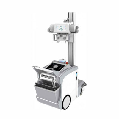
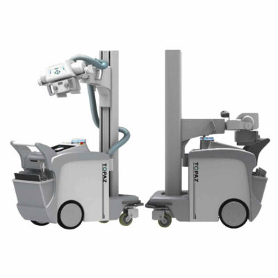
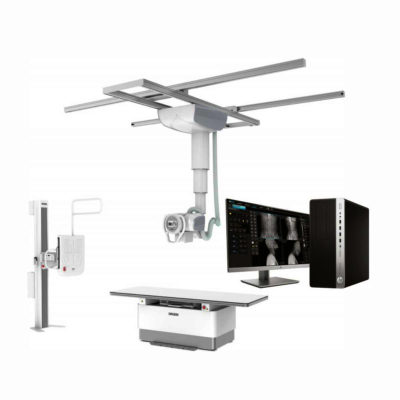
| Digital Optical Colposcope With Leica M60 Micro Optical Lens remove | Sonoscape S11 Ultrasound Machine remove | DrGem Floor Mounted Analogue X-ray remove | Sonoscape E2 Ultrasound Machine remove | Sonoscape S8 Exp Portable Ultrasound remove | Sonoscape P15 Ultrasound Machine With Four Probes remove | |||||||||||||||||||||||||||||
|---|---|---|---|---|---|---|---|---|---|---|---|---|---|---|---|---|---|---|---|---|---|---|---|---|---|---|---|---|---|---|---|---|---|---|
| Name | Digital Optical Colposcope With Leica M60 Micro Optical Lens remove | Sonoscape S11 Ultrasound Machine remove | DrGem Floor Mounted Analogue X-ray remove | Sonoscape E2 Ultrasound Machine remove | Sonoscape S8 Exp Portable Ultrasound remove | Sonoscape P15 Ultrasound Machine With Four Probes remove | ||||||||||||||||||||||||||||
| Image | 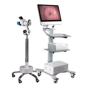 | 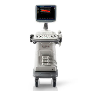 |  | 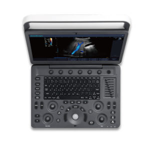 |  | 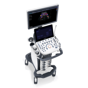 | ||||||||||||||||||||||||||||
| SKU | SF1033560012-1 | SF1033560074-6 | SF1033560012-17 | SF1033560012-15 | SF1033560012-8 | |||||||||||||||||||||||||||||
| Rating | ||||||||||||||||||||||||||||||||||
| Price |
|
|
|
|
|
| ||||||||||||||||||||||||||||
| Stock | ||||||||||||||||||||||||||||||||||
| Availability | ||||||||||||||||||||||||||||||||||
| Add to cart | ||||||||||||||||||||||||||||||||||
| Description | Shipped From Abroad
This high-definition digital optical colposcope integrates a Leica M60 micro-optical lens with 0.63×–4.0× optical amplification and a 2-megapixel digital CCD. It provides sharp cervical, vaginal, and vulva imaging with PACS connectivity for efficient clinical diagnostics.
Delivery & Availability:
Typically 10-21 working days – excluding furniture and heavy/bulky equipment. Please contact us for further information.
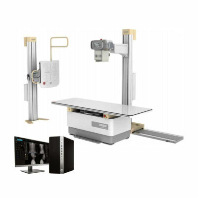
| In Stock A Value Choice beyond Your Expectation. SonoScape’s trolley color Doppler system S11 redefines price and performance with practical design. The S11 will go beyond your expectations but not your budget. Delivery & Availability: Typically 2 working days – excluding furniture and heavy/bulky equipment. Please contact us for further information. | In Stock GXR Analogue X-ray system matches with a radiographic room which perfectly fits your workow and can be easily upgraded to DR system with the help of DR interface and PC interface in GXR generator as well as Bucky suitable to Flat Panel Detector. GXR X-ray system is equipped with a high frequency X-ray generator which consistently produces high quality radiograph in favor of high quality X-ray output with a very small kV ripple and accurate mA and mAs. GXR X-ray system is designed to provide convenience to operator and comfort to patient. Delivery & Availability: Typically 21 working days – excluding furniture and heavy/bulky equipment. Please contact us for further information. | Shipped from Abroad Sonoscape E2 portable ultrasound machine is a color Doppler ultrasound system that reaches beyond your expectations due to its compact and fashionable appearance. It fulfills GI, OB/GYN, Cardiac and POC applications to fit your routine scanning needs while its color mode will help you for more accurate and efficient diagnosis of lesions. E2 provides a wide range of applications to assist users with routine scanning. E2 provides automatic calculations to enhance your diagnostic confidence and save you time for patient communication. Delivery & Availability: Typically 14 working days – excluding furniture and heavy/bulky equipment. Please contact us for further information. | Shipped from Abroad With ultra-modern innovative design and the clinically-proven technologies, S8 Exp is portable ultrasound scanner well equipped as a low-physical-effort and enhanced-image-quality ultrasound scanner, which not only provides optimized solutions for versatile applications, but does help to improve the user-experience for both routine and non-traditional challenges. Delivery & Availability: Typically 5-7 working days – excluding furniture and heavy/bulky equipment. Please contact us for further information. | In Stock A feature-rich system inheriting the Wi-Sono high-end platform, the P15 uses an array of advanced tools to help enhance the image quality. It's a cost-effective, simplified console with an intuitive user interface and multiple intelligent functions. Delivery & Availability: Typically 2 working days – excluding furniture and heavy/bulky equipment. Please contact us for further information. | ||||||||||||||||||||||||||||
| Content | Apparatus FeaturesSW-3300 series Digital Optical Colposcope is a high-resolution, high-definition color digital CCD imaging system with an optical microscope configuration. The development of high brightness luminous shadowless lighting sets, new shape, high speed image acquisition and processing system, image analysis and processing Microsoft, color printing report output device, rotary bracket, luxury car, etc, it collects a variety of advanced characteristics as a whole new type of digital optical colposcope imaging system. It can be connected with the PACS to store and transfer the data, sharing the patient's record sources.Technical parameters
Working PrincipleDigital optical colposcope merges high resolution electronic digital imaging system and high-tech computer technology, it suitable for the diagnosis of gynecological diseases, under light irradiation, with microscope and high-resolution color CCD camera system, magnify the cervix, vagina and vulva to the definite multiples, through the CCD to transfer the optical information into digital information and then the images formation, through images amplification, lessen, freeze, store, analysis we will see the nidus more accurate and clear, observe the invisible change of epithelium and vessel greatly enhance the accuracy in diagnosis of gynecology, so as to guide clinical treatment.Specifications
| DETAILS
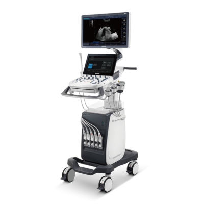
SonoScape’s trolley colour Doppler system S11 redefines price and performance with practical design. The S11 will go beyond your expectations but not your budget. As an easy-to-use ultrasound system, the S11 is integrated with a new software platform, especially optimized for a smooth workflow and convenient operation. The system speeds up the exam process and makes file management easier.
SPECIFICATION
- 15-inch high definition LCD monitor with articulating arm
- Compact and agile trolley design
- 3 active transducer sockets available for a wide range of applications
- Duplex, Color Doppler, DPI, PW Doppler, tissue harmonic imaging, μ-scan speckle reduction imaging, compound imaging, trapezoidal imaging
- Customized settings based on your own working style
- Full patient database and image management solutions
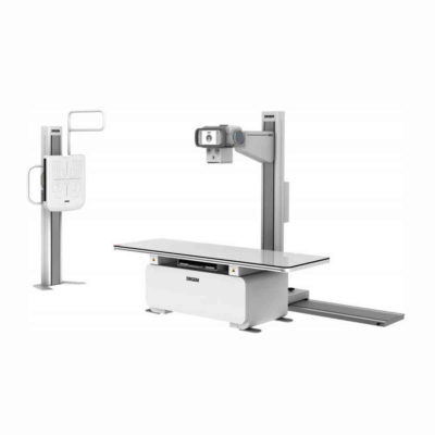
Click Here To Download Catalogue | DrGem GXR Floor Mounted Analogue X-ray system matches with a radiographic room which perfectly fits your workflow and can be easily upgraded to DR system with the help of DR interface and PC interface in GXR generator as well as Bucky suitable to Flat Panel Detector. GXR (Analogue X-ray)system is equipped with a high frequency X-ray generator which consistently produces high quality radiograph in favor of high quality X-ray output with a very small kV ripple and accurate mA and mAs. GXR (Analogue X-ray) system is designed to provide convenience to operator and comfort to patient.
Features of DrGem GXR Floor Mounted Analogue X-ray:
Click Here To Download Catalogue | SONOSCAPE E2 DETAILS
Auto Image Optimization
A portable ultrasound machine with the press of a button, the image is automatically adjusted and optimized, saving you time with parameter adjustments. Additionally, with Auto Focus on, the focus area follows the depth of the ROI box as it is moved in the scanning field, providing users with excellent image quality in the desired area of interest.
Automated Calculation
Auto IMT is used when determining the level of vascular sclerosis present in the patient by automatically tracing the thickness of the carotid vessels.
Auto trace provides users sensitive and accurate wave tracing, avoiding the error of manual trace and giving out calculation result in no time
In-Build Battery pack
This portable ultrasound machine was equipped with an in-build battery pack which enable the user to perform image scanning when AC power is not available.
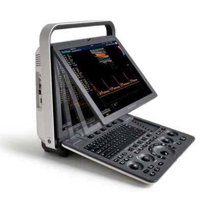
Click Here To Download Catalogue | Sonoscape S8 Exp Portable Ultrasound scannerDETAILS Agile and Versatile With ultra-modern innovative design and the clinically-proven technologies, S8 Exp Portable Ultrasound scanner is well equipped as a low-physical-effort and enhanced-image-quality ultrasound scanner, which not only provides optimized solutions for versatile applications but does help to improve the user experience for both routine and non-traditional challenges. Working with S8 Exp, it will trigger your unlimited reverie and endow you with endless charm. Carrying forward the classical design of SonoScape's portable ultrasound products, S8 Exp successfully combines the best ergonomics, attractive design and ease of use. This charismatic identity is also enhanced by a sophisticated color palette—with sedate grey as its interior paint color and pearl white as exterior cover, S8 Exp reveals a style of aristocrat and strong character among SonoScape's ultrasound systems. Workflow The S8 Exp is a portable ultrasound scanner that adapts to your workflow, whether you are in the consulting room, at the bedside, or at a remote location. With easy-to-use new platform designed for sonographers' needs and full connection interfaces for easy connectivity and data sharing, S8 Exp leads to improved user comfort and clinical outcome as well as patient throughput and working efficiency. Powerful Platform Embedded with SonoScape's core imaging technologies such as μ-scan, PHI and Spatial Compound, S8 Exp boasts exceptional 2D image, sensitive spectral, Color and Power Doppler, displaying well-defined anatomy and pathology and facilitating a highly optimized diagnostic user environment for conclusive diagnoses. Besides, S8 Exp offers a comprehensive selection of electronic probes to maximally extend its capabilities to meet a wide range of applications including the abdomen, pediatric, OB/GYN, cardiovascular, musculoskeletal, etc. The advanced probe technologies also effectively enhance the image quality and confidence in reaching clinical diagnoses even in difficult patients.Click Here To Download Catalogue | DETAILS
Super Wide-bandwidth Platform
Inheriting Wi-sono's ultra-wide system platform and with the advanced probe technology, high-resolution and deep penetration images are provided for precision medicine.
Spatial Compound Imaging
Spatial Compound Imaging utilizes several lines of sight for optimal contrast resolution, speckle reduction and border detection, with which P15 is ideal for superficial and abdominal imaging with better clarity and improved continuity of structures.
μ-Scan+
The new generation μ-Scan imaging technology gives you better image quality by reducing noise, improving signal strength and improving visualization.
Dynamic Color
Dynamic color improves upon already existing color Doppler technologies for a clearer capture of color flow and detailed visualization of even tiny veins with lower velocities.
Real-time Panoramic
With real-time panoramic, you can acquire an extended field of view for large organs or long vessels for easy measurement and diagnostic efficiency. Accomplished in real-time for the convenience of the sonographers, any mistake can also be easily back tracked and corrected without interrupting the scan.
3D/4D
Outstanding volume performance with speed and convenience makes P15 outshine others on volume imaging.
Tissue Doppler Imaging
Tissue Doppler Imaging allows clinical doctors to quantitatively evaluate local myocardial movements and functions, facilitating them with the ability to analyze and compare the motions of the different parts of the patient's heart.
Auto IMT
Quick measurement of intra-media vessel thickness ensures good reproducibility and high diagnostic efficiency.
Click Here To Download Catalogue | ||||||||||||||||||||||||||||
| Weight | N/A | N/A | N/A | N/A | N/A | N/A | ||||||||||||||||||||||||||||
| Dimensions | N/A | N/A | N/A | N/A | N/A | N/A | ||||||||||||||||||||||||||||
| Additional information |

Reviews
There are no reviews yet.