Nio Gray 5.8MP (MDNG‑6221)
$0.00
Shipped From Abroad
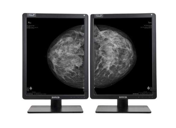
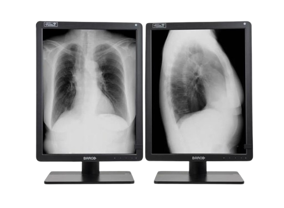
The Barco Nio Gray 5.8MP (MDNG-6221) is a high-performance grayscale medical display designed for mammography and diagnostic imaging. It ensures superior image clarity, consistent luminance, and advanced calibration for precise and reliable clinical interpretation in breast imaging and radiology.
Typically 10-21 working days – excluding furniture and heavy/bulky equipment. Please contact us for further information.
Description
The Barco Nio Gray 5.8MP (MDNG-6221) is a premium grayscale diagnostic display optimized for mammography and breast imaging. With a 5.8-megapixel resolution, exceptional luminance, and DICOM compliance, it delivers outstanding image quality for accurate detection and interpretation. Equipped with I-Guard for continuous quality assurance and QAWeb for remote management, this display ensures long-term consistency and reliability. Its ergonomic design and advanced grayscale rendering make it the ideal solution for radiologists requiring precise visualization in demanding diagnostic environments.
Bigger image, more details
Why 5.8MP? Well, in contrast to conventional 5.2MP display systems, you get 12% more pixels on your screen, which means that you can see more details at any given moment. Combine this with the tall 4:3 aspect ratio, which offers more room to view images in their entirety.
Reliable reading
The Nio Gray 5.8MP offers you more Just Noticeable Differences, thanks to its high brightness and contrast ratio. Our integrated stability, calibration, and uniformity technologies make sure that image quality, light output and DICOM compliance remain consistent over the years. The ambient light measurement sensor helps you stay in control of the environment you work in.
Efficient workflow
The Nio Gray 5.8MP is more than a grayscale display alone. It offers many ways to personalize settings to your liking, such as preferred tints of white or viewing angle. The KVM option helps you switch between workstations at the touch of a button.
On top of that, the display can help you improve your efficiency and speed, thanks to the set of Intuitive Workflow Tools included with our MXRT medical display controllers.
Did you know that SpotView, for example, makes it possible to make an area you choose twice as bright as it was originally? It’s been proven to help radiologists reduce their reading time by as much as 15.5%.
Long lifetime, clear view
Install our free and secure QAWeb Enterprise application, and rely on intervention-free, remote quality assurance.
To summarize, your Nio Gray 5.8MP is a functional, easy-to-use diagnostic display system, fully up to date with today’s innovations in general grayscale radiology, as well as 2D and 3D mammography. It comes with a 5-year warranty on all its components.
Ensuring diagnostic confidence with MDR Class IIa
Our radiology displays are MDR-certified as Class IIa. Their product information has been reviewed and cleared by independent medical and technical experts, and is audited yearly. In other words, we ensure diagnostic confidence and peace of mind for our users.
Technologies that enhance image quality:
- More detail on your screen, with 5.8MP resolution
- Designed to show breast images entirely, with a 3:4 aspect ratio
- Increased contrast, with a 1400:1 contrast ratio and 600 to 1000 cd/m² calibrated luminance
- Consistent brightness and grays, with Uniform Luminance Technology and SteadyGray
- Always stable DICOM images and auto QA, with I-Guard front sensor and optionally, QAWeb Enterprise
- Possibility to boost luminance, with I-Luminate and SpotView
- Optional settings and tools to adjust the display to your workflow, with Intuitive Workflow Tools
Ecolabel A for Nio Gray 5.8MP
The Nio Gray 5.8MP has been subjected to Barco’s ecoscoring protocol and has received an A rating. Some key factors that contributed to this rating are:
- Energy-efficient power supply, energy-efficient standby, and off modes
- Possibility to automatically switch to standby mode when the device is not in use
- Halogen-free cables and plastics
- Use of recycled cardboard in packaging (>85% recycled content)
- Product design optimized for disassembly with common tools
Features
-
5.8MP high-resolution grayscale medical display
-
Optimized for mammography and diagnostic imaging
-
High luminance and contrast for clinical accuracy
-
I-Guard sensor for real-time quality assurance
-
DICOM calibration for consistent grayscale performance
-
QAWeb for remote quality and compliance management
-
Ergonomic design for comfortable, extended use
Specifications
| Category | Specification |
|---|---|
| Screen Technology | LCD |
| Active Screen Size | 21.3″ (541 mm); 324 × 433 mm (12.77″ × 17″) |
| Aspect Ratio | 3:4 per display (portrait), 3:2 overall |
| Resolution | 5.8 MP (2100 × 2800 pixels) |
| Pixel Pitch | 0.1545 mm |
| Color Imaging | No |
| Gray Imaging | Yes |
| Bit Depth | 10-bit |
| Viewing Angle | 178° (H/V) |
| Uniformity Correction | ULT |
| SteadyGray | Yes |
| SteadyColor | N/A |
| I-Luminate | Yes |
| Ambient Light Presets | Yes, reading room selection |
| Ambient Light Sensor | Yes |
| Front Sensor | Yes (I-Guard) |
| Presence Sensor | N/A |
| Maximum Luminance | 1560 cd/m² |
| DICOM Calibrated Luminance | Factory default: 600 cd/m²; Warrantied max: 1000 cd/m² |
| Contrast Ratio | 1400:1 |
| Response Time | 12.5 ms ((Tr + Tf)/2) |
| Housing Color | Black (RAL 9004) / White (RAL 9003) |
| Video Input Signals | 2 × DisplayPort 1.4 |
| Video Output Signals | N/A |
| USB Ports | 2 × USB-B 2.0 upstream; 5 × USB-A 2.0 downstream (1 charging) |
| KVM Switch | Yes |
| Power Rating | 24 VDC, 5 A |
| Power Requirements | AdapterTech ATM160T-P240 (100–240 Vac, 50–60 Hz, 1.8–0.9 A; Output: 24 VDC, 6.6 A) |
| Power Consumption | 60 W (nominal); 0.4 W (hibernate/off) |
| Dimensions with Stand | Portrait: 378 × 528~628 × 235 mm; Landscape: 491 × 472~572 × 235 mm |
| Dimensions without Stand | Portrait: 378 × 491 × 84 mm; Landscape: 491 × 378 × 84 mm |
| Packaged Dimensions | 500 × 280 × 670 mm |
| Net Weight with Stand | With cover: 11.9 kg; Without cover: 10.6 kg |
| Net Weight without Stand | With cover: 6.9 kg; Without cover: 5.6 kg |
| Packaged Weight | With cover: 16.9 kg; Without cover: 15.6 kg |
| Tilt / Swivel / Pivot | Tilt: -10° to +30°; Swivel: ±45°; Pivot: 90° |
| Height Adjustment Range | 100 mm |
| Mounting Standard | VESA (100 mm) |
| Screen Protection | Optional protective, anti-reflective glass |
| Recommended Modalities | All digital images, including digital mammography |
| Certifications | FDA 510(K) K170476, CE0123, CCC, KC, INMETRO, BIS |
| Safety Standards | IEC/EN/AAMI/CSA 62368-1, 60601-1 |
| EMI Compliance | IEC/EN 60601-1-2, FCC Part 15 Class B, ICES-001 Level B, VCCI |
| Environmental Compliance | EU RoHS, China RoHS, REACH, Canada Health, WEEE, Packaging Directive |
| Supplied Accessories | User Guide, Documentation Disc, System Sheet, DisplayPort Cable, USB Cable, Mains Cable(s), External Power Supply |
| Optional Accessories | Graphics Board, QA Software, QAWeb Enterprise |
| Warranty | 5 years, including 40,000 hours backlight warranty |
| Operating Temperature | 0–40 °C (specs: 15–30 °C) |
| Storage Temperature | -20–60 °C |
| Operating Humidity | 8–80% (non-condensing) |
| Storage Humidity | 5–85% (non-condensing) |
| Operating Pressure | 70 kPa |
| Storage Pressure | 50–106 kPa |
Quick Comparison
| Nio Gray 5.8MP (MDNG‑6221) remove | Sonoscape P20 Ultrasound Machine remove | Sonoscape E1 Ultrasound Machine With Two Probes remove | DrGem Ceiling Analogue X-ray Machine remove | Sonoscape P10 Ultrasound Machine remove | Anke MRI Openmark 5000 Permanent System remove | ||||||||||||||||||||||||||||||||||||||||||||||||||||||||||||||||||||||||||||||||||||||||||||||||||||||||||||||||||||||||||||||||||||||||||||||||||||||||||||||||||||||||||||||||||||||||||||||||||||||||||||||||||||||||||||||||||||||||||||||||||||||||||||||||||||||||||||||||||||||||||||||||||||||||||||||||||||||||||||||||||||||||||||||||||||||||||||||||||||||||||||||||||||||||||||||||||||||||||||||||||||||||||||
|---|---|---|---|---|---|---|---|---|---|---|---|---|---|---|---|---|---|---|---|---|---|---|---|---|---|---|---|---|---|---|---|---|---|---|---|---|---|---|---|---|---|---|---|---|---|---|---|---|---|---|---|---|---|---|---|---|---|---|---|---|---|---|---|---|---|---|---|---|---|---|---|---|---|---|---|---|---|---|---|---|---|---|---|---|---|---|---|---|---|---|---|---|---|---|---|---|---|---|---|---|---|---|---|---|---|---|---|---|---|---|---|---|---|---|---|---|---|---|---|---|---|---|---|---|---|---|---|---|---|---|---|---|---|---|---|---|---|---|---|---|---|---|---|---|---|---|---|---|---|---|---|---|---|---|---|---|---|---|---|---|---|---|---|---|---|---|---|---|---|---|---|---|---|---|---|---|---|---|---|---|---|---|---|---|---|---|---|---|---|---|---|---|---|---|---|---|---|---|---|---|---|---|---|---|---|---|---|---|---|---|---|---|---|---|---|---|---|---|---|---|---|---|---|---|---|---|---|---|---|---|---|---|---|---|---|---|---|---|---|---|---|---|---|---|---|---|---|---|---|---|---|---|---|---|---|---|---|---|---|---|---|---|---|---|---|---|---|---|---|---|---|---|---|---|---|---|---|---|---|---|---|---|---|---|---|---|---|---|---|---|---|---|---|---|---|---|---|---|---|---|---|---|---|---|---|---|---|---|---|---|---|---|---|---|---|---|---|---|---|---|---|---|---|---|---|---|---|---|---|---|---|---|---|---|---|---|---|---|---|---|---|---|---|---|---|---|---|---|---|---|---|---|---|---|---|---|---|---|---|---|---|---|---|---|---|---|---|---|---|---|---|---|---|---|---|---|---|---|---|---|---|---|---|---|---|---|---|---|---|---|---|---|---|---|---|---|---|---|---|---|---|---|---|---|---|---|---|---|---|---|---|---|---|---|---|---|---|
| Name | Nio Gray 5.8MP (MDNG‑6221) remove | Sonoscape P20 Ultrasound Machine remove | Sonoscape E1 Ultrasound Machine With Two Probes remove | DrGem Ceiling Analogue X-ray Machine remove | Sonoscape P10 Ultrasound Machine remove | Anke MRI Openmark 5000 Permanent System remove | |||||||||||||||||||||||||||||||||||||||||||||||||||||||||||||||||||||||||||||||||||||||||||||||||||||||||||||||||||||||||||||||||||||||||||||||||||||||||||||||||||||||||||||||||||||||||||||||||||||||||||||||||||||||||||||||||||||||||||||||||||||||||||||||||||||||||||||||||||||||||||||||||||||||||||||||||||||||||||||||||||||||||||||||||||||||||||||||||||||||||||||||||||||||||||||||||||||||||||||||||||||||||||
| Image | 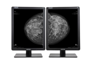 | 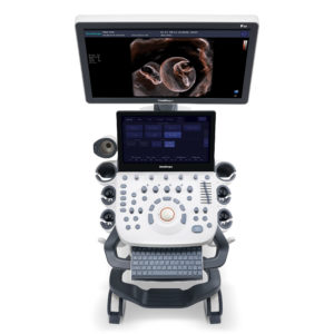 | 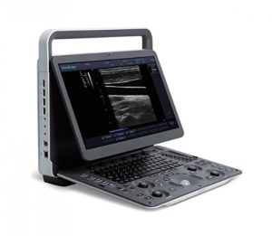 | 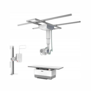 | 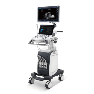 |  | |||||||||||||||||||||||||||||||||||||||||||||||||||||||||||||||||||||||||||||||||||||||||||||||||||||||||||||||||||||||||||||||||||||||||||||||||||||||||||||||||||||||||||||||||||||||||||||||||||||||||||||||||||||||||||||||||||||||||||||||||||||||||||||||||||||||||||||||||||||||||||||||||||||||||||||||||||||||||||||||||||||||||||||||||||||||||||||||||||||||||||||||||||||||||||||||||||||||||||||||||||||||||||
| SKU | SF1033560012-9 | SF1033560012-20 | SF1033560074-7 | SF1033560012-7 | SF1033560092-3 | ||||||||||||||||||||||||||||||||||||||||||||||||||||||||||||||||||||||||||||||||||||||||||||||||||||||||||||||||||||||||||||||||||||||||||||||||||||||||||||||||||||||||||||||||||||||||||||||||||||||||||||||||||||||||||||||||||||||||||||||||||||||||||||||||||||||||||||||||||||||||||||||||||||||||||||||||||||||||||||||||||||||||||||||||||||||||||||||||||||||||||||||||||||||||||||||||||||||||||||||||||||||||||||
| Rating | |||||||||||||||||||||||||||||||||||||||||||||||||||||||||||||||||||||||||||||||||||||||||||||||||||||||||||||||||||||||||||||||||||||||||||||||||||||||||||||||||||||||||||||||||||||||||||||||||||||||||||||||||||||||||||||||||||||||||||||||||||||||||||||||||||||||||||||||||||||||||||||||||||||||||||||||||||||||||||||||||||||||||||||||||||||||||||||||||||||||||||||||||||||||||||||||||||||||||||||||||||||||||||||||||
| Price |
|
|
|
|
|
| |||||||||||||||||||||||||||||||||||||||||||||||||||||||||||||||||||||||||||||||||||||||||||||||||||||||||||||||||||||||||||||||||||||||||||||||||||||||||||||||||||||||||||||||||||||||||||||||||||||||||||||||||||||||||||||||||||||||||||||||||||||||||||||||||||||||||||||||||||||||||||||||||||||||||||||||||||||||||||||||||||||||||||||||||||||||||||||||||||||||||||||||||||||||||||||||||||||||||||||||||||||||||||
| Stock | |||||||||||||||||||||||||||||||||||||||||||||||||||||||||||||||||||||||||||||||||||||||||||||||||||||||||||||||||||||||||||||||||||||||||||||||||||||||||||||||||||||||||||||||||||||||||||||||||||||||||||||||||||||||||||||||||||||||||||||||||||||||||||||||||||||||||||||||||||||||||||||||||||||||||||||||||||||||||||||||||||||||||||||||||||||||||||||||||||||||||||||||||||||||||||||||||||||||||||||||||||||||||||||||||
| Availability | |||||||||||||||||||||||||||||||||||||||||||||||||||||||||||||||||||||||||||||||||||||||||||||||||||||||||||||||||||||||||||||||||||||||||||||||||||||||||||||||||||||||||||||||||||||||||||||||||||||||||||||||||||||||||||||||||||||||||||||||||||||||||||||||||||||||||||||||||||||||||||||||||||||||||||||||||||||||||||||||||||||||||||||||||||||||||||||||||||||||||||||||||||||||||||||||||||||||||||||||||||||||||||||||||
| Add to cart | |||||||||||||||||||||||||||||||||||||||||||||||||||||||||||||||||||||||||||||||||||||||||||||||||||||||||||||||||||||||||||||||||||||||||||||||||||||||||||||||||||||||||||||||||||||||||||||||||||||||||||||||||||||||||||||||||||||||||||||||||||||||||||||||||||||||||||||||||||||||||||||||||||||||||||||||||||||||||||||||||||||||||||||||||||||||||||||||||||||||||||||||||||||||||||||||||||||||||||||||||||||||||||||||||
| Description | Shipped From Abroad
The Barco Nio Gray 5.8MP (MDNG-6221) is a high-performance grayscale medical display designed for mammography and diagnostic imaging. It ensures superior image clarity, consistent luminance, and advanced calibration for precise and reliable clinical interpretation in breast imaging and radiology.
Delivery & Availability:
Typically 10-21 working days – excluding furniture and heavy/bulky equipment. Please contact us for further information.
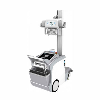
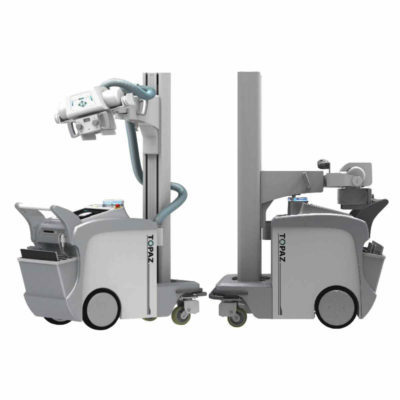
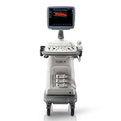
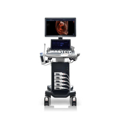
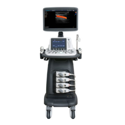
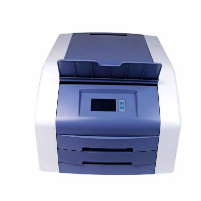
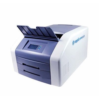
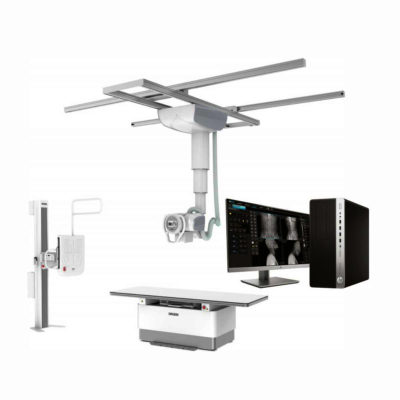
| Shipped from Abroad Incorporating innovative technologies, P20’s user-friendly design with a simple operation panel, intuitive user interface and a variety of intelligent auxiliary scanning tools, will significantly improve your daily examination experience. Besides general imaging applications, P20 has entitled with diagnostic 4D technology which has an extraordinary performance in obstetrics and gynecology applications. Delivery & Availability: Typically 5-7 working days – excluding furniture and heavy/bulky equipment. Please contact us for further information. | Shipped from Abroad SonoScape has developed a new probe and function for the E1 Exp. With these additions the E1 Exp will bring users a more efficient examination experience with satisfying image quality and a smooth workflow. Delivery & Availability: Typically 5-7 working days – excluding furniture and heavy/bulky equipment. Please contact us for further information. | Shipped from abroad The DrGem Ceiling Analogue X-ray Machine is a diagnostic radiography system that provides reliable high quality radiographic images with a reduced dose. The reliable high-frequency x-ray generators that are known worldwide for their excellent performance, lifetime and stability. Patient tables and wall stands are also offered. Delivery & Availability: Typically 21 working days – excluding furniture and heavy/bulky equipment. Please contact us for further information. | Shipped from Abroad The P10 color Doppler ultrasound system is a new generation product from SonoScape. It is designed to give high quality images, rich probe configurations, various clinical tools and automatic analysis software to provide you with comprehensive solutions for your growing demand for clinical applications. Delivery & Availability: Typically 5-7 working days – excluding furniture and heavy/bulky equipment. Please contact us for further information. | Shipped from Abroad
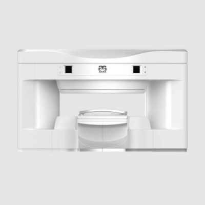
OPENMARK 5000 is 0.51T MRI. It's approved by FDA and has CE mark. It adopts two-pillar magnet design with 280 degree openness and equipped with powerful
RF and gradient system, together with advanced imaging technology, making it as a high-end system which is comparable to high-field MRI.
Delivery & Availability: Typically 90 working days – excluding furniture and heavy/bulky equipment. Please contact us for further information. | |||||||||||||||||||||||||||||||||||||||||||||||||||||||||||||||||||||||||||||||||||||||||||||||||||||||||||||||||||||||||||||||||||||||||||||||||||||||||||||||||||||||||||||||||||||||||||||||||||||||||||||||||||||||||||||||||||||||||||||||||||||||||||||||||||||||||||||||||||||||||||||||||||||||||||||||||||||||||||||||||||||||||||||||||||||||||||||||||||||||||||||||||||||||||||||||||||||||||||||||||||||||||||
| Content | The Barco Nio Gray 5.8MP (MDNG-6221) is a premium grayscale diagnostic display optimized for mammography and breast imaging. With a 5.8-megapixel resolution, exceptional luminance, and DICOM compliance, it delivers outstanding image quality for accurate detection and interpretation. Equipped with I-Guard for continuous quality assurance and QAWeb for remote management, this display ensures long-term consistency and reliability. Its ergonomic design and advanced grayscale rendering make it the ideal solution for radiologists requiring precise visualization in demanding diagnostic environments.
Bigger image, more details Why 5.8MP? Well, in contrast to conventional 5.2MP display systems, you get 12% more pixels on your screen, which means that you can see more details at any given moment. Combine this with the tall 4:3 aspect ratio, which offers more room to view images in their entirety. Reliable reading The Nio Gray 5.8MP offers you more Just Noticeable Differences, thanks to its high brightness and contrast ratio. Our integrated stability, calibration, and uniformity technologies make sure that image quality, light output and DICOM compliance remain consistent over the years. The ambient light measurement sensor helps you stay in control of the environment you work in. Efficient workflow The Nio Gray 5.8MP is more than a grayscale display alone. It offers many ways to personalize settings to your liking, such as preferred tints of white or viewing angle. The KVM option helps you switch between workstations at the touch of a button. On top of that, the display can help you improve your efficiency and speed, thanks to the set of Intuitive Workflow Tools included with our MXRT medical display controllers. Did you know that SpotView, for example, makes it possible to make an area you choose twice as bright as it was originally? It’s been proven to help radiologists reduce their reading time by as much as 15.5%. Long lifetime, clear view Install our free and secure QAWeb Enterprise application, and rely on intervention-free, remote quality assurance. To summarize, your Nio Gray 5.8MP is a functional, easy-to-use diagnostic display system, fully up to date with today’s innovations in general grayscale radiology, as well as 2D and 3D mammography. It comes with a 5-year warranty on all its components. Ensuring diagnostic confidence with MDR Class IIa Our radiology displays are MDR-certified as Class IIa. Their product information has been reviewed and cleared by independent medical and technical experts, and is audited yearly. In other words, we ensure diagnostic confidence and peace of mind for our users.Technologies that enhance image quality:
Ecolabel A for Nio Gray 5.8MPThe Nio Gray 5.8MP has been subjected to Barco’s ecoscoring protocol and has received an A rating. Some key factors that contributed to this rating are:
Features
Specifications
| DETAILS
Upgraded Images with More Clarity
SonoScape never stops making progress in improving the image quality of its ultrasound products to enhance the confidence of diagnosis for doctors. With extraordinary images provided by P20, the anatomy structures are clearer than ever.
C-Xlasto Imaging
With C-xlasto Imaging, P20 enables comprehensive quantitative elastic analysis. Meanwhile, C-xlasto on P20 is supported by linear, convex and transvaginal probes, to ensure good reproducibility and highly consistent quantitative elastic results.
S-Live
S-Live allows for detailed visualization of subtle anatomical features, thereby enabling intuitive diagnosis with real-time 3D images and enriching patient communication.
Pelvic Floor 4D
Transperineal 4D pelvic floor ultrasound can provide useful clinical values in assessing the vaginal delivery impact on the female anterior compartment, judging whether the pelvic organs are prolapsed or not and the extent, determining if the pelvic muscles were torn accurately.
Anatomic M Mode
Anatomic M Mode helps you observe the myocardial motion at different phases by freely placing sample lines. It accurately measures the myocardial thickness and the heart size of even difficult patients and supports the myocardial function and LV wall-motion assessment.
Tissue Doppler Imaging
P20 is endowed with Tissue Doppler Imaging which provides velocities and other clinical information on myocardial functions, facilitating clinical doctors with the ability to analyze and compare the motions of different parts of the patient's heart.
Click Here To Download Catalogue | DETAILS
Efficient Diagnosis
μ-Scan, Speckle Reduction & Edge Enhancement
Spatial Compound Imaging
PIH - Pure Inversion Harmonic
Wide Scan - Enlarged Image Area
Tissue-Specific Imaging
SR Flow
Ergonomic Designs
Up to 2 Transducer Ports
Light Weight and Compact
15.6 inch Anti-flickering HD LED Screen
Tilting Monitor Angle Adjustment
Backlit Keyboard and Intelligent Panel
Long-lasting Battery for 90 mins
Ease of Use
Quick Boot Up
Auto-Brightness Adjustment
Auto Image Optimization
Auto IMT
Auto Trace
Equipped Accessories
Wi-Fi and Bluetooth Available
DICOM
500GB Hard Disk
Height Adjustable Trolley
Durable, Carry-on Site Suitcase
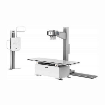
Click Here To Download Catalogue | DrGem Ceiling Analogue X-ray Machine is a diagnostic radiography system X-ray Machine that provides reliable high quality radiographic images with a reduced dose. The reliable high-frequency x-ray generators that are known worldwide for their excellent performance, lifetime and stability. Patient tables and wall stands are also offered.
Features of DrGem Ceiling Analogue X-ray Machine
Click Here To Download Catalogue | DETAILS
B + Compound
B + Compound utilizes several lines of sight for optimal contrast resolution, speckle reduction and border detection, with which P10 is ideal for superficial and abdominal imaging with better clarity and improved continuity of structures.
μ-Scan
The new generation μ-Scan imaging technology gives you better image quality by reducing noise, improving signal strength and improving visualization.
P10 offers a comprehensive selection of electronic probes to maximize its capabilities to meet a wide range of applications including abdomen, pediatric, OB/GYN, cardiovascular, musculoskeletal, etc. The advanced probe technologies also effectively enhance the image quality and confidence in reaching clinical diagnoses, even in difficult patients.
Convex Probe 3C-A
Ideal for an abundant of application such as abdomen, gynecology, obstetrics, urology and even abdomen biopsy.
Linear Probe L741
This linear probe is designed to satisfy vascular, breast, thyroid, and other small parts diagnosis, and its adjustable parameters could also present users a clear view of MSK and deep vessels.
Phase Array Probe 3P-A
For the purpose of adult and pediatric cardiology and emergency, the phase array probe provides elaborate presets for different exam modes, even for difficult patients.
Intracavitary Probe 6V1
Intracavitary probe could face application of gynecology, urology, prostate, and its temperature detection technology not only protects the patient but also extends the service life.
Click Here To Download Catalogue | OPENMARK 5000 is 0.51T MRI. It's approved by FDA and has CE mark. It adopts two-pillar magnet design with 280 degree openness and equipped with powerful
RF and gradient system, together with advanced imaging technology, making it as a high-end system which is comparable to high-field MRI.
Features:
Click Here To Download Catalogue | |||||||||||||||||||||||||||||||||||||||||||||||||||||||||||||||||||||||||||||||||||||||||||||||||||||||||||||||||||||||||||||||||||||||||||||||||||||||||||||||||||||||||||||||||||||||||||||||||||||||||||||||||||||||||||||||||||||||||||||||||||||||||||||||||||||||||||||||||||||||||||||||||||||||||||||||||||||||||||||||||||||||||||||||||||||||||||||||||||||||||||||||||||||||||||||||||||||||||||||||||||||||||||
| Weight | N/A | N/A | N/A | N/A | N/A | N/A | |||||||||||||||||||||||||||||||||||||||||||||||||||||||||||||||||||||||||||||||||||||||||||||||||||||||||||||||||||||||||||||||||||||||||||||||||||||||||||||||||||||||||||||||||||||||||||||||||||||||||||||||||||||||||||||||||||||||||||||||||||||||||||||||||||||||||||||||||||||||||||||||||||||||||||||||||||||||||||||||||||||||||||||||||||||||||||||||||||||||||||||||||||||||||||||||||||||||||||||||||||||||||||
| Dimensions | N/A | N/A | N/A | N/A | N/A | N/A | |||||||||||||||||||||||||||||||||||||||||||||||||||||||||||||||||||||||||||||||||||||||||||||||||||||||||||||||||||||||||||||||||||||||||||||||||||||||||||||||||||||||||||||||||||||||||||||||||||||||||||||||||||||||||||||||||||||||||||||||||||||||||||||||||||||||||||||||||||||||||||||||||||||||||||||||||||||||||||||||||||||||||||||||||||||||||||||||||||||||||||||||||||||||||||||||||||||||||||||||||||||||||||
| Additional information |
|


Reviews
There are no reviews yet.