
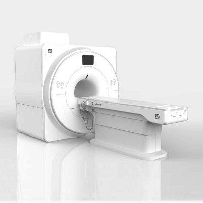
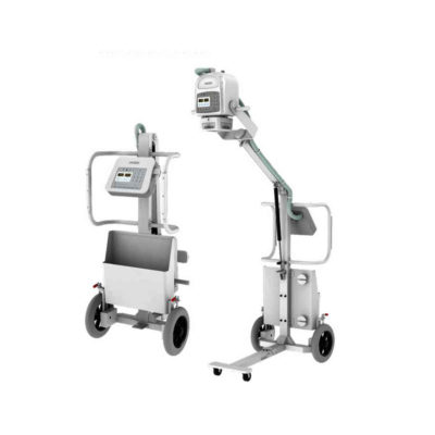
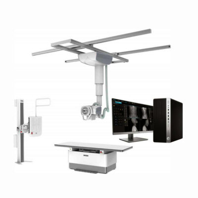
SHO-CMX01 Ceiling-Mounted X-Ray Machine
$0.00
The ceiling-mounted digital X-ray machine gives you versatile and high-quality imaging for head, chest, abdomen, lumbar spine, and limb examinations.
Shipped From China
Typically 10-21 working days – excluding furniture and heavy/bulky equipment. Please contact us for further information.
Description
The ceiling-mounted digital X-ray machine gives you versatile and high-quality imaging for head, chest, abdomen, lumbar spine, and limb examinations. Designed for hospitals of all sizes, it supports PA, LAT, and clinical static imaging, ensuring accurate diagnostics in a wide range of applications.
With its compact design and flexible movement, this system combines a floor-mounted table with a wall-mounted stand, making it easy for you to position patients efficiently while expanding your imaging capabilities. The motorized height-adjustable radiographic table enhances workflow, allowing for faster and more precise examinations.
This radiographic equipment is equipped with an 80kW, 1000mA high-voltage generator, a Siemens X-ray tube, Vieworks wireless flat panel detector, motorized collimator, and a 23.8-inch Dell workstation display. A 10-inch LCD touchscreen control panel simplifies operation, so you can focus on delivering the best care to your patients.
Specifications
| Model | SHO-CMX01 Series |
| Output power | 80 kW |
| Current | 10-1000 mA |
| Voltage | 40-150 kv |
| Exposure time | 0.001-10 s |
| Focus size | 0.6/1.2 mm |
| Flat Panel Detector (Wireless) | |
| Type | Amorphous Silicon |
| Scintillator | Cesium Iodide |
| Active Area | 17×17 inch (43×43 cm) |
| Active Pixel | 3072×3072 |
| Pixel Pitch | 140 um |
| A/D Conversion | 16 bits |
| DQE | ≧70% |
| Spatial Resolution | 37 lp/cm |
| Acquisition time | ≤2S |
| Collimator | |
| Operating mode | Electric |
| Max. Field of view | 45×45 cm |
| Illuminant | LED |
| Bucky stand | |
| Vertical travel | 1.4 m |
| Phototharapy Table | |
| Tabletop transverse travel | 260 mm |
| Tabletop longitudinal travel | 750 mm |
| Tabletop vertical travel | 210 mm |
| Workstation | |
| Monitor | 23.8’’ LCD Dell brand |
| CPU | ≥ 3.7GHz |
| Memory | ≥ 4 GB |
| HDD | ≥ 1T |
| DICOM3.0 | Query for integration with any PACS |
Quick Comparison
| SHO-CMX01 Ceiling-Mounted X-Ray Machine remove | Sonoscape S8 Exp Portable Ultrasound remove | SIGNERS SUPiA X-ray Digitizer ( CR Scanner) remove | Sonoscape E1 Ultrasound Machine With Two Probes remove | DrGem Ceiling Mounted Digital X-ray remove | Anke Supermark 1.5T MRI Machine remove | ||||||||||||||||||||||||||||||||||||||||||||||||||||||||||||||||||||||||||||||||||||||||||||||||||||||||||||||||||||||||||||||||||||||||||||||||||||||||||||||||||||||||||||||||||||||||||||||||||||||||||||||||||||||||||||||||||||||||||||||||||||||||||||||||||||||||||||||||||||||||||||||||||||||||||||||||||||||||||||||||||||||||||||||||||||||||||||||||||||||||||||||||||||||||||||||||||||||||
|---|---|---|---|---|---|---|---|---|---|---|---|---|---|---|---|---|---|---|---|---|---|---|---|---|---|---|---|---|---|---|---|---|---|---|---|---|---|---|---|---|---|---|---|---|---|---|---|---|---|---|---|---|---|---|---|---|---|---|---|---|---|---|---|---|---|---|---|---|---|---|---|---|---|---|---|---|---|---|---|---|---|---|---|---|---|---|---|---|---|---|---|---|---|---|---|---|---|---|---|---|---|---|---|---|---|---|---|---|---|---|---|---|---|---|---|---|---|---|---|---|---|---|---|---|---|---|---|---|---|---|---|---|---|---|---|---|---|---|---|---|---|---|---|---|---|---|---|---|---|---|---|---|---|---|---|---|---|---|---|---|---|---|---|---|---|---|---|---|---|---|---|---|---|---|---|---|---|---|---|---|---|---|---|---|---|---|---|---|---|---|---|---|---|---|---|---|---|---|---|---|---|---|---|---|---|---|---|---|---|---|---|---|---|---|---|---|---|---|---|---|---|---|---|---|---|---|---|---|---|---|---|---|---|---|---|---|---|---|---|---|---|---|---|---|---|---|---|---|---|---|---|---|---|---|---|---|---|---|---|---|---|---|---|---|---|---|---|---|---|---|---|---|---|---|---|---|---|---|---|---|---|---|---|---|---|---|---|---|---|---|---|---|---|---|---|---|---|---|---|---|---|---|---|---|---|---|---|---|---|---|---|---|---|---|---|---|---|---|---|---|---|---|---|---|---|---|---|---|---|---|---|---|---|---|---|---|---|---|---|---|---|---|---|---|---|---|---|---|---|---|---|---|---|---|---|---|---|---|---|---|---|---|---|---|---|---|---|---|---|---|---|---|---|---|---|---|---|---|---|---|---|---|---|---|---|---|---|---|---|---|---|---|---|---|---|---|---|
| Name | SHO-CMX01 Ceiling-Mounted X-Ray Machine remove | Sonoscape S8 Exp Portable Ultrasound remove | SIGNERS SUPiA X-ray Digitizer ( CR Scanner) remove | Sonoscape E1 Ultrasound Machine With Two Probes remove | DrGem Ceiling Mounted Digital X-ray remove | Anke Supermark 1.5T MRI Machine remove | |||||||||||||||||||||||||||||||||||||||||||||||||||||||||||||||||||||||||||||||||||||||||||||||||||||||||||||||||||||||||||||||||||||||||||||||||||||||||||||||||||||||||||||||||||||||||||||||||||||||||||||||||||||||||||||||||||||||||||||||||||||||||||||||||||||||||||||||||||||||||||||||||||||||||||||||||||||||||||||||||||||||||||||||||||||||||||||||||||||||||||||||||||||||||||||||||||||||
| Image | 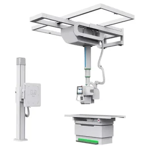 |  |  |  | 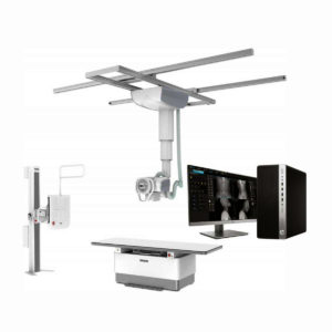 |  | |||||||||||||||||||||||||||||||||||||||||||||||||||||||||||||||||||||||||||||||||||||||||||||||||||||||||||||||||||||||||||||||||||||||||||||||||||||||||||||||||||||||||||||||||||||||||||||||||||||||||||||||||||||||||||||||||||||||||||||||||||||||||||||||||||||||||||||||||||||||||||||||||||||||||||||||||||||||||||||||||||||||||||||||||||||||||||||||||||||||||||||||||||||||||||||||||||||||
| SKU | SF1033560012-15 | SF1033560050-01 | SF1033560012-20 | SF1033560074-4 | SF1033560092-4 | ||||||||||||||||||||||||||||||||||||||||||||||||||||||||||||||||||||||||||||||||||||||||||||||||||||||||||||||||||||||||||||||||||||||||||||||||||||||||||||||||||||||||||||||||||||||||||||||||||||||||||||||||||||||||||||||||||||||||||||||||||||||||||||||||||||||||||||||||||||||||||||||||||||||||||||||||||||||||||||||||||||||||||||||||||||||||||||||||||||||||||||||||||||||||||||||||||||||||
| Rating | |||||||||||||||||||||||||||||||||||||||||||||||||||||||||||||||||||||||||||||||||||||||||||||||||||||||||||||||||||||||||||||||||||||||||||||||||||||||||||||||||||||||||||||||||||||||||||||||||||||||||||||||||||||||||||||||||||||||||||||||||||||||||||||||||||||||||||||||||||||||||||||||||||||||||||||||||||||||||||||||||||||||||||||||||||||||||||||||||||||||||||||||||||||||||||||||||||||||||||||
| Price |
|
|
|
|
|
| |||||||||||||||||||||||||||||||||||||||||||||||||||||||||||||||||||||||||||||||||||||||||||||||||||||||||||||||||||||||||||||||||||||||||||||||||||||||||||||||||||||||||||||||||||||||||||||||||||||||||||||||||||||||||||||||||||||||||||||||||||||||||||||||||||||||||||||||||||||||||||||||||||||||||||||||||||||||||||||||||||||||||||||||||||||||||||||||||||||||||||||||||||||||||||||||||||||||
| Stock | |||||||||||||||||||||||||||||||||||||||||||||||||||||||||||||||||||||||||||||||||||||||||||||||||||||||||||||||||||||||||||||||||||||||||||||||||||||||||||||||||||||||||||||||||||||||||||||||||||||||||||||||||||||||||||||||||||||||||||||||||||||||||||||||||||||||||||||||||||||||||||||||||||||||||||||||||||||||||||||||||||||||||||||||||||||||||||||||||||||||||||||||||||||||||||||||||||||||||||||
| Availability | |||||||||||||||||||||||||||||||||||||||||||||||||||||||||||||||||||||||||||||||||||||||||||||||||||||||||||||||||||||||||||||||||||||||||||||||||||||||||||||||||||||||||||||||||||||||||||||||||||||||||||||||||||||||||||||||||||||||||||||||||||||||||||||||||||||||||||||||||||||||||||||||||||||||||||||||||||||||||||||||||||||||||||||||||||||||||||||||||||||||||||||||||||||||||||||||||||||||||||||
| Add to cart | |||||||||||||||||||||||||||||||||||||||||||||||||||||||||||||||||||||||||||||||||||||||||||||||||||||||||||||||||||||||||||||||||||||||||||||||||||||||||||||||||||||||||||||||||||||||||||||||||||||||||||||||||||||||||||||||||||||||||||||||||||||||||||||||||||||||||||||||||||||||||||||||||||||||||||||||||||||||||||||||||||||||||||||||||||||||||||||||||||||||||||||||||||||||||||||||||||||||||||||
| Description | The ceiling-mounted digital X-ray machine gives you versatile and high-quality imaging for head, chest, abdomen, lumbar spine, and limb examinations.
Shipped From China
Delivery & Availability:
Typically 10-21 working days – excluding furniture and heavy/bulky equipment. Please contact us for further information.
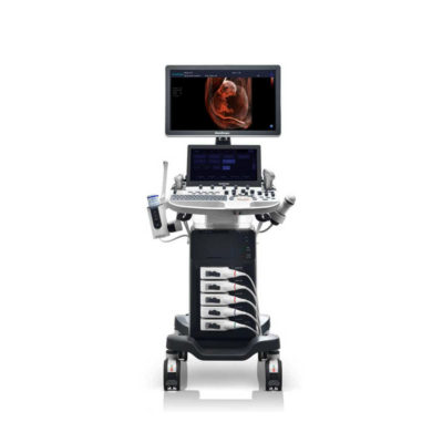
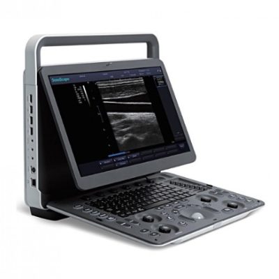
| Shipped from Abroad With ultra-modern innovative design and the clinically-proven technologies, S8 Exp is portable ultrasound scanner well equipped as a low-physical-effort and enhanced-image-quality ultrasound scanner, which not only provides optimized solutions for versatile applications, but does help to improve the user-experience for both routine and non-traditional challenges. Delivery & Availability: Typically 5-7 working days – excluding furniture and heavy/bulky equipment. Please contact us for further information. | Shipped from Abroad SUPiA made by Signers offers such a better clinic environment with no chemicals, ideal space, high-resolution image quality, and affordability. Delivery & Availability: Typically 14 working days – excluding furniture and heavy/bulky equipment. Please contact us for further information. | Shipped from Abroad SonoScape has developed a new probe and function for the E1 Exp. With these additions the E1 Exp will bring users a more efficient examination experience with satisfying image quality and a smooth workflow. Delivery & Availability: Typically 5-7 working days – excluding furniture and heavy/bulky equipment. Please contact us for further information. | In Stock The GXR-SD is a diagnostic digital radiography system that provides reliable high quality digital radiographic images with a reduced dose. The GXR-SD DR systems offer comprehensive digital solutions to all radiography needs, featuring ACQUIDR digital imaging system with stationary or portable digital flat-panel detectors as well as reliable high-frequency x-ray generators that are known worldwide for their excellent performance, lifetime and stability. Patient tables and wall stands are also offered. Delivery & Availability: Typically 21 working days – excluding furniture and heavy/bulky equipment. Please contact us for further information. | Shipped from Abroad
SuperMark 1.5T is a new generation superconducting MRI system based on years of experience in production and research. It's applicable to whole body scan, such as, nervous system, spine, joint soft tissue, pelvic and abdominal cavity, etc
Delivery & Availability: Typically 90 working days – excluding furniture and heavy/bulky equipment. Please contact us for further information. | |||||||||||||||||||||||||||||||||||||||||||||||||||||||||||||||||||||||||||||||||||||||||||||||||||||||||||||||||||||||||||||||||||||||||||||||||||||||||||||||||||||||||||||||||||||||||||||||||||||||||||||||||||||||||||||||||||||||||||||||||||||||||||||||||||||||||||||||||||||||||||||||||||||||||||||||||||||||||||||||||||||||||||||||||||||||||||||||||||||||||||||||||||||||||||||||||||||||
| Content | The ceiling-mounted digital X-ray machine gives you versatile and high-quality imaging for head, chest, abdomen, lumbar spine, and limb examinations. Designed for hospitals of all sizes, it supports PA, LAT, and clinical static imaging, ensuring accurate diagnostics in a wide range of applications.
With its compact design and flexible movement, this system combines a floor-mounted table with a wall-mounted stand, making it easy for you to position patients efficiently while expanding your imaging capabilities. The motorized height-adjustable radiographic table enhances workflow, allowing for faster and more precise examinations.
This radiographic equipment is equipped with an 80kW, 1000mA high-voltage generator, a Siemens X-ray tube, Vieworks wireless flat panel detector, motorized collimator, and a 23.8-inch Dell workstation display. A 10-inch LCD touchscreen control panel simplifies operation, so you can focus on delivering the best care to your patients.
Specifications
| Sonoscape S8 Exp Portable Ultrasound scannerDETAILS Agile and Versatile With ultra-modern innovative design and the clinically-proven technologies, S8 Exp Portable Ultrasound scanner is well equipped as a low-physical-effort and enhanced-image-quality ultrasound scanner, which not only provides optimized solutions for versatile applications but does help to improve the user experience for both routine and non-traditional challenges. Working with S8 Exp, it will trigger your unlimited reverie and endow you with endless charm. Carrying forward the classical design of SonoScape's portable ultrasound products, S8 Exp successfully combines the best ergonomics, attractive design and ease of use. This charismatic identity is also enhanced by a sophisticated color palette—with sedate grey as its interior paint color and pearl white as exterior cover, S8 Exp reveals a style of aristocrat and strong character among SonoScape's ultrasound systems. Workflow The S8 Exp is a portable ultrasound scanner that adapts to your workflow, whether you are in the consulting room, at the bedside, or at a remote location. With easy-to-use new platform designed for sonographers' needs and full connection interfaces for easy connectivity and data sharing, S8 Exp leads to improved user comfort and clinical outcome as well as patient throughput and working efficiency. Powerful Platform Embedded with SonoScape's core imaging technologies such as μ-scan, PHI and Spatial Compound, S8 Exp boasts exceptional 2D image, sensitive spectral, Color and Power Doppler, displaying well-defined anatomy and pathology and facilitating a highly optimized diagnostic user environment for conclusive diagnoses. Besides, S8 Exp offers a comprehensive selection of electronic probes to maximally extend its capabilities to meet a wide range of applications including the abdomen, pediatric, OB/GYN, cardiovascular, musculoskeletal, etc. The advanced probe technologies also effectively enhance the image quality and confidence in reaching clinical diagnoses even in difficult patients.Click Here To Download Catalogue | SUPiA X-ray Digitizer made by Signers offers such a better clinic environment with no chemicals, ideal space, high-resolution image quality, and affordability
FEATURE
Rigid Type
• No damage or scratch on image plates during scanning & erasing
• Scanning & Erasing without a roller
• No cut-off image during winter and cold period
Durability
• Extremely simple structure design
• Strong aluminum base plate
• Flip covers preventing dust from inside scanner
Barcode System
• Automatically recognising cassette sizes(14x17", 10x12", 18x24cm) by barcode reader
Compact & lightweight design
Click Here To Download Catalogue | DETAILS
Efficient Diagnosis
μ-Scan, Speckle Reduction & Edge Enhancement
Spatial Compound Imaging
PIH - Pure Inversion Harmonic
Wide Scan - Enlarged Image Area
Tissue-Specific Imaging
SR Flow
Ergonomic Designs
Up to 2 Transducer Ports
Light Weight and Compact
15.6 inch Anti-flickering HD LED Screen
Tilting Monitor Angle Adjustment
Backlit Keyboard and Intelligent Panel
Long-lasting Battery for 90 mins
Ease of Use
Quick Boot Up
Auto-Brightness Adjustment
Auto Image Optimization
Auto IMT
Auto Trace
Equipped Accessories
Wi-Fi and Bluetooth Available
DICOM
500GB Hard Disk
Height Adjustable Trolley
Durable, Carry-on Site Suitcase
Click Here To Download Catalogue | DrGem Ceiling Mounted Digital X-ray is a diagnostic digital radiography system that provides reliable high quality digital radiographic images with a reduced dose. The GXR-SD DR systems offer comprehensive digital solutions to all radiography needs, featuring ACQUIDR digital imaging system with stationary or portable digital flat-panel detectors as well as reliable high-frequency x-ray generators that are known worldwide for their excellent performance, lifetime and stability. Patient tables and wall stands are also offered.
Features:
Click Here To Download Catalogue | SuperMark 1.5T is a new generation superconducting MRI system based on years of experience in production and research. It's applicable to whole body scan, such as, nervous system, spine, joint soft tissue, pelvic and abdominal cavity, etc. SuperMark 1.5T provides not only conventional pulse sequences and clinical diagnosis functions, but also provides advanced functional applications, for instance, 3D angiography and water imaging. It adopts brand new ANKE APEX operating system which ensures easy operation and fast diagnosis.
Technical Advantages:
Click Here To Download Catalogue | |||||||||||||||||||||||||||||||||||||||||||||||||||||||||||||||||||||||||||||||||||||||||||||||||||||||||||||||||||||||||||||||||||||||||||||||||||||||||||||||||||||||||||||||||||||||||||||||||||||||||||||||||||||||||||||||||||||||||||||||||||||||||||||||||||||||||||||||||||||||||||||||||||||||||||||||||||||||||||||||||||||||||||||||||||||||||||||||||||||||||||||||||||||||||||||||||||||||
| Weight | N/A | N/A | N/A | N/A | N/A | N/A | |||||||||||||||||||||||||||||||||||||||||||||||||||||||||||||||||||||||||||||||||||||||||||||||||||||||||||||||||||||||||||||||||||||||||||||||||||||||||||||||||||||||||||||||||||||||||||||||||||||||||||||||||||||||||||||||||||||||||||||||||||||||||||||||||||||||||||||||||||||||||||||||||||||||||||||||||||||||||||||||||||||||||||||||||||||||||||||||||||||||||||||||||||||||||||||||||||||||
| Dimensions | N/A | N/A | N/A | N/A | N/A | N/A | |||||||||||||||||||||||||||||||||||||||||||||||||||||||||||||||||||||||||||||||||||||||||||||||||||||||||||||||||||||||||||||||||||||||||||||||||||||||||||||||||||||||||||||||||||||||||||||||||||||||||||||||||||||||||||||||||||||||||||||||||||||||||||||||||||||||||||||||||||||||||||||||||||||||||||||||||||||||||||||||||||||||||||||||||||||||||||||||||||||||||||||||||||||||||||||||||||||||
| Additional information |
| ||||||||||||||||||||||||||||||||||||||||||||||||||||||||||||||||||||||||||||||||||||||||||||||||||||||||||||||||||||||||||||||||||||||||||||||||||||||||||||||||||||||||||||||||||||||||||||||||||||||||||||||||||||||||||||||||||||||||||||||||||||||||||||||||||||||||||||||||||||||||||||||||||||||||||||||||||||||||||||||||||||||||||||||||||||||||||||||||||||||||||||||||||||||||||||||||||||||||||||

Reviews
There are no reviews yet.