Barco MDPC-8127
$0.00
Shipped From Abroad
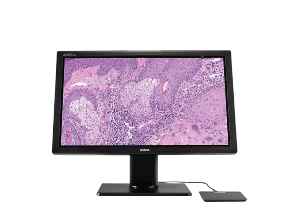
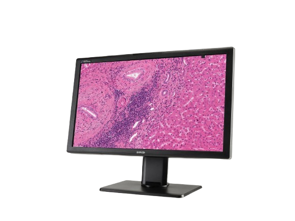
The Barco MDPC-8127 is a 27-inch, 8 MP (3840×2160) diagnostic display cleared for digital pathology. Built for rapid slide viewing with color fidelity, uniformity correction, and integrated quality calibration, it supports high-confidence primary diagnosis in pathology workflows.
Typically 10-21 working days – excluding furniture and heavy/bulky equipment. Please contact us for further information.
Description
The Barco MDPC-8127 is a medical-grade display purpose-built for digital pathology. It delivers ultra-high definition 8 MP imaging at up to 120 Hz, allowing pathologists to pan and zoom with minimal blur. The IPS LCD panel offers rich color and grayscale rendering, wide viewing angles, and a host of calibration and uniformity features (PPU, SteadyColor, I-Guard). With regulatory clearances as an IVD device (FDA and CE), it can be integrated into validated whole-slide imaging systems for use in primary diagnosis and clinical workflows.
Meet MDPC-8127, our ultra-high definition medical-grade display designed exclusively for digital pathology. With regulatory clearances as an IVD device (Europe and FDA) for use in digital pathology, including primary diagnosis, it’s the first display that you can confidently integrate into your digital pathology workflow with multiple whole slide imaging systems. Work with superior imaging technology and analyze your histological samples confidently, with unprecedented visual richness.
*In the USA, the MDPC-8127 can be used with WSI scanners and viewing software that have been validated for use with the display.
The device may be used for primary diagnosis in the following validated FDA-cleared WSI systems and digital pathology viewing software:
Barco’s MDPC-8127 510(k) clearance:
- Philips Intellisite Pathology Solution with Philips Image Management System viewing software, cleared under K192259
- Philips Intellisite Pathology Solution with Paige.AI Inc. FullFocus DX viewing software, cleared under K201005
- Leica Aperio AT2 DX System with ImageScope DX viewing software, cleared under K190332
- Leica Aperio AT2 DX scanner with Sectra Digital Pathology Module, cleared under K193054
FDA- Cleared WSI systems and Digital Pathology Viewing Software with MDPC-8127:
- Hamamatsu NanoZoomer S360MD slide scanner system, cleared under K233027
- Leica Aperio GT 450 DX scanner with Aperio WebViewer DX, cleared under K232202
- Philips Intellisite Pathology Solution with JelloX MetaLite Dx Digital Pathology Software, cleared under K240303
- Philips IntelliSite Pathology Solution with Philips Image Management System and Galen™ Second Read™ AI, cleared under K241232
- Epredia E1000 Dx scanner with E1000 Dx IMS, cleared under K241717
- Hamamatsu NanoZoomer S360MD scanner with Lumea Viewer+, cleared under K242244
MDPC-8127 Validation protocol:
- Leica Aperio GT 450 DX scanner with Sectra Digital Pathology Module (3.3)
- Roche Ventana DP200 scanner with Roche uPath Enterprise Software
- Hamamatsu NanoZoomer S360MD Slide scanner with Proscia Concentriq® AP-Dx Digital Pathology software
A+ ecolabel for MDPC-8127
The MDPC-8127 has been subjected to Barco’s ecoscoring protocol and has received an A+ rating. Some key factors that contributed to this rating are:
- High energy efficiency
- Possibility to switch to standby mode when the device is not in use
- Use of halogen-free materials on all levels: cables, PCBs, plastics
- The number of screws and screw types is reduced significantly to improve ease of disassembly
- Unpainted large plastic parts
Features
Quick Comparison
| Barco MDPC-8127 remove | AMR 400 Manual Rotary Microtome remove | TEC2800 Tissue Embedding Center remove | AEC380 Tissue Embedding Center remove | AEM450 Semi-automatic Rotary Microtome remove | Urinary Tract Infection Test Strips remove | |||||||||||||||||||||||||||||||||||||||||||||||||||||||||||||||||||||||||||||||||||||||||||||||||||||||||||||||||||||||||||||||||||||||||||||||||||||||||||||||||||||||||||||||||||||||||||||||||||||||||||||||||||||||||||||||||||||||||||||||||||||||||||||||||||||||||||||||||||||||||||||||||||||||||||||||||||||||||||||||||||||||||||||||||||||||||||||||||||||||||||||||||||||||||||||||||||||||||||||||||||||||||||||||||||
|---|---|---|---|---|---|---|---|---|---|---|---|---|---|---|---|---|---|---|---|---|---|---|---|---|---|---|---|---|---|---|---|---|---|---|---|---|---|---|---|---|---|---|---|---|---|---|---|---|---|---|---|---|---|---|---|---|---|---|---|---|---|---|---|---|---|---|---|---|---|---|---|---|---|---|---|---|---|---|---|---|---|---|---|---|---|---|---|---|---|---|---|---|---|---|---|---|---|---|---|---|---|---|---|---|---|---|---|---|---|---|---|---|---|---|---|---|---|---|---|---|---|---|---|---|---|---|---|---|---|---|---|---|---|---|---|---|---|---|---|---|---|---|---|---|---|---|---|---|---|---|---|---|---|---|---|---|---|---|---|---|---|---|---|---|---|---|---|---|---|---|---|---|---|---|---|---|---|---|---|---|---|---|---|---|---|---|---|---|---|---|---|---|---|---|---|---|---|---|---|---|---|---|---|---|---|---|---|---|---|---|---|---|---|---|---|---|---|---|---|---|---|---|---|---|---|---|---|---|---|---|---|---|---|---|---|---|---|---|---|---|---|---|---|---|---|---|---|---|---|---|---|---|---|---|---|---|---|---|---|---|---|---|---|---|---|---|---|---|---|---|---|---|---|---|---|---|---|---|---|---|---|---|---|---|---|---|---|---|---|---|---|---|---|---|---|---|---|---|---|---|---|---|---|---|---|---|---|---|---|---|---|---|---|---|---|---|---|---|---|---|---|---|---|---|---|---|---|---|---|---|---|---|---|---|---|---|---|---|---|---|---|---|---|---|---|---|---|---|---|---|---|---|---|---|---|---|---|---|---|---|---|---|---|---|---|---|---|---|---|---|---|---|---|---|---|---|---|---|---|---|---|---|---|---|---|---|---|---|---|---|---|---|---|---|---|---|---|---|---|---|---|---|---|---|---|---|---|---|---|---|---|---|---|---|---|---|---|---|---|---|---|---|---|---|
| Name | Barco MDPC-8127 remove | AMR 400 Manual Rotary Microtome remove | TEC2800 Tissue Embedding Center remove | AEC380 Tissue Embedding Center remove | AEM450 Semi-automatic Rotary Microtome remove | Urinary Tract Infection Test Strips remove | ||||||||||||||||||||||||||||||||||||||||||||||||||||||||||||||||||||||||||||||||||||||||||||||||||||||||||||||||||||||||||||||||||||||||||||||||||||||||||||||||||||||||||||||||||||||||||||||||||||||||||||||||||||||||||||||||||||||||||||||||||||||||||||||||||||||||||||||||||||||||||||||||||||||||||||||||||||||||||||||||||||||||||||||||||||||||||||||||||||||||||||||||||||||||||||||||||||||||||||||||||||||||||||||||||
| Image | 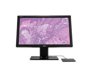 | 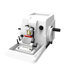 |  | 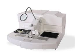 | 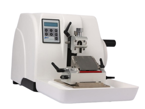 |  | ||||||||||||||||||||||||||||||||||||||||||||||||||||||||||||||||||||||||||||||||||||||||||||||||||||||||||||||||||||||||||||||||||||||||||||||||||||||||||||||||||||||||||||||||||||||||||||||||||||||||||||||||||||||||||||||||||||||||||||||||||||||||||||||||||||||||||||||||||||||||||||||||||||||||||||||||||||||||||||||||||||||||||||||||||||||||||||||||||||||||||||||||||||||||||||||||||||||||||||||||||||||||||||||||||
| SKU | SF1033560130185-5 | |||||||||||||||||||||||||||||||||||||||||||||||||||||||||||||||||||||||||||||||||||||||||||||||||||||||||||||||||||||||||||||||||||||||||||||||||||||||||||||||||||||||||||||||||||||||||||||||||||||||||||||||||||||||||||||||||||||||||||||||||||||||||||||||||||||||||||||||||||||||||||||||||||||||||||||||||||||||||||||||||||||||||||||||||||||||||||||||||||||||||||||||||||||||||||||||||||||||||||||||||||||||||||||||||||||||
| Rating | ||||||||||||||||||||||||||||||||||||||||||||||||||||||||||||||||||||||||||||||||||||||||||||||||||||||||||||||||||||||||||||||||||||||||||||||||||||||||||||||||||||||||||||||||||||||||||||||||||||||||||||||||||||||||||||||||||||||||||||||||||||||||||||||||||||||||||||||||||||||||||||||||||||||||||||||||||||||||||||||||||||||||||||||||||||||||||||||||||||||||||||||||||||||||||||||||||||||||||||||||||||||||||||||||||||||||
| Price |
|
| $5,520.00 | $7,590.00 | $6,095.00 |
| ||||||||||||||||||||||||||||||||||||||||||||||||||||||||||||||||||||||||||||||||||||||||||||||||||||||||||||||||||||||||||||||||||||||||||||||||||||||||||||||||||||||||||||||||||||||||||||||||||||||||||||||||||||||||||||||||||||||||||||||||||||||||||||||||||||||||||||||||||||||||||||||||||||||||||||||||||||||||||||||||||||||||||||||||||||||||||||||||||||||||||||||||||||||||||||||||||||||||||||||||||||||||||||||||||
| Stock | ||||||||||||||||||||||||||||||||||||||||||||||||||||||||||||||||||||||||||||||||||||||||||||||||||||||||||||||||||||||||||||||||||||||||||||||||||||||||||||||||||||||||||||||||||||||||||||||||||||||||||||||||||||||||||||||||||||||||||||||||||||||||||||||||||||||||||||||||||||||||||||||||||||||||||||||||||||||||||||||||||||||||||||||||||||||||||||||||||||||||||||||||||||||||||||||||||||||||||||||||||||||||||||||||||||||||
| Availability | ||||||||||||||||||||||||||||||||||||||||||||||||||||||||||||||||||||||||||||||||||||||||||||||||||||||||||||||||||||||||||||||||||||||||||||||||||||||||||||||||||||||||||||||||||||||||||||||||||||||||||||||||||||||||||||||||||||||||||||||||||||||||||||||||||||||||||||||||||||||||||||||||||||||||||||||||||||||||||||||||||||||||||||||||||||||||||||||||||||||||||||||||||||||||||||||||||||||||||||||||||||||||||||||||||||||||
| Add to cart | ||||||||||||||||||||||||||||||||||||||||||||||||||||||||||||||||||||||||||||||||||||||||||||||||||||||||||||||||||||||||||||||||||||||||||||||||||||||||||||||||||||||||||||||||||||||||||||||||||||||||||||||||||||||||||||||||||||||||||||||||||||||||||||||||||||||||||||||||||||||||||||||||||||||||||||||||||||||||||||||||||||||||||||||||||||||||||||||||||||||||||||||||||||||||||||||||||||||||||||||||||||||||||||||||||||||||
| Description | Shipped From Abroad
The Barco MDPC-8127 is a 27-inch, 8 MP (3840×2160) diagnostic display cleared for digital pathology. Built for rapid slide viewing with color fidelity, uniformity correction, and integrated quality calibration, it supports high-confidence primary diagnosis in pathology workflows.
Delivery & Availability:
Typically 10-21 working days – excluding furniture and heavy/bulky equipment. Please contact us for further information.
| Shipped From Abroad
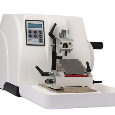
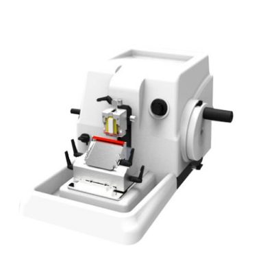
The AMR 400 is a manual rotary microtome engineered for high-precision sectioning of biological specimens. It produces reproducible, ultra-thin slices for microscopic examination in histology and pathology labs.
Delivery & Availability:
Typically 10-21 working days – excluding furniture and heavy/bulky equipment. Please contact us for further information.
| Shipped From Abroad
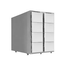
The TEC2800 Tissue Embedding Center optimizes paraffin embedding with a large 6 L reservoir, adjustable paraffin control, LCD display, cold spot cooling, and multi-forceps heating for efficient, consistent histology embedding workflows in pathology and research labs.
Delivery & Availability:
Typically 10-21 working days – excluding furniture and heavy/bulky equipment. Please contact us for further information.
| Shipped From Abroad
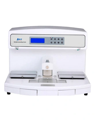
The AEC380 Tissue Embedding Center is a dual-platform histology workstation with touchscreen control, integrated cold spot, forceps heating, and precise paraffin flow for efficient, ergonomic embedding of tissue samples in pathology and research labs.
Delivery & Availability:
Typically 10-21 working days – excluding furniture and heavy/bulky equipment. Please contact us for further information.
| Shipped From Abroad
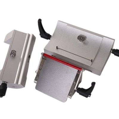
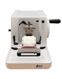
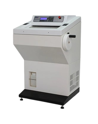
The AEM 450 is a semi-automatic rotary microtome engineered for precise, reliable thin-sectioning of biological specimens, enabling routine histology and pathology sample preparation with excellent control and consistency.
Delivery & Availability:
Typically 10-21 working days – excluding furniture and heavy/bulky equipment. Please contact us for further information.
| Shipped From Abroad
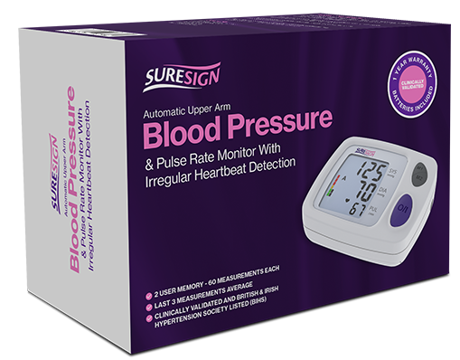
The Urinary Tract Infection Test Strips are a fast, disposable diagnostic tool that detects key UTI markers such as nitrites, leukocyte esterase, and pH changes in urine. With results in just 60 seconds, they offer an accurate, convenient solution for both home and clinical use.
Delivery & Availability:
Typically 10-21 working days – excluding furniture and heavy/bulky equipment. Please contact us for further information.
| ||||||||||||||||||||||||||||||||||||||||||||||||||||||||||||||||||||||||||||||||||||||||||||||||||||||||||||||||||||||||||||||||||||||||||||||||||||||||||||||||||||||||||||||||||||||||||||||||||||||||||||||||||||||||||||||||||||||||||||||||||||||||||||||||||||||||||||||||||||||||||||||||||||||||||||||||||||||||||||||||||||||||||||||||||||||||||||||||||||||||||||||||||||||||||||||||||||||||||||||||||||||||||||||||||
| Content | https://youtu.be/BBEp30jpeBk?si=uX5iI48ddzdmHAXt
The Barco MDPC-8127 is a medical-grade display purpose-built for digital pathology. It delivers ultra-high definition 8 MP imaging at up to 120 Hz, allowing pathologists to pan and zoom with minimal blur. The IPS LCD panel offers rich color and grayscale rendering, wide viewing angles, and a host of calibration and uniformity features (PPU, SteadyColor, I-Guard). With regulatory clearances as an IVD device (FDA and CE), it can be integrated into validated whole-slide imaging systems for use in primary diagnosis and clinical workflows.
Meet MDPC-8127, our ultra-high definition medical-grade display designed exclusively for digital pathology. With regulatory clearances as an IVD device (Europe and FDA) for use in digital pathology, including primary diagnosis, it’s the first display that you can confidently integrate into your digital pathology workflow with multiple whole slide imaging systems. Work with superior imaging technology and analyze your histological samples confidently, with unprecedented visual richness. *In the USA, the MDPC-8127 can be used with WSI scanners and viewing software that have been validated for use with the display. The device may be used for primary diagnosis in the following validated FDA-cleared WSI systems and digital pathology viewing software: Barco’s MDPC-8127 510(k) clearance:
FDA- Cleared WSI systems and Digital Pathology Viewing Software with MDPC-8127:
MDPC-8127 Validation protocol:
The MDPC-8127 has been subjected to Barco’s ecoscoring protocol and has received an A+ rating. Some key factors that contributed to this rating are:
Features | This mechanical microtome enables hand-driven sectioning of tissue blocks by rotating the handwheel to advance the specimen toward a blade. It offers adjustable section thickness from very thin (microns) to thicker cuts, supports both low and high-profile disposable blades, and includes built-in safety and ergonomic design features. It is widely used in clinical pathology, research histology, veterinary labs, and teaching facilities to prepare tissue sections that can be stained and analyzed for disease diagnosis or scientific study.
Features of AMR 400 Manual Rotary Microtome
Specifications of AMR 400 Manual Rotary Microtome
| The TEC2800 Tissue Embedding Center is a high-performance histology workstation designed to streamline paraffin embedding prior to microtome sectioning. Featuring a large 6 L paraffin tank, user-friendly LCD interface, adjustable paraffin flow via touchpad or foot pedal, and generous working surface, it supports high throughput. An integrated Peltier cold spot cools samples to −5 °C, while the built-in forceps heating station accommodates up to six forceps simultaneously. Ideal for clinical, research, and diagnostic labs, it enhances precision and efficiency in tissue block preparation.
Features of TEC2800 Tissue Embedding CenterEmbedding Dispensing Console
Specifications of TEC2800 Tissue Embedding Center
| The AEC380 Tissue Embedding Center is a high-performance histology instrument designed to streamline paraffin embedding prior to microtome sectioning. With a smart color touchscreen, dual embedding platforms for simultaneous work, and six independently heated zones, it delivers precise temperature control. Integrated cold spot and paraffin trimmer modules support consistent embedding quality. Manual or foot-switch paraffin release enhances usability, while heated forceps holders and constant-temperature trays facilitate wax removal and mold reuse. Ideal for clinical, research, and diagnostic labs.
Features of AEC380 Tissue Embedding Center
Specifications of AEC380 Tissue Embedding Center
| https://youtu.be/MGm_pHdrpDw?si=CrAqO26VkvGpU7ek
This semi-automatic rotary microtome improves traditional manual sectioning by incorporating assisted feed control and an intuitive control panel that monitors key functions such as section thickness, trimming, and section count. It integrates an ergonomic handwheel, an adjustable specimen clamp, and effective retraction features to help produce uniform tissue slices from embedded specimens. Designed for pathology and research labs, it simplifies the workflow for preparing tissue sections for microscopic analysis while maintaining safety and precision.
Features of AEM450 Semi-automatic Rotary Microtome
Specifications of AEM450 Semi-automatic Rotary Microtome
| The Urinary Tract Infection Test Strips are a sensitive, rapid, and user-friendly diagnostic tool designed for early detection of urinary tract infections (UTIs). Engineered for both clinical and at-home use, these disposable test strips detect key biomarkers—such as nitrites, leukocyte esterase, and urine pH—that indicate the presence of a bacterial infection. With a simple dip-and-read process, the test strips provide results in about 60 seconds, allowing for quick screening and timely intervention. Key Features:
Technical Specifications:
Applications & Benefits:
| ||||||||||||||||||||||||||||||||||||||||||||||||||||||||||||||||||||||||||||||||||||||||||||||||||||||||||||||||||||||||||||||||||||||||||||||||||||||||||||||||||||||||||||||||||||||||||||||||||||||||||||||||||||||||||||||||||||||||||||||||||||||||||||||||||||||||||||||||||||||||||||||||||||||||||||||||||||||||||||||||||||||||||||||||||||||||||||||||||||||||||||||||||||||||||||||||||||||||||||||||||||||||||||||||||
| Weight | N/A | N/A | 26 kg | N/A | 34 kg | N/A | ||||||||||||||||||||||||||||||||||||||||||||||||||||||||||||||||||||||||||||||||||||||||||||||||||||||||||||||||||||||||||||||||||||||||||||||||||||||||||||||||||||||||||||||||||||||||||||||||||||||||||||||||||||||||||||||||||||||||||||||||||||||||||||||||||||||||||||||||||||||||||||||||||||||||||||||||||||||||||||||||||||||||||||||||||||||||||||||||||||||||||||||||||||||||||||||||||||||||||||||||||||||||||||||||||
| Dimensions | N/A | 52.5 × 41.7 × 28.0 cm | 56.0 × 64.0 × 40.0 cm | 126.0 × 64.0 × 52.0 cm | 57.5 × 40.5 × 32.5 cm | N/A | ||||||||||||||||||||||||||||||||||||||||||||||||||||||||||||||||||||||||||||||||||||||||||||||||||||||||||||||||||||||||||||||||||||||||||||||||||||||||||||||||||||||||||||||||||||||||||||||||||||||||||||||||||||||||||||||||||||||||||||||||||||||||||||||||||||||||||||||||||||||||||||||||||||||||||||||||||||||||||||||||||||||||||||||||||||||||||||||||||||||||||||||||||||||||||||||||||||||||||||||||||||||||||||||||||
| Additional information |
|
|
|
| ||||||||||||||||||||||||||||||||||||||||||||||||||||||||||||||||||||||||||||||||||||||||||||||||||||||||||||||||||||||||||||||||||||||||||||||||||||||||||||||||||||||||||||||||||||||||||||||||||||||||||||||||||||||||||||||||||||||||||||||||||||||||||||||||||||||||||||||||||||||||||||||||||||||||||||||||||||||||||||||||||||||||||||||||||||||||||||||||||||||||||||||||||||||||||||||||||||||||||||||||||||||||||||||||||||

Reviews
There are no reviews yet.