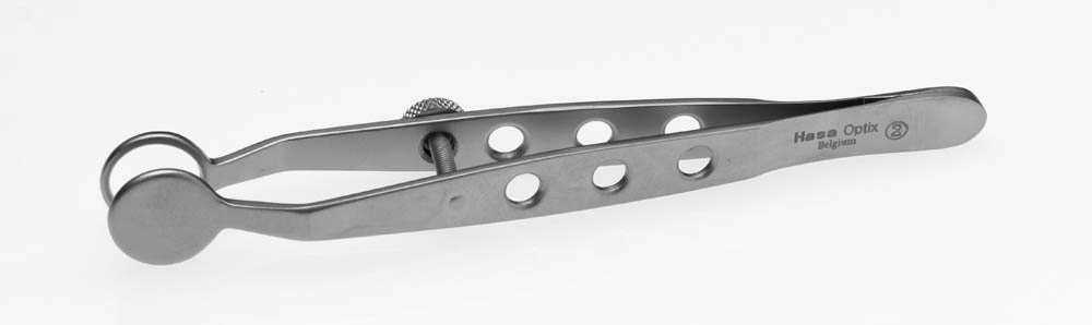
| Description | Shipped From Abroad
11mm Diameter, Round Plate
Delivery & Availability:
Typically 10-21 working days – excluding furniture and heavy/bulky equipment. Please contact us for further information.
| Shipped from abroad
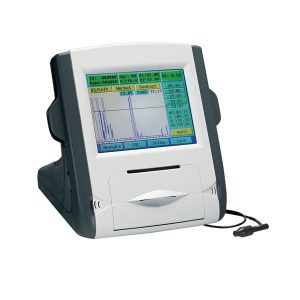
- Large color liquid-crystal screen
- Touch screen input, easy operation
- Curve freezing: Manual/Auto mode, controlled by pedal
- Built-in speed thermal printer
- Enter the name & ID; easy to check archive
Delivery & Availability:
Typically 14 working days – excluding furniture and heavy/bulky equipment. Please contact us for further information.
| Shipped from abroad
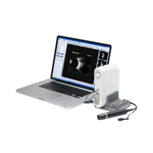
- Software image workstation
- B, B+B, B+A, A modes
- Video review for 100 images
- PDF report output
- Optional 20MHz B Probe: vitreous plus function
Delivery & Availability:
Typically 14 working days – excluding furniture and heavy/bulky equipment. Please contact us for further information.
| Shipped from abroad
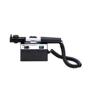
The product can quickly and precisely measure the astigmatism axis and is one of the necessary instruments in optometry inspection.
Delivery & Availability:
Typically 14 working days – excluding furniture and heavy/bulky equipment. Please contact us for further information.
| Shipped from abroad
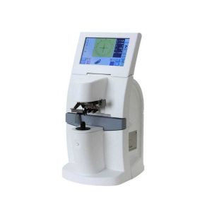
- 7.0-inch color LCD touch panel.
- Hartman sensor with 108 multiple measure points.
- Green measurement light beam.
- Dyeing lens, sunglasses measured easily.
Delivery & Availability:
Typically 14 working days – excluding furniture and heavy/bulky equipment. Please contact us for further information.
| Shipped from abroad
- Top Optical System.
- High eyepoint, comfort.
- Five steps drum zoom design, easier to use 6x, 10x, 16x, 25x, 40x magnification.
- High Definition Eyepieces, More Comfortable for Viewing.
Delivery & Availability:
Typically 14 working days – excluding furniture and heavy/bulky equipment. Please contact us for further information.
|
| Content | | Feature:
- Large color liquid-crystal screen
- Touch screen input, easy operation
- Curve freezing: Manual/Auto mode, controlled by a pedal
- Built-in speed thermal printer
- Enter the name & ID; easy to check the archive
Technical Specifications:
| Model |
SW-1000 Ophthalmic A Scan Biometer |
| A Scan |
| A scan probe |
10MHz import small size probe, built-in luminotron |
| Measuring range |
15mm-40mm |
| Measurement precision |
±0. 05mm; with macula lutea trace function |
| Measurement |
Anterior chamber depth, lens thickness, vitreous body length, total length and average |
| Method of measurement |
immersion and contact |
| Eye mode |
Phakic/ Aphakic/Dense/ various IOL |
| IOL formula |
SRK-II, SRK-T, BINKHORST- Ⅱ, HOLLADAY, HOFFER-Q, HAIGIS |
| Storage |
10 cases, 5 readings each case |
| Output |
A scan waveform and IOL calculation sheet
|
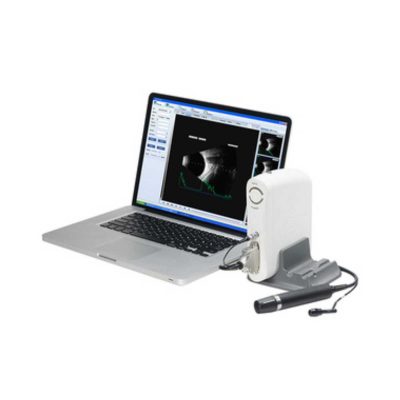
| Functions of Ophthalmic AB Scan Machine:
- Software image workstation
- B, B+B, B+A, A modes
- Video review for 100 images
- PDF report output
- Optional 20MHz B Probe: vitreous plus function
Technical Specifications:
| A scan |
1.Probe: 10MHz frequencies, with LED
2.Depth: 40mm
3.Precision: ±0.05mm
4.Eye mode: Phakic / Aphakic / Dense / Various IOL
5.Measurement: Anterior chamber depth, lens thickness, vitreous body length, total length and average
6.IOL Formula: SRK-II, SRK-T, BINKHORST, HOLLADAY, HOFFER-Q, HAIGIS, Stat.
7.Calculation: Average and standard deviation
8.Store: 10 Scanning results for each eye |
| B scan |
1.Probe: 10MHz/20MHz (optional), Magnetic driven, noiseless
2.Scanning Mode: Sector Scanning
3.Resolution: Lateral ≤0.3mm; Vertical≤0.2mm
4.Geometric Location Precision: Lateral≤10%; Vertical≤5%
5.Depth: 60mm
6.Enhance the part of vitreous body and retina
7.Gain of probe:30dB-105dB
8.Scanning Angle : 53°
9.Gray Scale: 256
10.False Color: Multi colors OTC
11.Measure Mode: distances, perimeter and area
12.Movies: 100 images movie review,AVI ZIP JPG format image output
13.Output: PDF format case report, connect to normal printer |
| Others |
1.Display Mode :B, B+B, B+A, A
2.Hint: preset keyword
3.Case Search: Multi-keywords
4.Working Platform: Windows XP, VISTA, WINDOWS7
5.User-defined report template |
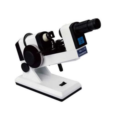
| The product can quickly and precisely measure the astigmatism axis and is one of the necessary instruments in optometry inspection.
Features:
- The filament can be rotated for 360° and move upward and downward. The brightness of the streak can be adjusted.
- Quickly and precisely measure the astigmatism axis.
- The light can be converged, radiated, and paralleled.
- Automatic Power Off function effectively protects the bulb.
- Two-steps adjustable brightness for bulb: S(Bright) and W(Dark).
Technical Specifications:
| Working Distance |
1 m |
| Streak |
3 mm-20 mm |
| Streak Rotation |
360° |
| Illumination Source |
3V/2W, halogen bulb |
| Input Voltage |
AC 220V±10%, 50Hz±1Hz |
| Power Consumption |
4.5 VA |
| Features:
- 7.0-inch color LCD touch panel.
- Hartman sensor with 108 multiple measure points.
- Green measurement light beam.
- Dyeing lens, sunglasses measured easily.
Technical Specifications:
- Sphere lenses: 0~±25D
- Cylinder lenses: 0~±10D
- Cylinder Axis Angle: 0°~180°
- Add.:0~10D
- Power supply: 100~240V, 50/60HZ, 30W
- Weight: 5kg
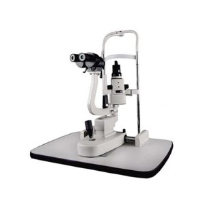
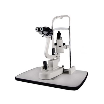
| Slit Lamp Features:
- Top Optical System.
- High eyepoint, comfort.
- Five steps drum zoom design, easier to use 6x, 10x, 16x, 25x, 40x magnification.
- High Definition Eyepieces, More Comfortable for Viewing.
Technical Specifications:
| Microscope Type |
Galileo Parallel |
| Optics |
Super Optic System |
| Magnification Change Way |
Drum Rotation |
| Eyepiece Magnification |
12.5x |
| Total Magnifications |
6x 10x, 16x, 25x, 40x |
| Diopter Adjustment |
-5D ~+5D |
| Slit Width |
0-14MM Continuous |
| Slit Height |
1-14MM Continuous |
| Slit Angle |
0°- 180° Adjustable |
| Light Source |
LED |
| Light Spot Diameter |
0.2mm, 2mm, 3mm, 5mm, 10mm, 14mm |
| Filter |
Heat Absorption; Grey; Redfree; Cobalt Blue |
| Fixation |
Red LED |
| Electrical |
|
| Illumination Bulb |
LED |
| Input Voltage |
110V/220V (±10%) |
| Applanation Tonometer Interface |
Included |
|


Reviews
There are no reviews yet.