DIN 1.5 mm EEG Cable
$0.00
Shipped From Abroad
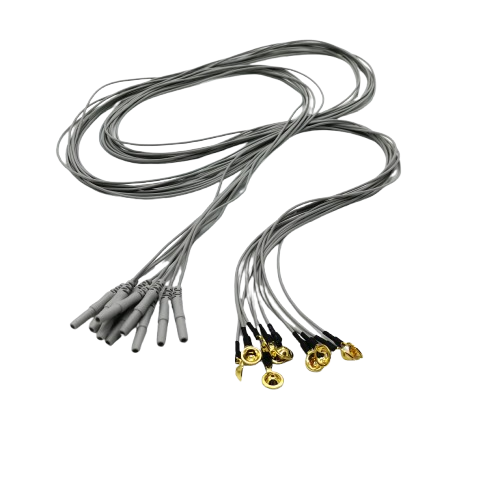
High-performance EEG cable with DIN 1.5 mm connector designed for accurate brainwave signal transmission. Made from flexible, durable TPU material with color-coded leads, it’s compatible with standard EEG systems for neurodiagnostic and sleep study applications.
Typically 10-21 working days – excluding furniture and heavy/bulky equipment. Please contact us for further information.
Description
Key Features
-
DIN 1.5 mm Connector: Standard size for compatibility with most EEG machines
-
High Signal Fidelity: Minimizes signal loss for accurate EEG readings
-
Flexible TPU Cable: Durable, tangle-free, and latex-free for patient comfort
-
Color-Coded Leads: Easy identification and quick setup
-
Reusable Design: Long-lasting and cost-effective
Technical Specifications
| Specification | Details |
|---|---|
| Connector Type | DIN 1.5 mm |
| Electrode Type | Cup electrode (silver- or gold-plated) |
| Lead Wire Material | Multi-strand silver-plated copper with TPU insulation |
| Cable Jacket | Thermoplastic Polyurethane (TPU) |
| Color Coding | AHA/IEC standard, multicolor set |
| Cable Length | 1.5 – 2.0 meters (customizable) |
| Reusability | Yes |
| Compatibility | EEG machines with 1.5 mm DIN input |
| Certifications | CE, ISO 13485 |
| Packaging | Single lead or full set, individually sealed |
| Latex-Free | Yes |
Quick Comparison
| DIN 1.5 mm EEG Cable remove | Portable Wireless EEG/PSG Recorder remove | 20 Lead Colorful Brain Wires remove | Neuron Spectrum 61..65 EEG remove | Neuron-Spectrum-1/BFB remove | Neuron Spectrum 65 Video EEG Monitoring System remove | |||||||||||||||||||||||||||||||||||||||||||||||||||||||||||||||||||||||||||||||||
|---|---|---|---|---|---|---|---|---|---|---|---|---|---|---|---|---|---|---|---|---|---|---|---|---|---|---|---|---|---|---|---|---|---|---|---|---|---|---|---|---|---|---|---|---|---|---|---|---|---|---|---|---|---|---|---|---|---|---|---|---|---|---|---|---|---|---|---|---|---|---|---|---|---|---|---|---|---|---|---|---|---|---|---|---|---|---|
| Name | DIN 1.5 mm EEG Cable remove | Portable Wireless EEG/PSG Recorder remove | 20 Lead Colorful Brain Wires remove | Neuron Spectrum 61..65 EEG remove | Neuron-Spectrum-1/BFB remove | Neuron Spectrum 65 Video EEG Monitoring System remove | ||||||||||||||||||||||||||||||||||||||||||||||||||||||||||||||||||||||||||||||||
| Image | 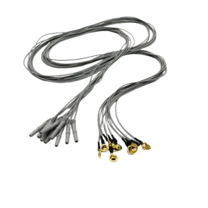 | 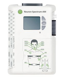 | 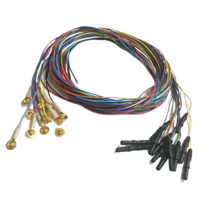 | 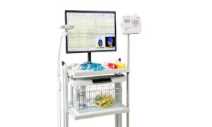 | 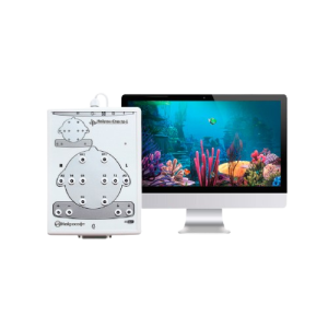 | 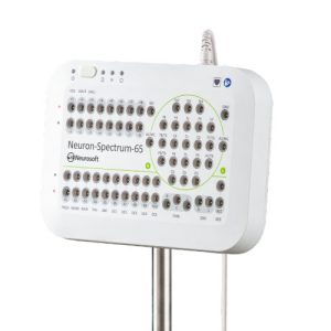 | ||||||||||||||||||||||||||||||||||||||||||||||||||||||||||||||||||||||||||||||||
| SKU | SF1033560130187-4 | SF1033560130187-1 | SF1033560130187-6 | SF1033560130187-2 | ||||||||||||||||||||||||||||||||||||||||||||||||||||||||||||||||||||||||||||||||||
| Rating | ||||||||||||||||||||||||||||||||||||||||||||||||||||||||||||||||||||||||||||||||||||||
| Price |
|
|
|
|
|
| ||||||||||||||||||||||||||||||||||||||||||||||||||||||||||||||||||||||||||||||||
| Stock | ||||||||||||||||||||||||||||||||||||||||||||||||||||||||||||||||||||||||||||||||||||||
| Availability | ||||||||||||||||||||||||||||||||||||||||||||||||||||||||||||||||||||||||||||||||||||||
| Add to cart | ||||||||||||||||||||||||||||||||||||||||||||||||||||||||||||||||||||||||||||||||||||||
| Description | Shipped From Abroad
High-performance EEG cable with DIN 1.5 mm connector designed for accurate brainwave signal transmission. Made from flexible, durable TPU material with color-coded leads, it's compatible with standard EEG systems for neurodiagnostic and sleep study applications.
Delivery & Availability:
Typically 10-21 working days – excluding furniture and heavy/bulky equipment. Please contact us for further information.
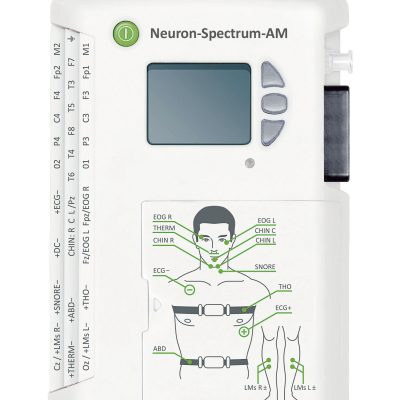
| Shipped From Abroad
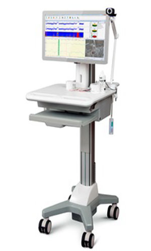
This Portable Wireless EEG/PSG Recorder is a compact, battery-powered device that wirelessly monitors brain activity (EEG) and sleep patterns (PSG). It enables real-time data transmission via Bluetooth or Wi-Fi, making it ideal for sleep studies, neuroscience research, brain-computer interfaces, and remote patient monitoring.
Delivery & Availability:
Typically 10-21 working days – excluding furniture and heavy/bulky equipment. Please contact us for further information.
| Shipped From Abroad
This 20-lead colorful EEG wire set features silver/gold-plated cup electrodes and TPU cables for durability and precision. Compatible with standard EEG systems, the reusable leads ensure optimal signal quality and easy application in neurodiagnostic and sleep monitoring environments.
Delivery & Availability:
Typically 10-21 working days – excluding furniture and heavy/bulky equipment. Please contact us for further information.
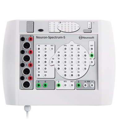
| Shipped From Abroad
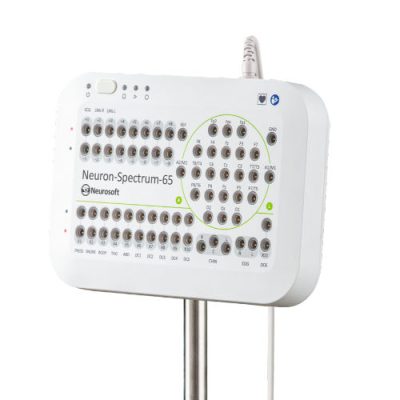
The Neuron-Spectrum 61–65 EEG series offers advanced neurophysiological diagnostic systems with 11 to 39 channels, supporting ICU monitoring, routine EEG, long-term video EEG, and polysomnography. These systems feature real-time impedance monitoring, multimodal stimulation, and high-quality data acquisition for clinical and research applications.
Delivery & Availability:
Typically 10-21 working days – excluding furniture and heavy/bulky equipment. Please contact us for further information.
| Shipped From Abroad
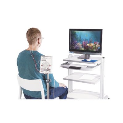
The Neuron-Spectrum-1/BFB by Neurosoft is a comprehensive system designed for biofeedback and neurofeedback training. It enables the creation of individualized training profiles using various physiological parameters, including EEG, heart rate (HR), electromyography (EMG), respiratory rate, galvanic skin response (GSR), blood oxygen saturation (SpO₂), and temperature.
Delivery & Availability:
Typically 10-21 working days – excluding furniture and heavy/bulky equipment. Please contact us for further information.
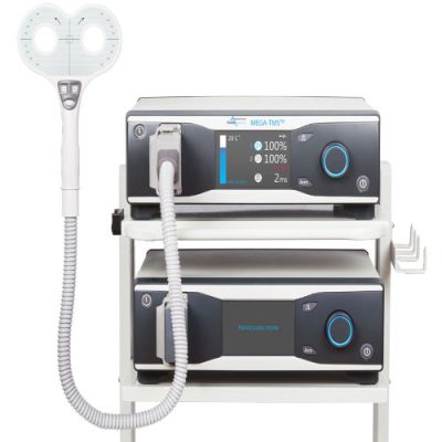
| Shipped From Abroad
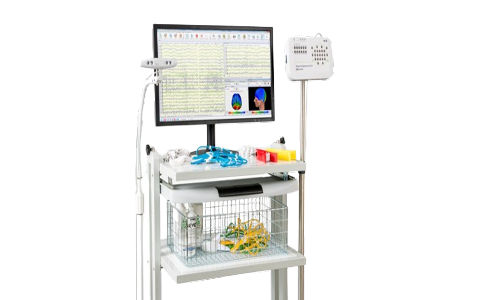
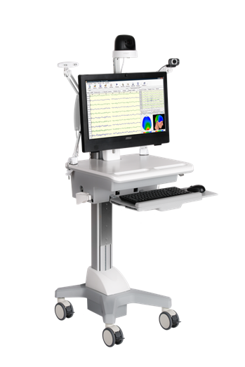
The Neuron-Spectrum-65 Video EEG Monitoring System by Neurosoft is an advanced 39-channel EEG system designed for comprehensive neurophysiological diagnostics, including video EEG monitoring, polysomnography (PSG), and long-term studies. It features high-quality signal acquisition, real-time impedance monitoring, and multimodal data integration for clinical and research applications.
Delivery & Availability:
Typically 10-21 working days – excluding furniture and heavy/bulky equipment. Please contact us for further information.
| ||||||||||||||||||||||||||||||||||||||||||||||||||||||||||||||||||||||||||||||||
| Content | Key Features
Technical Specifications
| https://youtu.be/iKK7jb3M9Bo?list=PLU2BvNtfeStNd_cVOfahLwAr4ucZS1fpm
Features
ApplicationsFree moving during examination Thanks to the wireless interface to transfer data to PC, Neuron-Spectrum-AM allows patients to feel comfortable and move freely within the place they are examined. A small unit is placed on the patient’s body and records EEG signal even when the patient is walking or moving from one room to another. The recorded data is always under the control of specialists. This is especially important if the patient is a child. The range of wireless interface is 50 meters and it can be increased by using several Wi-Fi access points. Neuron-Spectrum-AM is a portable EEG and PSG recorder. Use the display and control buttons to measure impedance, start and stop examinations. With the built-in Wi-Fi module, the functionality of the system is increased. The data can be transferred to PC for review and analysis directly during the acquisition. Out-patient EEG studies and video EEG monitoring (up to several days) Due to modern energy management technologies, Neuron-Spectrum-AM can be used for long-term EEG monitoring up to several days. The hot battery replacement makes it possible to perform examinations practically unlimited in duration. A 32 GB memory card can store more than 30 days of continuous recording. Using such functionality, you can perform synchronous video EEG recording in ambulatory setting continuously for several days. Control of electrode placement during acquisition The quality of recorded signals depends directly on the quality of placed electrodes. Using Neuron-Spectrum-AM, you can measure impedance before the acquisition and directly during it. You can quickly see the electrodes applied improperly and correct their placement. The impedance of each electrode is saved in the examination. So, when you review and analyze the recorded data, you can see the signal quality for each channel. It is especially important during long-term video EEG monitoring. EEG review and analysis during acquisition Via Internet, the operator can connect to the laptop in the patient’s room and check the quality of EEG signal and electrode placement, as well as the patient’s condition. If the recorded data is transferred to the cloud storage, the operator can view and analyze the obtained information directly during the acquisition. After examination, all data are available in the cloud database Download Brochure | Features
Technical Specifications
| https://youtu.be/JDAsx3pO4U8?si=fzekZEwG6_K2pHgL
FeaturesFlexible software The advantage of the Neuron-Spectrum.NET software is its wide functionality. You can use the simple interface, or you can customize the software using the special settings. Before the study, you may choose the pre-defined electrode montage or create your own one depending on the type of test you perform. Using the convenient navigation tools, you can quickly view the recorded EEG curves, highlight artifacts, and arrange epochs. For the advanced analysis of recorded data, the contemporary tools for mathematical analysis of EEG are used. The automatic report is generated using the preset report templates and integrated glossary. The report is easily edited, saved to PDF, printed or sent automatically. Electrode impedance indication at the lead inputs The impedance indicators on the amplifier ensure the continuous impedance monitoring during the acquisition, timely correction of electrode placement and good quality of EEG recording. Operation mode can be switched with just one button on the front panel. The LED indicator next to the button shows the mode: acquisition, EEG monitoring or impedance measurement. It makes the EEG study much easier. Electrodes and caps of various manufacturers During the study, the disk, cup and bridge electrodes as well as the electrode caps of different sizes can be applied. They are connected to the amplifier unit via touch-proof connectors and standard plug. Operation in unshielded environments The EEG systems of Neuron-Spectrum line can be used in any suitable room, which is convenient for staff and comfortable for patients. The costs for preparing a specialist’s workplace are also reduced significantly. The device is small and USB powered, so you can establish your EEG lab anywhere. Download Brochure | https://youtu.be/zAow7-WVqes?list=PLU2BvNtfeStNd_cVOfahLwAr4ucZS1fpm
System for Biofeedback TrainingsFeatures
ApplicationsReliable system with necessary accessories in base delivery set The delivery set of Neuron-Spectrum-1/BFB includes 8-channel EEG system, special accessories for biofeedback trainings and also Neuron-Spectrum.NET software module. It is only one configuration and we are always ready to customize the delivery set for you and meet your exact needs. Trainings using various physiological parameters Using Neuron-Spectrum-1/BFB, you can perform the following trainings: Neurofeedback — trainings using parameters of EEG. Biofeedback — trainings using HR, EMG, respiratory rate, GSR, SpO2, temperature. Standard alpha-theta training and beta-training are implemented in the Neuron-Spectrum-1/BFB software, but you can always create the individual training program for each patient using various physiological parameters. Extensive interactive content for biofeedback The Neuron-Spectrum-1/BFB software has integrated training protocols where the obtained physiological signal can be associated with the images, photos, films, games and music. The specialist can choose from the variety of offered options or even use its own one. Monitoring of training efficiency You can monitor the training process and change the parameters in real time. There is no need to stop the training. All training sessions are stored in the database where you can review the history of patients and analyze their training efficiency. Automatic examination report When the training is finished, the software generates the report that can be viewed simultaneously with EEG traces. The report can be edited and printed and its soft copy can be saved in the database. If you have already purchased the Neurosoft EEG system and would like to perform training with biological feedback, just buy the software module with accessories depending on the training type you would like to arrange. The Neuron-Spectrum.NET software is compatible with all Neurosoft EEG systems. Download Brochure | https://youtu.be/JDAsx3pO4U8
Features
AdvantagesLong-term video EEG monitoring Neuron-Spectrum-64 provides synchronous long-term video and EEG monitoring even for several days. The high-quality EEG signal and frame-accurate video synchronization are guaranteed. You can also connect the recording unit to a local area network (LAN) to place the patient in one room, and the analysis and data processing unit to another one. EEG workstation and mobile system Depending on your needs, we can offer you stationary or mobile system for video EEG monitoring. If you have a special room, you can place EEG workstation here. In this case, the patient’s room and the room for EEG review and analysis can be removed from each other at any distance. If there is no such room, the mobile system for video EEG monitoring is a perfect choice that allows you to perform examinations in any suitable room of your medical facility. Up to 3 Full HD video cameras In your EEG lab, you can perform continuous synchronous video EEG recording using up to 3 network cameras with maximum resolution of 1920*1080 (Full HD), automatic day and night video recording with infrared lighting, and a built-in microphone. With such functionality, you will never miss a single detail. The storage and review of recorded video is ensured by modern video compression algorithms and editing tools. Special electrode caps The special electrode caps with built-in electrodes are a perfect choice for long-term EEG monitoring. There is no need to use adapters or other devices, just connect the cap to the standard connector on the EEG system. Our systems are delivered with special extension cables for electrode caps for the patients to feel comfortable and move freely within the place they are examined. The quality of electrode placement is very important during the whole acquisition process. The function of continuous impedance monitoring is implemented in Neuron-Spectrum-64. If the impedance of any electrode increases, the software informs the operator immediately. Throughout the examination, the impedance of each electrode is saved and displayed as a trend. So, you can always see the impedance value at the required time moment. EEG analysis during acquisition To save your time on routine operations, EEG search and analysis algorithms are built into Neuron-Spectrum.NET. And you can focus on really important things. During the acquisition, the software is already recording suspicious phenomena and making calculations and analysis of EEG signal. So, you can view the recorded video and EEG traces directly during the acquisition. By the time the examination is finished, you can create a report generated automatically according to a preset template. Pre-surgical monitoring to localize pathologic activity in the brain Before surgical therapy of pharmacoresistant epilepsy, it is important to localize the pathological activity in the brain. Using Neuron-Spectrum-64, you can perform the pre-surgical monitoring with both scalp and invasive (cortical or subcutaneous deep) EEG electrodes and calculate the location of pathological areas with a special 3D localization software. During the acquisition, the monopolar, bipolar or mixed montages can be used in the 10-20 or 10-10 systems. You can change the montage at any moment: before or during the acquisition or during the review and analysis of EEG record. You can also change the channel settings. For example, if you cannot remove the EEG baseline in frontal leads, specify the higher values for high-pass filter only for them. Change the parameters of any channel directly during the acquisition. Data storage on removable media When the exam is finished, you can record it on a removable media and give it to the patient or send to another medical facility. Using Neuron-Spectrum.NET, you can save the study to DVD or a flash card with a special viewer program and view the examination on any computer without installation.Specifications
| ||||||||||||||||||||||||||||||||||||||||||||||||||||||||||||||||||||||||||||||||
| Weight | N/A | N/A | N/A | N/A | N/A | N/A | ||||||||||||||||||||||||||||||||||||||||||||||||||||||||||||||||||||||||||||||||
| Dimensions | N/A | N/A | N/A | N/A | N/A | N/A | ||||||||||||||||||||||||||||||||||||||||||||||||||||||||||||||||||||||||||||||||
| Additional information |


Reviews
There are no reviews yet.