| Description | Shipped From Abroad
Delivery & Availability:
Typically 10-21 working days – excluding furniture and heavy/bulky equipment. Please contact us for further information.
| Ship from abroad
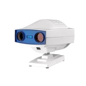
- Easy installation and maintenance: It is quite easy to replace light bulks, and unnecessary to adjust its focus and position.
- Customer Programmable: Icons can be displayed in predefined sequence by programming.
- Display Single Icon: Display single icon by multi-function mask plate.
- Tilted Placement: Perfect image position can be obtained by adjusting the horizontal axis of projector.
- Standards parts: Metal plate with polarized coating (410x 410mm)
Delivery & Availability:
Typically 14 working days – excluding furniture and heavy/bulky equipment. Please contact us for further information.
| 83Shipped from abroad
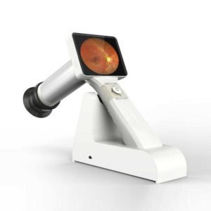
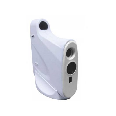
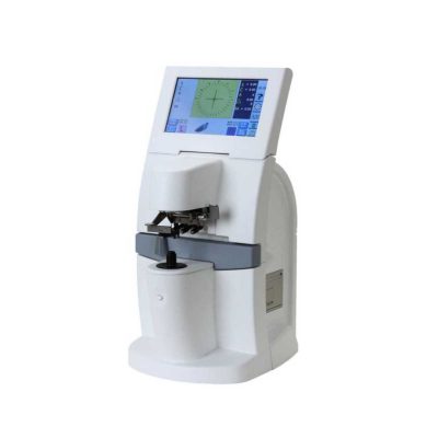
This is a portable medical camera for fundus imaging, diagnosis, and especially for fundus disease screening. It's compact, easy to obtain high definition fundus image. It can be conveniently applied to rapid screening, out diagnosis, bedside diagnosis and remote medical treatment, etc.
Delivery & Availability:
Typically 14 working days – excluding furniture and heavy/bulky equipment. Please contact us for further information.
| Shipped from abroad
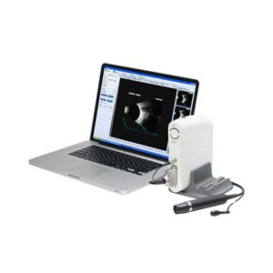
- Software image workstation
- B, B+B, B+A, A modes
- Video review for 100 images
- PDF report output
- Optional 20MHz B Probe: vitreous plus function
Delivery & Availability:
Typically 14 working days – excluding furniture and heavy/bulky equipment. Please contact us for further information.
| Shipped from abroad
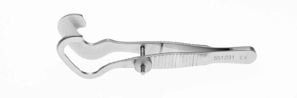
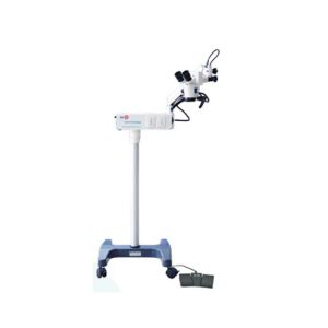
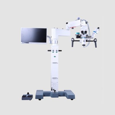
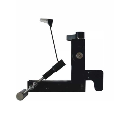
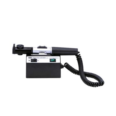
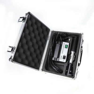
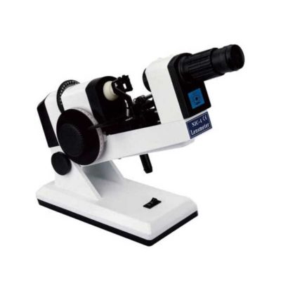
YZ20P5 Operation Microscope is a simple binocular coaxial microscope for a single man. The Operation Microscope is small, light, and convenient. It has high agility and can meet the requirements for general ophthalmology operation. The microscope fits mobile medical treatment.
Delivery & Availability:
Typically 14 working days – excluding furniture and heavy/bulky equipment. Please contact us for further information.
| In Stock
Features:
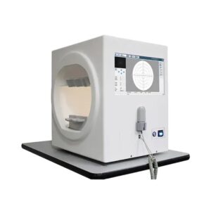
The Bio-1000 automated perimeter absorbs the advantages of international advanced perimetry devices. It comprises the highly integrated computer, optics, machinery and electronics systems.
Delivery & Availability:
Typically 7-14 working days – excluding furniture and heavy/bulky equipment. Please contact us for further information.
|
| Content | | Digital Chart Projector-Features:
- Easy installation and maintenance: It is quite easy to replace light bulks, and unnecessary to adjust its focus and position.
- Customer Programmable: Icons can be displayed in predefined sequence by programming.
- Display Single Icon: Display single icon by multi-function mask plate.
- Tilted Placement: Perfect image position can be obtained by adjusting the horizontal axis of projector.
- Standards parts: Metal plate with polarized coating (410x 410mm)
Technical Specifications of Digital Chart Projector:
|
Projection Distance |
1.5m~6m |
|
Projection Magnification |
30x (at 5m) |
|
Projection Size |
330x270mm (at 5 m) |
|
Chart Switching Speed |
One chart per 0.03s |
|
Mask Switching Speed |
1 open, 5 horizontal different lines, 8 vertical lines, 21 single letters, 1 red/green |
|
Program |
2 sets programs, each program contains up to 30 steps |
|
Speed of mask conversion |
One mask per 0.03s |
|
Lamp |
LED lamp |
|
Auto-off function |
After 10 minutes idle time |
|
Power Source |
AC 220V, 50Hz or 110V, 60Hz |
|
Power consumption |
40W |
|
Accessories |
Remote control, polarized metal screen, halogen lamp, polarized glasses, fuses (2), batteries (2) |
| Portable Fundus Camera is a portable medical camera for fundus imaging, diagnosis, and especially for fundus disease screening. It's compact, easy to obtain high definition fundus image. It can be conveniently applied to rapid screening, out diagnosis, bedside diagnosis and remote medical treatment, etc.
Features of Portable Fundus Camera:
- Images real-time display, one-button magnifying
- Non-mydriatic and high definition imaging.
- Micro SD memory card up to 32GB for 80,000 images
- USB and WiFi connection
- Low weight 450g, movable and portable
- A rechargeable battery provides up to more than 4 hours of continuous operation.
- Optional imaging modules for examination: posterior ophthalmoscope, anterior segment ophthalmoscope, otoscope, rhinoscope, laryngoscope and dermatoscope
- High definition and stable image
- Easy one-handed operation and excellent portability
- Large data service and a variety of image acquisition mode
Technical Specifications of Portable Fundus Camera:
| Model |
MC-600 |
| FOV |
45° |
| Minimum pupil |
2.5mm |
| Refractive compensation |
-20D~+20D |
| Light source |
White LED/IR |
| Image resolution |
1920×1080 |
| Focus Mode |
Manual |
| Screen |
3.5" color |
| Power |
≤6VA |
| Storage |
8GB Micro SD card |
| Power supply |
3.7VLithium Battery |
| Interface |
Mini USB/Wifi |
| N. Weight |
450g(Typical) |
| G. Weight |
2.5kg |
| Size |
160mm*90mm*190mm |
| Packing size |
360mm*310mm*160mm |
| Functions of Ophthalmic AB Scan Machine:
- Software image workstation
- B, B+B, B+A, A modes
- Video review for 100 images
- PDF report output
- Optional 20MHz B Probe: vitreous plus function
Technical Specifications:
| A scan |
1.Probe: 10MHz frequencies, with LED
2.Depth: 40mm
3.Precision: ±0.05mm
4.Eye mode: Phakic / Aphakic / Dense / Various IOL
5.Measurement: Anterior chamber depth, lens thickness, vitreous body length, total length and average
6.IOL Formula: SRK-II, SRK-T, BINKHORST, HOLLADAY, HOFFER-Q, HAIGIS, Stat.
7.Calculation: Average and standard deviation
8.Store: 10 Scanning results for each eye |
| B scan |
1.Probe: 10MHz/20MHz (optional), Magnetic driven, noiseless
2.Scanning Mode: Sector Scanning
3.Resolution: Lateral ≤0.3mm; Vertical≤0.2mm
4.Geometric Location Precision: Lateral≤10%; Vertical≤5%
5.Depth: 60mm
6.Enhance the part of vitreous body and retina
7.Gain of probe:30dB-105dB
8.Scanning Angle : 53°
9.Gray Scale: 256
10.False Color: Multi colors OTC
11.Measure Mode: distances, perimeter and area
12.Movies: 100 images movie review,AVI ZIP JPG format image output
13.Output: PDF format case report, connect to normal printer |
| Others |
1.Display Mode :B, B+B, B+A, A
2.Hint: preset keyword
3.Case Search: Multi-keywords
4.Working Platform: Windows XP, VISTA, WINDOWS7
5.User-defined report template |
| Operation Microscope(YZ20P5) is a simple binocular coaxial microscope for a single man. The Microscope is small, light, and convenient. It has high agility and can meet the requirements for general ophthalmology operation. The microscope fits mobile medical treatment.
Features of Operation Microscope:
- Multi-layer coating technology is used on optical lenses to enhance the transmission rate and prevent mildew.
- Foot-controlled focus, three-step magnification change, good depth of field of view, and good binocular fusion to meet the need of cataract surgery.
- Using apochromatic technology to make different wavelengths of the light focus near the focal point behind the lens, so as to make the operator's vision more clearly.
- The machine weighs only 41Kg to be light and compact and is especially applicable for mobile medical.
- Optional desktop components to make the machine more portable and can also be customized according to special requirements to meet the needs in ophthalmology, ENT and other surgery.
- Optional F250/F300/F400 lens.
Technical Specifications of Operation Microscope:
|
Eyepiece Magnification
|
12.5×
|
|
Focal Length of Objective Lens
|
F=200
|
|
Working Distance
|
190 mm
|
|
Magnification for Main Microscope
|
5.3×, 8×, 16×
|
|
Diameter of Field
|
37 mm, 25 mm, 16.7 mm
|
|
Diopter Adjustment
|
±5D
|
|
Pupillary Distance
|
50 mm ~ 70 mm
|
|
Maximum Resolution
|
100 LP/mm
|
|
Light Source
|
12V/100W halogen lamp for medical use
|
|
Illumination Type
|
6° coaxial illumination of the cold light source
|
|
Coaxial Illuminance
|
≥30,000 Lx
|
|
Reaching Radius of Arm
|
870 mm
|
|
Adjustable Vertical Range
|
700 mm~1100 mm
|
| Fine Focusing Range |
30 mm |
|
Input Voltage
|
AC 220V/110V±10%, 50Hz/60Hz
|
|
Power Consumption
|
120 VA
|
|
Fuse
|
AC 250V T4.0A, AC 125V T8.0A
|
|
Electrical Safety Standard
|
IEC 60601-1, Class I
|
|
Packing Volume
|
0.2 m3,1 carton
|
|
Total Weight
|
41 kg
|
| The Bio-1000 automated perimeter absorbs the advantages of international advanced perimetry devices. It comprises the highly integrated computer, optics, machinery and electronics systems. Incorporated with the advanced configuration, comprehensive software inspection categories, and strictly in accordance with international Goldman standard, it provide scientific means for glaucoma, fundus disease, visual pathway injury and neurological diseases.
Feature:
* Comprehensive real-time monitoring,Heiji-krakau physiological blind spot monitoring,gaze tracking/head position tracking,automatic measurement of pupil diameter, reduce the impact of pupil effect on visual field detection.
* Personalized design,accurate clinical analysis,accurate and repid examination strategy.
* Under international Goldman standard,providing a variety of classic test procedures and report analysis.
Technical Specification:
| Inspection range |
90° |
| Inspection distance |
30cm |
| Background light brightness |
White 31.5abs |
| Visual target brightness |
1abs --- 10000abs |
| Visual target size |
Goldmann III |
| Visual target interval time |
200ms |
| Visual target interval time |
Stardard or adjustment according to patient reflection |
| Threshold test model |
Center5°- 16°,center10°- 68, 24-2, 30-2, 30°- 60°,nasal step |
| Upper threshold test model |
30°-40,30°-76,P-60,60°-81,60°-120,190°-135,nasal side |
| Special detection strategy |
Esterman monocular,Esterman binocular,user-customized test,150°driver monocular fast detection, 150°driver monocular standard detection, upper36°detection, blind spot detection, 150° horizontal straightness detection |
| Pupil size test |
Automatic |
| Fixation monitoring |
Blind spot monitoring, eye position deviation alarm, eyedeviation curve |
| Enviroment light detection |
Automatic |
| Analysis software |
Reliability analysis,single vision report,triple visionreport,GPA half vision analysis, GPA glaucoma developing analysis, VFI vision index analysis |
| Operation system |
Win7 above |
|


Reviews
There are no reviews yet.