NEVRO MICROSCOPE
$0.00
Shipped From Abroad
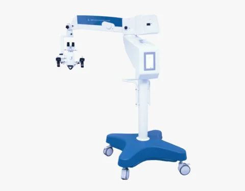
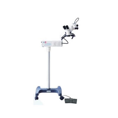
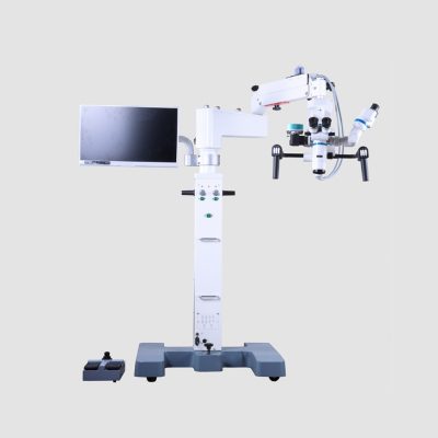
NEVRO Microscope, a line of equipment specially designed in detail, to meet the most demanding standards of surgical procedures,
for the areas of Neurosurgery and ENT, head and neck.
All functions can be activated via Gand Grip, with total comfort and lightness. This ensures less effort and ease during the procedures.
It has electromagnetic brakes, which control any and all positioning accurately, ensuring comfort, ease and time optimization.
Through the LCD touch screen, the same controls made via Hand Grip can also be performed with a simple touch.
Microsurgeries currently require cutting-edge technology with the ability to perform extremely complex procedures in an agile and simple way.
For optimizing surgical processes, the NEVRO microscope is the most efficient and advanced.
Delivery & Availability:
Typically 10-21 working days – excluding furniture and heavy/bulky equipment. Please contact us for further information.
Typically 10-21 working days – excluding furniture and heavy/bulky equipment. Please contact us for further information.
Description
Description
Control via Hand Grip
All microscope functions can be activated via Hand Grip, with total accuracy, comfort and lightness. This ensures less effort and ease during all surgical procedures.
Electromagnetic Brakes
Through the electromagnetic brakes, any and all positioning of the microscope can be done with total precision and comfort. Guaranteeing less effort, ease and time optimization.
LCD screen
The touch screen LCD display is used to view and activate all equipment settings. With its touch screen, the same controls made via Hand Grip can also be performed with a simple touch.
Quick Comparison
| NEVRO MICROSCOPE remove | Binocular Indirect Ophthalmoscope remove | Slit Lamp with Workstation remove | SW-1000 Ophthalmic Ultrasound Scanner-A-Scan remove | Retinoscope remove | Manual lensmeter remove | |||||||||||||||||||||||||||||||||||||||||||||||||||||||||||||||||||||||||||||||||||||
|---|---|---|---|---|---|---|---|---|---|---|---|---|---|---|---|---|---|---|---|---|---|---|---|---|---|---|---|---|---|---|---|---|---|---|---|---|---|---|---|---|---|---|---|---|---|---|---|---|---|---|---|---|---|---|---|---|---|---|---|---|---|---|---|---|---|---|---|---|---|---|---|---|---|---|---|---|---|---|---|---|---|---|---|---|---|---|---|---|---|---|
| Name | NEVRO MICROSCOPE remove | Binocular Indirect Ophthalmoscope remove | Slit Lamp with Workstation remove | SW-1000 Ophthalmic Ultrasound Scanner-A-Scan remove | Retinoscope remove | Manual lensmeter remove | ||||||||||||||||||||||||||||||||||||||||||||||||||||||||||||||||||||||||||||||||||||
| Image |  | 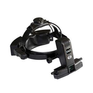 | 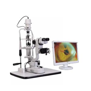 | 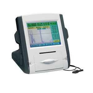 | 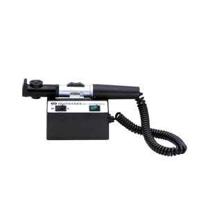 | 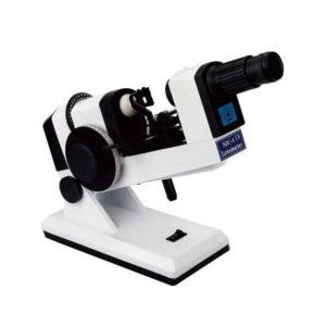 | ||||||||||||||||||||||||||||||||||||||||||||||||||||||||||||||||||||||||||||||||||||
| SKU | SF103356013091-12 | SF1033560107-4 | SF1033560107-7 | SF1033560107-27 | SF1033560107-12 | SF1033560107-22 | ||||||||||||||||||||||||||||||||||||||||||||||||||||||||||||||||||||||||||||||||||||
| Rating | ||||||||||||||||||||||||||||||||||||||||||||||||||||||||||||||||||||||||||||||||||||||||||
| Price |
| $880.00 | $3,740.00 | $1,815.00 | $165.00 |
| ||||||||||||||||||||||||||||||||||||||||||||||||||||||||||||||||||||||||||||||||||||
| Stock | ||||||||||||||||||||||||||||||||||||||||||||||||||||||||||||||||||||||||||||||||||||||||||
| Availability | ||||||||||||||||||||||||||||||||||||||||||||||||||||||||||||||||||||||||||||||||||||||||||
| Add to cart | ||||||||||||||||||||||||||||||||||||||||||||||||||||||||||||||||||||||||||||||||||||||||||
| Description | Shipped From Abroad
NEVRO Microscope, a line of equipment specially designed in detail, to meet the most demanding standards of surgical procedures,
for the areas of Neurosurgery and ENT, head and neck.
All functions can be activated via Gand Grip, with total comfort and lightness. This ensures less effort and ease during the procedures.
It has electromagnetic brakes, which control any and all positioning accurately, ensuring comfort, ease and time optimization.
Through the LCD touch screen, the same controls made via Hand Grip can also be performed with a simple touch.
Microsurgeries currently require cutting-edge technology with the ability to perform extremely complex procedures in an agile and simple way.
For optimizing surgical processes, the NEVRO microscope is the most efficient and advanced.
Delivery & Availability:
Typically 10-21 working days – excluding furniture and heavy/bulky equipment. Please contact us for further information.
| Shipped from abroad
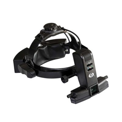
Super lightweight design, reduce fatigue, operation is very convenient.
| Shipped from abroad
| Shipped from abroad
| Shipped from abroad
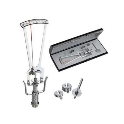
The product can quickly and precisely measure the astigmatism axis and is one of the necessary instruments in optometry inspection.
| Shipped from abroad
The NJC-4 Lensmeter is used to measure the diopters of spherical lens and cylinder lens, the axis of cylinder lens, the strength and the baseline direction of shuttle lens, it can also stamp the optical center of lens, the axis of cylinder len and the base direction of shuttle lens.
| ||||||||||||||||||||||||||||||||||||||||||||||||||||||||||||||||||||||||||||||||||||
| Content | DescriptionControl via Hand Grip All microscope functions can be activated via Hand Grip, with total accuracy, comfort and lightness. This ensures less effort and ease during all surgical procedures. Electromagnetic Brakes Through the electromagnetic brakes, any and all positioning of the microscope can be done with total precision and comfort. Guaranteeing less effort, ease and time optimization. LCD screen The touch screen LCD display is used to view and activate all equipment settings. With its touch screen, the same controls made via Hand Grip can also be performed with a simple touch. | Ophthalmoscope Features:
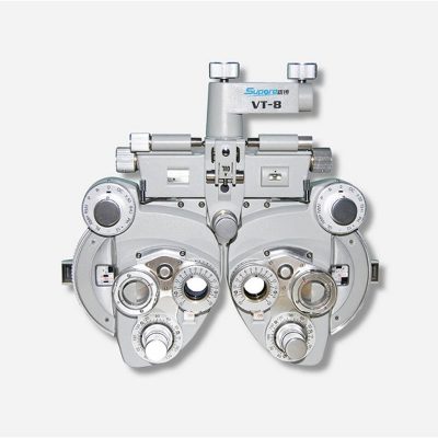
| Slit Lamp with Workstation Features:
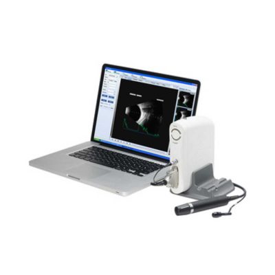
| Feature:
| The product can quickly and precisely measure the astigmatism axis and is one of the necessary instruments in optometry inspection.
Features:
| The NJC-4 Lensmeter is used to measure the diopters of the spherical lens and cylinder lens, the axis of cylinder lens, the strength and the baseline direction of shuttle lens, it can also stamp the optical center of the lens, the axis of cylinder lens, and the base direction of shuttle lens. This instrument (Both Ac and Dc are permitted Two cells When Dc) has clear readings and graduations as well as high objective precision and reliabilities except that it can be operated easily and conveniently, AU the lenses and the made glasses can be measured by it, therefore, it is a required and ideal measuring instrument for glasses manufactures, glasses stores and ophthalmological hospitals.
Technical Specifications:
| ||||||||||||||||||||||||||||||||||||||||||||||||||||||||||||||||||||||||||||||||||||
| Weight | N/A | N/A | N/A | N/A | N/A | N/A | ||||||||||||||||||||||||||||||||||||||||||||||||||||||||||||||||||||||||||||||||||||
| Dimensions | N/A | N/A | N/A | N/A | N/A | N/A | ||||||||||||||||||||||||||||||||||||||||||||||||||||||||||||||||||||||||||||||||||||
| Additional information | ||||||||||||||||||||||||||||||||||||||||||||||||||||||||||||||||||||||||||||||||||||||||||


Reviews
There are no reviews yet.