


3B Scientific Gallstone Model – 3B Smart Anatomy
$0.00
Ship from abroad
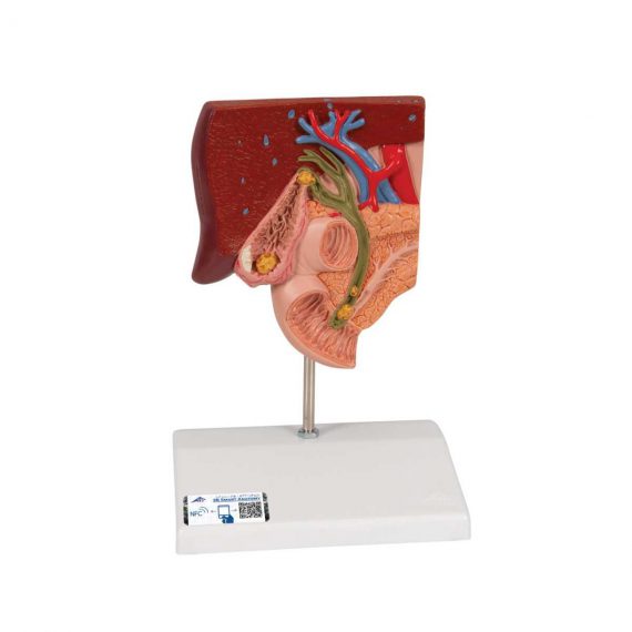
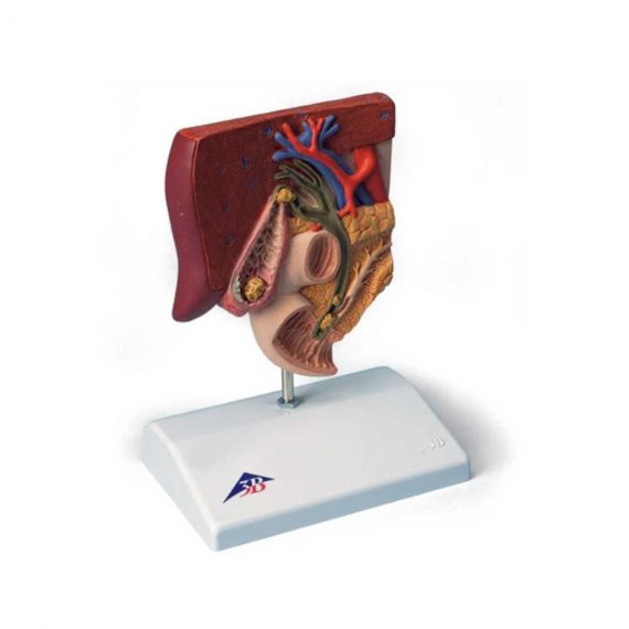
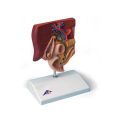
This graphic gallstone model for patient education shows the anatomy of the biliary system and its surroundings in half natural size. Both acute inflammation (cholecystitis) and the tissue changes caused by chronic inflammation can be identified in the gallbladder wall.
Delivery & Availability:
Typically 5-7 working days – excluding furniture and heavy/bulky equipment. Please contact us for further information.
Description
This graphic gallstone model for patient education shows the anatomy of the biliary system and its surroundings in half natural size. Both acute inflammation (cholecystitis) and the tissue changes caused by chronic inflammation can be identified in the gallbladder wall. Gallstones can be found in the following typical locations:
- In the fundus area of the gall bladder
- In the area of the spiral valve
- In the area of the common bile duct
- In the papillary opening to the small intestine
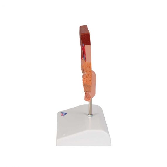
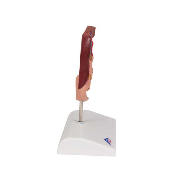
Gallstone model mounted on base.
Quick Comparison
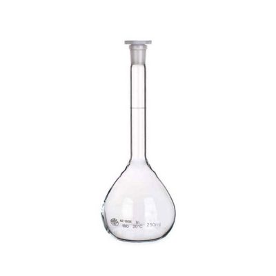
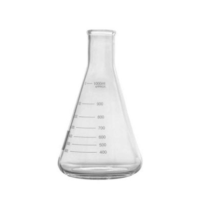
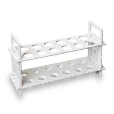
| 3B Scientific Gallstone Model - 3B Smart Anatomy remove | Conical Glass Flask 500ml remove | Plastic Test Tube Rack remove | Measuring Beaker 250ml remove | Bunsen Burner remove | Petri Dish Per Pack remove | |
|---|---|---|---|---|---|---|
| Name | 3B Scientific Gallstone Model - 3B Smart Anatomy remove | Conical Glass Flask 500ml remove | Plastic Test Tube Rack remove | Measuring Beaker 250ml remove | Bunsen Burner remove | Petri Dish Per Pack remove |
| Image |  |  |  | 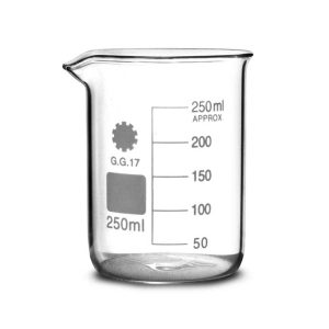 | 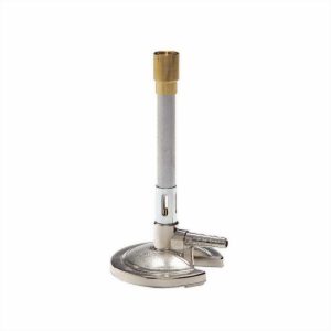 | 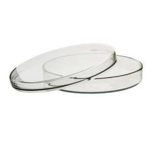 |
| SKU | SF1033560099-37 | SF1033560084-159 | SF1033560084-174 | SF1033560084-153 | SF1033560084-155 | SF1033560084-172 |
| Rating | ||||||
| Price |
| $6.00 | $3.00 | $4.95 | $4.25 |
|
| Stock | ||||||
| Availability | ||||||
| Add to cart | ||||||
| Description | Ship from abroad
This graphic gallstone model for patient education shows the anatomy of the biliary system and its surroundings in half natural size. Both acute inflammation (cholecystitis) and the tissue changes caused by chronic inflammation can be identified in the gallbladder wall.
| In stock
| In stock
| In stock
• Height: 95mm
• Width: 85mm
• Diameter: 75mm
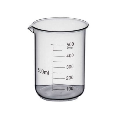
• Volume: 250ml
| In stock
| In stock
Premium petri dishes made to withstand wet or dry repeated sterilisation. The heavy borosilicate glass and can handle direct flames.
|
| Content | This graphic gallstone model for patient education shows the anatomy of the biliary system and its surroundings in half natural size. Both acute inflammation (cholecystitis) and the tissue changes caused by chronic inflammation can be identified in the gallbladder wall. Gallstones can be found in the following typical locations:
|
|
| Measuring beaker • Lined with metric increments up to 200ml • Material: Borosilicate glass • Pouring lip • Great for molecular mixology and molecular gastronomy • Height: 95mm • Width: 85mm • Diameter: 75mm • Volume: 250ml |
| Premium petri dishes made to withstand wet or dry repeated sterilisation. The heavy borosilicate glass and can handle direct flames.
Features:
|
| Weight | N/A | N/A | N/A | N/A | N/A | N/A |
| Dimensions | N/A | N/A | N/A | N/A | N/A | N/A |
| Additional information |

Reviews
There are no reviews yet.