
BONE DENSITOMETER (SONOST-2000)
$4,950.00
Shipped From Abroad
SONOST-2000 is a compact quantitative ultrasound(QUS) bone mineral densitometer (BMD) with an easy, quick, simple and safe measurement.
Typically 10-21 working days – excluding furniture and heavy/bulky equipment. Please contact us for further information.
Description
Description
Quantitative Ultrasound (QUS) Bone Mineral Densitometer
Easy & Quick & Simple & Safe Measurement
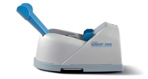
SONOST-2000 is a dry mode ultrasound bone densitometry that boasts a quick measuring speed of 15 seconds, easy operation and high durability. Its compact design makes it portable enough to use with a laptop anywhere.
Features
-
Dry-type Ultrasound BMD with No Radiation
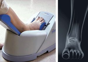
SONOST-2000 is a dry-type ultrasound bone densitometry which is free of the radiation. It enables a swift and easy measurement of Calcaneus by calculating the sound of speed (SOS) and Broadband Ultrasound Attenuation (BUA). Due to its compact design, you can use SONOST-2000 with a laptop for your portable measurement.

-
Dry-type Ultrasound BMD with No Radiation
SONOST-2000 is a dry-type ultrasound bone densitometry which is free of the radiation. It enables a swift and easy measurement of Calcaneus by calculating the sound of speed (SOS) and Broadband Ultrasound Attenuation (BUA). Due to its compact design, you can use SONOST-2000 with a laptop for your portable measurement.
Specification
| Measurement Type | Ultrasound |
| Measurement site | Calcaneus (Heel) |
| Measured parameter | T-score, Z-score, SOS, BUA, BQI index |
| Reproducibility | SOS – ≤ 1% CV BUA – ≤ 2% CV |
| QC check | Daily QC phantom |
| Dimension | (W)523mm × (D)286mm × (H)254mm (Calf supporter folded) (W)523mm × (D)286mm × (H)254mm |
| Weight | 7.8kg |
Click here to download Manual
Click here to download Catalogue
Quick Comparison
| BONE DENSITOMETER (SONOST-2000) remove | DrGem Diamond All-In-One Digital X-ray Machine remove | Sonoscape S22 Ultrasound Machine remove | Sonoscape P15 Ultrasound Machine With Four Probes remove | Jade Mobile X-ray machine (Analogue) remove | Topaz Digital X-ray Machine remove | |||||||||||||||
|---|---|---|---|---|---|---|---|---|---|---|---|---|---|---|---|---|---|---|---|---|
| Name | BONE DENSITOMETER (SONOST-2000) remove | DrGem Diamond All-In-One Digital X-ray Machine remove | Sonoscape S22 Ultrasound Machine remove | Sonoscape P15 Ultrasound Machine With Four Probes remove | Jade Mobile X-ray machine (Analogue) remove | Topaz Digital X-ray Machine remove | ||||||||||||||
| Image | 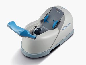 |  |  |  |  |  | ||||||||||||||
| SKU | SF1033560130103-4 | SF1033560074-3 | SF1033560012-3 | SF1033560012-8 | SF1033560074-2 | SF1033560074-1 | ||||||||||||||
| Rating | ||||||||||||||||||||
| Price | $4,950.00 |
| $9,350.00 | $13,900.00 |
|
| ||||||||||||||
| Stock | ||||||||||||||||||||
| Availability | ||||||||||||||||||||
| Add to cart | ||||||||||||||||||||
| Description | Shipped From Abroad
SONOST-2000 is a compact quantitative ultrasound(QUS) bone mineral densitometer (BMD) with an easy, quick, simple and safe measurement.
Delivery & Availability:
Typically 10-21 working days – excluding furniture and heavy/bulky equipment. Please contact us for further information.
| Shipped from Abroad DrGem Diamond All-In-One Digital X-ray Machine is a fully automatic digital radiography system providing state-of-the-art image quality, image processing and user interface. With a wide selection of anatomical studies on the imaging software, DIAMOND automatically sets up the x-ray generator’s preprogrammed exposure technique settings, motorized radiographic stand positioning, x-ray collimation and post-image processing for the selected study. Specifically designed to increase workflow, this fully digital system offers convenient auto-positioning and advanced image processing to achieve big performance with little effort. Delivery & Availability: Typically 21 working days – excluding furniture and heavy/bulky equipment. Please contact us for further information. | Shipped from Abroad As SonoScape steps forward to add value and efficiency to ultrasound, the latest S22 was designed in a user-friendly platform to address current and future demanding needs. It represents an excellent mix in performance and price. Delivery & Availability: Typically 5-7 working days – excluding furniture and heavy/bulky equipment. Please contact us for further information. | In Stock A feature-rich system inheriting the Wi-Sono high-end platform, the P15 uses an array of advanced tools to help enhance the image quality. It's a cost-effective, simplified console with an intuitive user interface and multiple intelligent functions. Delivery & Availability: Typically 2 working days – excluding furniture and heavy/bulky equipment. Please contact us for further information. | In Stock JADE is one of the lightest portable X-ray systems on the market, allowing it to be used in any imaginable way including bedside, operating rooms, intensive care units and in veterinary fields. With a simple, easy-to-use operator console, three-way control, two-step foldable stand and auto lock system, JADE is a user-friendly portable X-ray system. Delivery & Availability: Typically 21 working days – excluding furniture and heavy/bulky equipment. Please contact us for further information. | In Stock DRGEM’s TOPAZ X-ray machine is a state-of-the-art mobile digital radiography system, designed with maximum comfort for patients and users in mind. From its user-friendly software to smooth movements, TOPAZ is made to improve your workflow and provide you with high-quality images. Delivery & Availability: Typically 21 working days – excluding furniture and heavy/bulky equipment. Please contact us for further information. | ||||||||||||||
| Content | Description
https://youtu.be/kXoZ1-3LrpQ?si=YTIRiaLkeHQX6c2G
Quantitative Ultrasound (QUS) Bone Mineral DensitometerEasy & Quick & Simple & Safe Measurement
SONOST-2000 is a dry mode ultrasound bone densitometry that boasts a quick measuring speed of 15 seconds, easy operation and high durability. Its compact design makes it portable enough to use with a laptop anywhere.
Features
Specification
Click here to download ManualClick here to download Catalogue | DrGem Diamond All-In-One Digital X-ray Machine is a fully automatic digital radiography system providing state-of-the-art image quality, image processing and user interface. With a wide selection of anatomical studies on the imaging software, DIAMOND automatically sets up the x-ray generator’s pre-programmed exposure technique settings, motorized radiographic stand positioning, x-ray collimation and post-image processing for the selected study. Specifically designed to increase workflow, this fully digital system offers convenient auto-positioning and advanced image processing to achieve big performance with little effort.
Features of DrGem Diamond All-In-One Digital X-ray Machine:
Outstanding Image Quality -
Digital radiography via at panel detector improves your workflow, exam speed and comfort with efficiency. Digital at panel detector with Csl screen provides excellent spatial resolution, MTF, DQE and stability based on ne pixel pitch. A 3-field ion-chamber is provided for AEC function.
Automatic Collimation –
Automatic x-ray eld size control of the motorized collimator corresponds to dierent SIDs. Includes user adjustable lamp timer with on/oswitch.
Automatic Positioning –
Click Here To Download Catalogue | DETAILS
As SonoScape steps forward to add value and efficiency to ultrasound, the latest S22 was designed in a user-friendly platform to address current and future demanding needs. It represents an excellent mix in performance and price.
S22, is a shared service ultrasound system with a slim and elegant package that has combined mobility with utility to fit in specific clinical situations including emergency department, ICU, operating room and so on. Furthermore, its ergonomic design, easy operating and flexible data management will give you a memorable experience.
SPECIFICATION
• Large high-resolution widescreen LED
• Sensitive touch screen
• Four transducer sockets plus one socket for pencil probe
• A comprehensive selection of probes: linear, Convex, Micro-convex, Volumetric, Endocavity, Bi-plane, Phased Array, TEE, Intraoperative, Pencil
• Premium application technology: 4D, μ-scan speckle reduction, compound imaging, Pulse Inversion Harmonic Imaging, Color M-Mode, Steer M-Mode, PDI, TDI, Real-time Panoramic Imaging, Trapezoid Imaging, Auto-IMT…
• Full patient database and image management solutions: DICOM 3.0, AVI/JPG, USB 2.0, HDD, DVD, PDF report
• Multi-Language Input Keyboard
• Built-in battery
Click Here To Download Catalogue | DETAILS
Super Wide-bandwidth Platform
Inheriting Wi-sono's ultra-wide system platform and with the advanced probe technology, high-resolution and deep penetration images are provided for precision medicine.
Spatial Compound Imaging
Spatial Compound Imaging utilizes several lines of sight for optimal contrast resolution, speckle reduction and border detection, with which P15 is ideal for superficial and abdominal imaging with better clarity and improved continuity of structures.
μ-Scan+
The new generation μ-Scan imaging technology gives you better image quality by reducing noise, improving signal strength and improving visualization.
Dynamic Color
Dynamic color improves upon already existing color Doppler technologies for a clearer capture of color flow and detailed visualization of even tiny veins with lower velocities.
Real-time Panoramic
With real-time panoramic, you can acquire an extended field of view for large organs or long vessels for easy measurement and diagnostic efficiency. Accomplished in real-time for the convenience of the sonographers, any mistake can also be easily back tracked and corrected without interrupting the scan.
3D/4D
Outstanding volume performance with speed and convenience makes P15 outshine others on volume imaging.
Tissue Doppler Imaging
Tissue Doppler Imaging allows clinical doctors to quantitatively evaluate local myocardial movements and functions, facilitating them with the ability to analyze and compare the motions of the different parts of the patient's heart.
Auto IMT
Quick measurement of intra-media vessel thickness ensures good reproducibility and high diagnostic efficiency.
Click Here To Download Catalogue | JADE Mobile X-ray machine is one of the lightest portable X-ray systems on the market, allowing it to be used in any imaginable way including bedside, operating rooms, intensive care units and veterinary fields. With a simple, easy-to-use operator console, three-way control, two-step foldable stand and auto-lock system, the JADE Mobile X-ray machine is a user-friendly portable X-ray system.
Convenient & Intuitive Operation:
JADE is one of the lightest portable X-ray systems on the market, allowing it to be used in any imaginable way including bedside, operating rooms, intensive care units and in veterinary fields. With a simple, easy-to-use operator console, three-way control, two-step foldable stand and auto-lock system, JADE is a user-friendly portable X-ray system.
Compact & Powerful Design:
JADE Mobile X-ray machine is an innovative, highly versatile portable X-ray system suitable for a variety of clinical uses. Utilizing the unique technology used in DRGEM’s universally recognized X-ray generators, JADE is a compact but powerful unit with a 4kW output and thoughtfully designed components to increase efficiency and maximize workflow. The core part of X-ray source adopts high-quality tube assembly, X-ray collimator and high frequency X-ray generator with excellent performance, lifetime and stability.
Features:
Click Here To Download Catalogue | TOPAZ X-ray machine is among the high end X-ray machine manufactured by DRGEM, a digital X-ray system that provides quality images with little or no effort.
It begins with Advanced Technology
Integrating high technology and over a decade of experience in conventional and digital radiography systems, DRGEM’s TOPAZ X-ray machine is a state-of-the-art mobile digital radiography system, designed with maximum comfort for patients and users. From its user-friendly software to smooth movements, TOPAZ X-ray machine is made to improve your workflow and provide you with high-quality images.
Full Featured Imaging Software & Excellent Digital Image Processing
With a high-performance, built-in touchscreen, TOPAZ X-ray machine offers a user-friendly interface and powerful software for easy operation and increased workflow. The anatomical view-based digital image processing, automatically optimizes and enhances the quality of the image. it also comes with automatic image storage and print with DICOM 3.0 networking capability. additionally, the system offers increasing exam throughput while decreasing examination time.
Click Here To Download Catalogue | ||||||||||||||
| Weight | N/A | N/A | N/A | N/A | N/A | N/A | ||||||||||||||
| Dimensions | N/A | N/A | N/A | N/A | N/A | N/A | ||||||||||||||
| Additional information |


Reviews
There are no reviews yet.