| Description | Shipped From Abroad
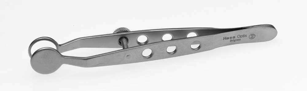
11mm Diameter, Round Plate
Delivery & Availability:
Typically 10-21 working days – excluding furniture and heavy/bulky equipment. Please contact us for further information.
| Shipped from abroad
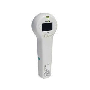
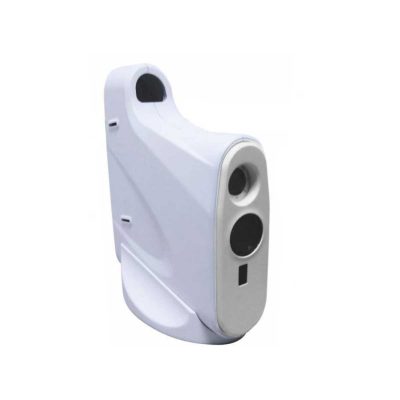
Ultra-small corneal curvature tester, is mainly used to measure the corneal curvature radius and diopter, wireless output print data.
Delivery & Availability:
Typically 14 working days – excluding furniture and heavy/bulky equipment. Please contact us for further information.
| Shipped from abroad
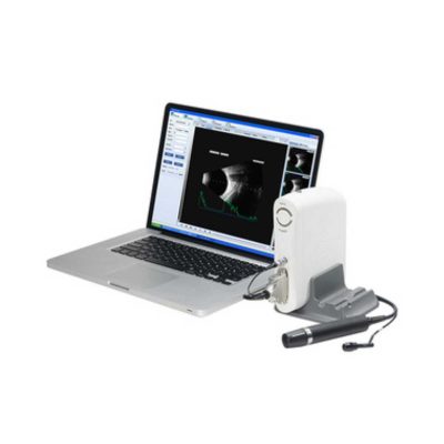
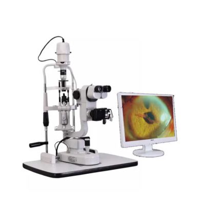
- Digital Slit lamp system,Galilean optical system,Five magnifications.
- Independent image process and management software system
- More than 10 million pixel image resolution
Delivery & Availability:
Typically 14 working days – excluding furniture and heavy/bulky equipment. Please contact us for further information.
| Shipped from abroad
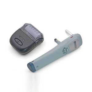
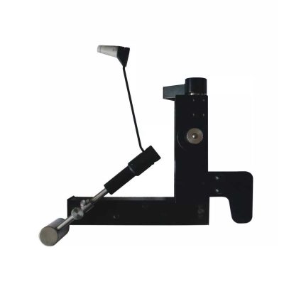
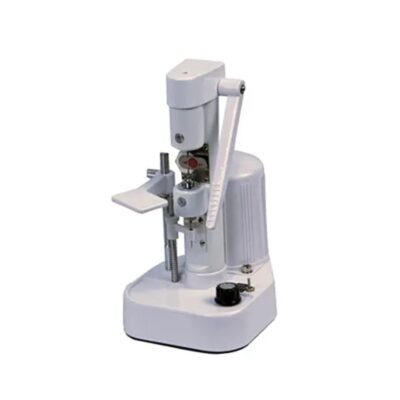
Tonometer SW-500 with vertical and horizontal two working modes, wireless output print data.
Delivery & Availability:
Typically 14 working days – excluding furniture and heavy/bulky equipment. Please contact us for further information.
| Ship from abroad
- Equipped with comprehensive measuring functions, it provides SPH, CYL, AXIS and pupil distance optometry
- Durable and easy to operate
- Easily and intuitively read the sphere focal scale value
- High eco-friendly materials
- Design fitting the face curve and no stimulation
Delivery & Availability:
Typically 14 working days – excluding furniture and heavy/bulky equipment. Please contact us for further information.
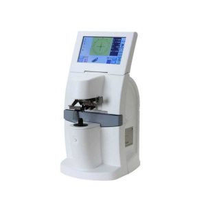
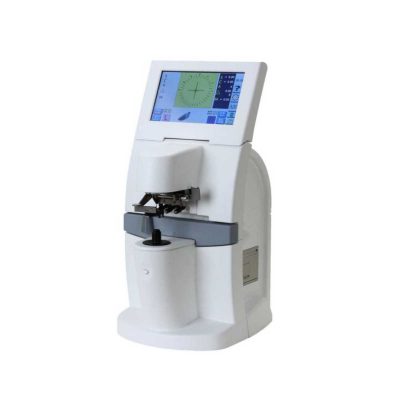
| Shipped from abroad
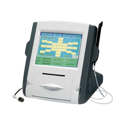
- 7.0-inch color LCD touch panel.
- Hartman sensor with 108 multiple measure points.
- Green measurement light beam.
- Dyeing lens, sunglasses measured easily.
Delivery & Availability:
Typically 14 working days – excluding furniture and heavy/bulky equipment. Please contact us for further information.
|
| Content | | Portable Keratometer Features:
Ultra-small corneal curvature tester, is mainly used to measure the corneal curvature radius and diopter, wireless output print data.
Technical Specifications:
| Model |
SW-100 |
| Measuring Range |
6.5mm~9.5mm: ±0.05mm: 0.01mm |
| Precision |
± 0.05mm |
| Resolution of Curvature Radius of Cornea |
0.01mm |
| Single Measuring Time |
0.03s |
| Output |
Wireless Infrared Thermal Printer |
| Can observe the eye directly through the screen. |
| Weight |
0.5Kg(with batteries) |
| Dimension |
240mmx90mmx60mm |
| Power |
500mW+15% |
| Slit Lamp with Workstation Features:
- Digital Slit lamp system, Galilean optical system, Five magnifications
- Independent image process and management software system
- More than 10 million pixel image resolution
Technical Specifications:
| Microscope Type |
Galileo Parallel |
| Digital System |
External Digital Slit Lamp with 10 Mega pixels |
| Magnification Change Way |
Drum Five Magnifications |
| Eyepiece Magnification |
12.5x |
| Total Magnifications |
6x, 10x, 16x, 25x,40x |
| Diopter Adjustment |
-5D ~+5D |
| Slit Width |
0-14MM Continuous |
| Slit Height |
1-14MM Continuous |
| Slit Angle |
0°- 180° Adjustable |
| Slit Inclination Angle |
5°,10°,15°, 20° |
| Light Spot Diameter |
0.2mm, 2mm, 3mm, 5mm, 10mm, 14mm |
| Filter |
Heat Absorption; Grey; Red free; Cobalt Blue |
| Fixation |
Red LED |
| Illumination Bulb |
12V/50W German OSRAM Halogen Tungsten Lamp |
| Tonometer SW-500 with vertical and horizontal two working modes, wireless output print data. The equipment is used to measure intraocular pressure, using the principle of: the probe hits the surfaces of different hardness at a certain speed, has a different reaction when the probe rebounds. Be of advantages of high accuracy, portable, without anesthesia, without the cross-infection, etc.
Features:
- Measurement can go along both in standing and lying posture
- Wireless data printing
- Easy to learn, easy to use
- Lightweight and handy
- No anesthesia, avoiding discomfort reaction
Technical Specifications:
| Performance Index: |
| Measuring Range : |
3mmHg~70mmHg |
| Measuring Error: |
±1.5mmHg(3mmHg≤intraocular pressure≤25mmHg) |
| ±2.5mmHg(25mmHg<intraocular pressure≤70mmHg) |
| Environment Requirement: |
| Transport and Storage: |
Ambient temperature:-20℃~+55℃ |
| Relative moisture:≦95% |
| Atmosphere pressure:500hPa~1060hPa |
| Running: |
Ambient temperature:+5℃~+40℃ |
| Relative moisture:≦80% |
| Atmosphere pressure:700hPa~1060hPa |
| Rating voltage: DC3V(2 AA Batteries) |
| Rating input power:1VA |
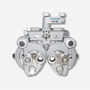
| Features:
- Equipped with comprehensive measuring functions, it provides SPH, CYL, AXIS and pupil distance optometry
- Durable and easy to operate
- Easily and intuitively read the sphere focal scale value
- High eco-friendly materials
- Design fitting the face curve and no stimulation
- Easy to take and clean
- Free switch between the cross-cylindrical lens and the rotary prism
- When the rotating risk is turning by the sphere, it can make sphere power adjust 3.00D for big scope.
- It is designed expediently and smartly for a particular cross cylinder. Supporting supplementary lens could increase scope of measurement.
Technical Specifications:
|
Sphere |
Range:-19.00~+16.75m-1 Step: 0.25m-1, 3.00m-1 |
|
Cylinder |
Range: 0.00~-6.00m-1(Measuring Range With Accessories0.00~-8.00m-1) Step: 0.25m-1 |
|
Cylinder Axis |
Range: 0~180°, Step:5° |
|
Distance of Optical center (also known as Pupil) |
Range: 50~75mmStep: 1mm |
|
Sight Switch |
Range:∞~380mm (distance of Optical center is64mm) |
|
Front Chin Test |
Range: 0~16mm |
|
Distance (from cornea vertex to the lens surface) |
16mm |
|
Standard Accessories Lens |
two pieces of Auxiliary Cylinder -2.00m-1 and -0.12m-1 respectively |
|
Standard Accessories |
one piece of M2 Hexagon wrench , one piece of a Myopia Standard Card, two piece of Myopia Standard Card , one piece of standard card holder , a dust cover |
|
Auxiliary Lens |
“O”:Open aperture
“R”:Retinoscope lens
“R”:Retinoscope lens
“R”:Retinoscope lens
*Lens of +1.50m-1 ,It is suit for the distance of 67 centimeters
“P”:Polaroid
* it is used for examining the dioptric balance of eyes , Implicit strabismus and stereo vision
“RMV”:Red Vertical maddox
*Be used to examine Implicit strabismus
“RMH”:Red horizontal maddox
*Be used to examine Implicit strabismus
“WMV”:Plane Vertical Maddox
*Be used to examine Implicit strabismus
“WMH”:Plane horizontal maddox
*Be used to examine Implicit strabismus
“RL”:Red lens
*Be used to examine eye function, Blending function and Implicit strabismus
“GL”:Green lens
*Be used to examine eye function, Blending function and Implicit strabismus
“+”:Test mark of optical center adjustment
“+.12”:Dioptric of the Spherical Lens is +0.12m-1
*Be used for the semi-adjustment of sphere lens, 0.25m-1
“PH”:1mmPinhole lens
*Be used to exclude visual non-refractive errors of the tested eye
“6ΔU”:6ΔBottom-up prism
*Be used to examine the rotating prism with the detection of nearly horizontal squint
“10ΔI”:10ΔBottom-up prism
*Be used to examine the rotating prism with the detection of nearly horizontal squint
“±0.50”:Cross-cylindrical lens
*Be used to examine the corrected dioptric of the Presbyopia and spherical lens
“OC”:Black lens
|
|
size |
338(L)×99(W)×292(H)mm |
|
NW |
about 5kg |
| Features:
- 7.0-inch color LCD touch panel.
- Hartman sensor with 108 multiple measure points.
- Green measurement light beam.
- Dyeing lens, sunglasses measured easily.
Technical Specifications:
- Sphere lenses: 0~±25D
- Cylinder lenses: 0~±10D
- Cylinder Axis Angle: 0°~180°
- Add.:0~10D
- Power supply: 100~240V, 50/60HZ, 30W
- Weight: 5kg
|


Reviews
There are no reviews yet.