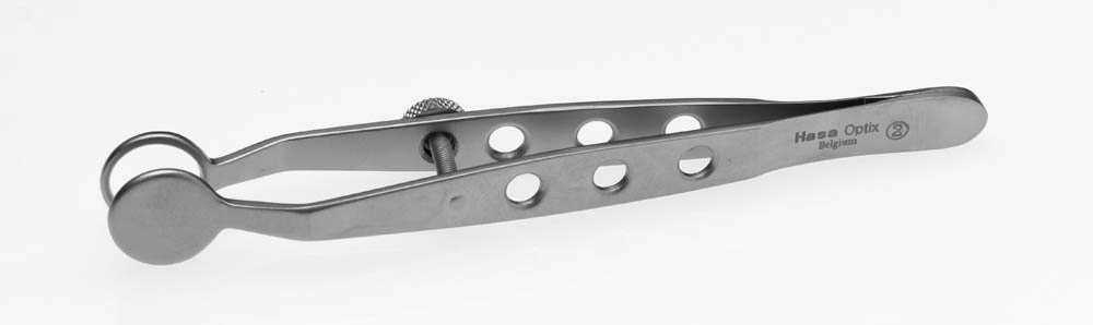
Chalazion Forceps
$0.00
Shipped From Abroad
11mm Diameter, Round Plate
Delivery & Availability:
Typically 10-21 working days – excluding furniture and heavy/bulky equipment. Please contact us for further information.
Typically 10-21 working days – excluding furniture and heavy/bulky equipment. Please contact us for further information.
Quick Comparison
| Chalazion Forceps remove | Ophthalmic Ultrasound Pachymeter remove | SW-1000 Ophthalmic Ultrasound Scanner-A-Scan remove | Tonometer remove | Ear Irrigation and acumen removal remove | Digital Chart Projector remove | |||||||||||||||||||||||||||||||||||||||||||||||||||||||||||||||||||||||||||||||
|---|---|---|---|---|---|---|---|---|---|---|---|---|---|---|---|---|---|---|---|---|---|---|---|---|---|---|---|---|---|---|---|---|---|---|---|---|---|---|---|---|---|---|---|---|---|---|---|---|---|---|---|---|---|---|---|---|---|---|---|---|---|---|---|---|---|---|---|---|---|---|---|---|---|---|---|---|---|---|---|---|---|---|---|---|
| Name | Chalazion Forceps remove | Ophthalmic Ultrasound Pachymeter remove | SW-1000 Ophthalmic Ultrasound Scanner-A-Scan remove | Tonometer remove | Ear Irrigation and acumen removal remove | Digital Chart Projector remove | ||||||||||||||||||||||||||||||||||||||||||||||||||||||||||||||||||||||||||||||
| Image |  |  |  |  | 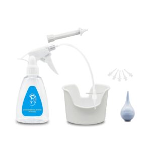 | 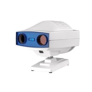 | ||||||||||||||||||||||||||||||||||||||||||||||||||||||||||||||||||||||||||||||
| SKU | SF103356013094-31 | SF1033560107-18 | SF1033560107-27 | SF1033560107-9 | SF103356013012 | SF1033560107-25 | ||||||||||||||||||||||||||||||||||||||||||||||||||||||||||||||||||||||||||||||
| Rating | ||||||||||||||||||||||||||||||||||||||||||||||||||||||||||||||||||||||||||||||||||||
| Price |
| $2,365.00 | $1,815.00 | $220.00 |
|
| ||||||||||||||||||||||||||||||||||||||||||||||||||||||||||||||||||||||||||||||
| Stock | ||||||||||||||||||||||||||||||||||||||||||||||||||||||||||||||||||||||||||||||||||||
| Availability | ||||||||||||||||||||||||||||||||||||||||||||||||||||||||||||||||||||||||||||||||||||
| Add to cart | ||||||||||||||||||||||||||||||||||||||||||||||||||||||||||||||||||||||||||||||||||||
| Description | Shipped From Abroad
11mm Diameter, Round Plate
Delivery & Availability:
Typically 10-21 working days – excluding furniture and heavy/bulky equipment. Please contact us for further information.
| Shipped from abroad
| Shipped from abroad
| Shipped from abroad
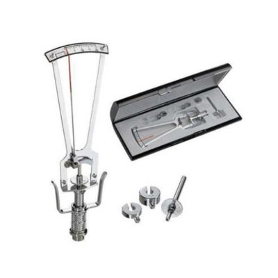
This product is used to measure the intraocular pressure (IOP) by measuring the depth produced on the surface of the cornea by a load of a known weight. Each division on the scale corresponds to 1/20mm corneal depth.
| In Stock
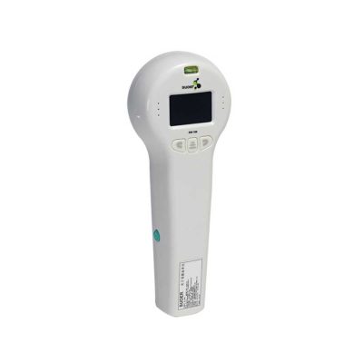
Features:
●Professional
Same ear wax removal tool as those used by doctors, you can easily eliminate ear wax buildup at home, really save your money and time on medical visiting. Safe and Environmentally Friendly.
●Quick & Easy
This ear wax removal kit is a quick, effective treatment for excess ear wax buildup. Fill the bottle with solution, Twist on the disposable tip, Use the trigger handle to spray solution into the ear canal. So Easy.
Delivery & Availability:
Typically 7-14 working days – excluding furniture and heavy/bulky equipment. Please contact us for further information.
| Ship from abroad
| ||||||||||||||||||||||||||||||||||||||||||||||||||||||||||||||||||||||||||||||
| Content | Features:
| Feature:
| This product is used to measure the intraocular pressure (IOP) by measuring the depth produced on the surface of the cornea by a load of a known weight. Each division on the scale corresponds to 1/20mm corneal depth.
Features:
| Features: ●Professional Same ear wax removal tool as those used by doctors, you can easily eliminate ear wax buildup at home, really save your money and time on medical visiting. Safe and Environmentally Friendly. ●Quick & Easy This ear wax removal kit is a quick, effective treatment for excess ear wax buildup. Fill the bottle with solution, Twist on the disposable tip, Use the trigger handle to spray solution into the ear canal. So Easy. ●Standard Capacity of the ear cleaner solution bottle is 10.6Oz, it has the most suitable size to hold in hand. Working at condition 32-122℉(0-50℃). Recommend to fill 1/5 of the bottle with OTC hydrogen peroxide, and 4/5 with very warm water. ●Complete Ear Washer System Our earwax removal kit comes with 1× Ear Washer Bottle, 1× Wash Basin, 1× Rubber Bulb, 1× Short Injection Head, 1× Long Hose Injection Head, 5× Disposable Tip, 1× User Manual. | Digital Chart Projector-Features:
| |||||||||||||||||||||||||||||||||||||||||||||||||||||||||||||||||||||||||||||||
| Weight | N/A | N/A | N/A | N/A | N/A | N/A | ||||||||||||||||||||||||||||||||||||||||||||||||||||||||||||||||||||||||||||||
| Dimensions | N/A | N/A | N/A | N/A | N/A | N/A | ||||||||||||||||||||||||||||||||||||||||||||||||||||||||||||||||||||||||||||||
| Additional information | ||||||||||||||||||||||||||||||||||||||||||||||||||||||||||||||||||||||||||||||||||||


Reviews
There are no reviews yet.