
Colonoscopy Training Model
$0.00
Shipped From Abroad
The Colonoscopy Training Model is a realistic medical simulation model for training lower gastrointestinal endoscopic procedures.
Typically 10-21 working days – excluding furniture and heavy/bulky equipment. Please contact us for further information.
Description
The Colonoscopy Training Model (Model: PLXH1001) by Preclinic is a realistic medical simulation model for training lower gastrointestinal endoscopic procedures. It’s designed from adult lower digestive tract CT data, using soft silica gel (hardness ~18A-30A), to mimic the colon anatomy including the ascending, transverse, and descending colon. Trainers can practice colonoscopy, biopsy, polypectomy, and other procedures using real endoscopic tools. The model includes fixed parts (colon, base, upper cover) and replaceable simulated polyp modules, providing realistic tactile feedback and versatility for different training scenarios.
Key Features & Specifications
| Feature | Details |
|---|---|
| Model | PLXH1001 Colonoscopy Training Model |
| Material | Highly soft silica gel, hardness ~ 18A-30A |
| Size | Length: ~68 cm (26.8 in); Width: ~23 cm (9.1 in); Height: ~25 cm (9.8 in) |
| Fixed Components | Colon model, base, upper cover |
| Replaceable Components | Simulated polyps; multiple lesion modules so you can simulate different pathologies (tumors, polyps, inflammation) |
| Procedures Supported | Colonoscopy, GI biopsy, polypectomy |
| Anatomical Accuracy | Includes anatomical parts: anus, rectum, sigmoid colon, descending/transverse/ascending colon, cecum, appendix and opening of small intestine. Based on adult CT data. |
| Compatibility | Works with most brands of colonoscopes and biopsy / polypectomy tools (forceps, snares, injection needles) |
| Lead Time | ~15 days |
| Port | Shanghai, China |
Quick Comparison
| Colonoscopy Training Model remove | 3B Scientific Life-Size Human Heart Model, 5 Parts with Representation of Systole - 3B Smart Anatomy remove | 3B Scientific Ovaries and Fallopian Tubes Model with Stages of Fertilization, 2-times Magnified - 3B Smart Anatomy remove | 3B Scientific 3/4 Life-Size Dual Sex Human Muscle Model on Metal Stand, 45-Part - 3B Smart Anatomy remove | 3B Scientific Human Heart Model, 7 Part - 3B Smart Anatomy remove | 3B Scientific Human Skull with Facial Muscles - 3B Smart Anatomy remove | |||||||||||||||||||||||
|---|---|---|---|---|---|---|---|---|---|---|---|---|---|---|---|---|---|---|---|---|---|---|---|---|---|---|---|---|
| Name | Colonoscopy Training Model remove | 3B Scientific Life-Size Human Heart Model, 5 Parts with Representation of Systole - 3B Smart Anatomy remove | 3B Scientific Ovaries and Fallopian Tubes Model with Stages of Fertilization, 2-times Magnified - 3B Smart Anatomy remove | 3B Scientific 3/4 Life-Size Dual Sex Human Muscle Model on Metal Stand, 45-Part - 3B Smart Anatomy remove | 3B Scientific Human Heart Model, 7 Part - 3B Smart Anatomy remove | 3B Scientific Human Skull with Facial Muscles - 3B Smart Anatomy remove | ||||||||||||||||||||||
| Image | 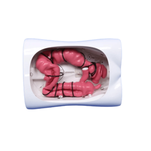 | 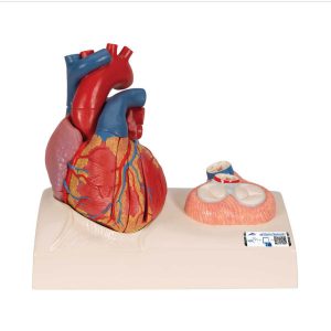 |  | 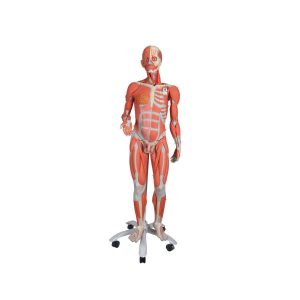 | 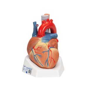 | 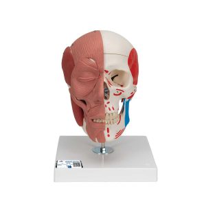 | ||||||||||||||||||||||
| SKU | SF1033560099-11 | SF1033560099-23 | SF1033560099-12 | SF1033560099-8 | SF1033560099-10 | |||||||||||||||||||||||
| Rating | ||||||||||||||||||||||||||||
| Price |
|
|
|
|
|
| ||||||||||||||||||||||
| Stock | ||||||||||||||||||||||||||||
| Availability | ||||||||||||||||||||||||||||
| Add to cart | ||||||||||||||||||||||||||||
| Description | Shipped From Abroad
The Colonoscopy Training Model is a realistic medical simulation model for training lower gastrointestinal endoscopic procedures.
Delivery & Availability:
Typically 10-21 working days – excluding furniture and heavy/bulky equipment. Please contact us for further information.
| Ship from abroad
This model is cast from a real human heart and didactically prepared to facilitate a better understanding of the anatomy and blood flow of the heart. It shows the cardiac valves during diastole and on the base the valves are shown in systole. A dissection through the median plane makes an easy demonstration possible.
Its attention to detail and high quality craftsmanship makes it definitely the top of the line heart model.
| Ship from abroad
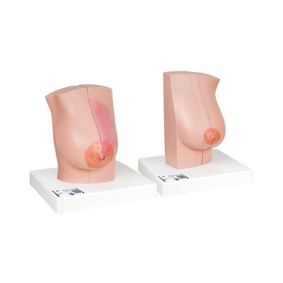
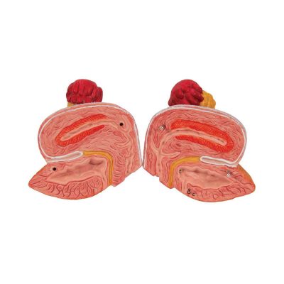
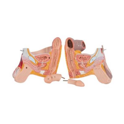
The model illustrates schematically how the ovum matures, how ovulation and fertilization occur and how the fertilized ovum develops to the stage where it embeds itself in the womb wall to begin the growth into an embryo. The various stages are shown in larger-than-life model form in an ovary, fallopian tube, and womb. An even more enlarged illustration of each is also printed on the base. Supplied on a base.
| Ship from abroad
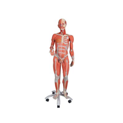
This muscle figure is the finest teaching tool available! Standing over 4 1/2 feet tall, this 3/4 life-size human replica depicts deep and superficial musculature in addition to the body's major nerves, vessels, tissues and organs in exquisite detail. The internal organs are removable (45 pieces in all) to reveal the fundamental interrelationships of human morphology. Remove the calvarium to view the 3-part removable brain. Look beneath the liver to reveal the gallbladder and bile duct. Peer inside the appendix, stomach lungs, heart or kidney. Remove and view the details of 13 different muscles of the arms and legs.
| Ship from abroad
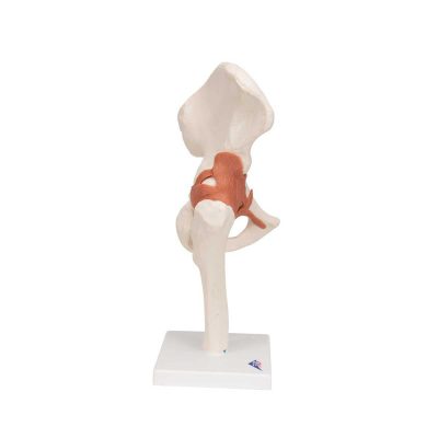
This heart model shows the anatomy of the heart model and is horizontally sectioned at the level of the valve plane.
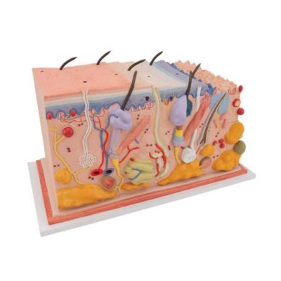
| Ship from abroad
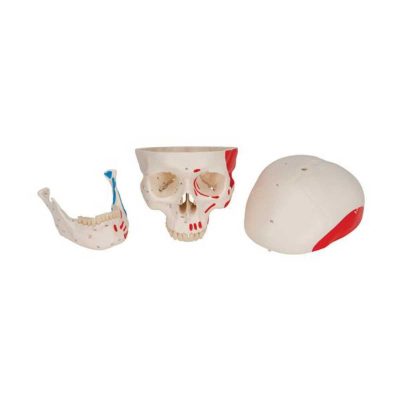
This model of the face musculature by 3B Scientific is used to easily demonstrate the causes of temporomandibular disorders and other dysfunctional disturbances of the TMJ and masticatory muscles.
| ||||||||||||||||||||||
| Content | The Colonoscopy Training Model (Model: PLXH1001) by Preclinic is a realistic medical simulation model for training lower gastrointestinal endoscopic procedures. It’s designed from adult lower digestive tract CT data, using soft silica gel (hardness ~18A-30A), to mimic the colon anatomy including the ascending, transverse, and descending colon. Trainers can practice colonoscopy, biopsy, polypectomy, and other procedures using real endoscopic tools. The model includes fixed parts (colon, base, upper cover) and replaceable simulated polyp modules, providing realistic tactile feedback and versatility for different training scenarios.
Key Features & Specifications
| This model is cast from a real human heart and didactically prepared to facilitate a better understanding of the anatomy and blood flow of the heart. It shows the cardiac valves during diastole and on the base the valves are shown in systole. A dissection through the median plane makes an easy demonstration possible. Its attention to detail and high quality craftsmanship makes it definitely the top of the line heart model. The following feature set makes it stand out from the crowd and a must have for any health professional.
| The model illustrates schematically how the ovum matures, how ovulation and fertilization occur and how the fertilized ovum develops to the stage where it embeds itself in the womb wall to begin the growth into an embryo. The various stages are shown in larger-than-life model form in an ovary, fallopian tube, and womb. An even more enlarged illustration of each is also printed on the base. Supplied on a base. | This muscle figure is the finest teaching tool available! Standing over 4 1/2 feet tall, this 3/4 life-size human replica depicts deep and superficial musculature in addition to the body's major nerves, vessels, tissues and organs in exquisite detail. The internal organs are removable (45 pieces in all) to reveal the fundamental interrelationships of human morphology. Remove the calvarium to view the 3-part removable brain. Look beneath the liver to reveal the gallbladder and bile duct. Peer inside the appendix, stomach lungs, heart or kidney. Remove and view the details of 13 different muscles of the arms and legs.
This dual sex muscle figure version has interchangeable genital inserts and a female mammary gland as well as a detailed multilingual product manual identifying over 600 hand-numbered structures. Hand-painted and mounted on a convenient roller base.
Includes the following features:
| This heart model shows the anatomy of the heart model and is horizontally sectioned at the level of the valve plane. The following parts can be removed from the heart:
|
| ||||||||||||||||||||||
| Weight | N/A | N/A | N/A | N/A | N/A | N/A | ||||||||||||||||||||||
| Dimensions | N/A | N/A | N/A | N/A | N/A | N/A | ||||||||||||||||||||||
| Additional information |


Reviews
There are no reviews yet.