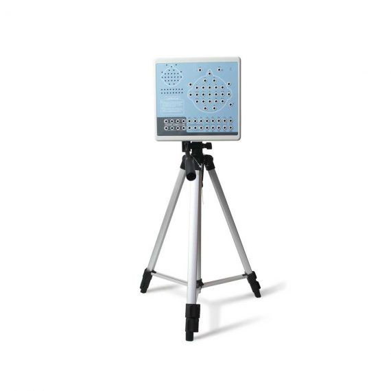
Contec KT88-3200 Electroencephalogram (EEG) Machine
$1,320.00
Shipped From Abroad
KT88-3200 Digital Brain Electric Activity Mapping collects EEG signal with electrodes, via integrated amplification, A/D transformation, PC auto-analysis, FFT, to form an electroencephalogram that displays with color depth. The product is applicable for checking such diseases as epilepsy, intracranial inflammation, cerebrovascular diseases, and brain tumors.
Delivery & Availability:
Typically 5-7 working days – excluding furniture and heavy/bulky equipment. Please contact us for further information.
Description
KT88-3200 Digital Brain Electric Activity Mapping collects EEG signal with electrodes, via integrated amplification, A/D transformation, PC auto-analysis, FFT, to form electroencephalogram that displays with color depth. The product is applicable for checking such diseases as epilepsy, intracranial inflammation, cerebrovascular diseases and brain tumors.
Features:
- Method of 10/20 electrodes placement under international standard system, leads can be changed during replaying. Support different types of combined leads during sampling.
- Adopt bioelectricity amplifier for distilling brainwave, continuous recording time can be up to 24 hours, integrated full-automatic calibration system.
- Powerful playback function: amplitude and display speed are adjustable. The special subdividing time line divides the waveform in one second into 5 parts, which is easy for doctors to look over the waveform.
- Digital filter system can be set as required, providing different window types
- EEG signal clipping function, analyze and store any section of EEG wave, and select several waveform segments for automatically analyzing and distilling to different parameters.
- Electronic frequency ruler, convenient to measure the basic information of any appointed EEG waveform. With partial enlarging window, accurate measurement of EEG period, amplitude and frequency, which can be adjusted according to personnel judgment.
- Mark EEG wave under the events of opening eyes, closing eyes and flashing with different colors, and user-defined events can be added, waveform color for evoked event can also be set freely, ensures that the waveform in corresponding time can be rapidly found by event name during case playback.
- Powerful automatic analysis function, can carry through the power spectrum analysis and pathologic wave detection for appointed waveform. Many graphs can be displayed in the same screen, including kinds of BEAM, numerical BEAM, compressed spectrum graph, trend graph, and so on.
- Professional isolation transformer, dual power supply isolation system and optoelectronic data transmission to ensure security. Use USB interface to transmit data which just need to be inserted.
- Multifunctional flash stimulator of USB interface, and flashing can be controlled manually or automatically. A flash stimulation scheme can be set and performed in the process of sampling.
- Perfect case management function, provides many means for research and quick statistic information; convenient case export and import function, and stores with MO or CD-RW disk, which is easy for data research.
- Integrative image and character report, report can be edited in mode and switched to Word document.
- Case files can be transformed into EDF and BDF data format, convenient for data exchange, academic exchange and further analysis.
- System parameters and display modes can be set as required, which meets different user’s requirement.
- Add marks and annotations to the waveform designated, which can rapidly find the waveform in that time by marks.
- Optional video function: USB camera is easy to install, convenient to use and exact to record. With flexible playback function, which can browse the waveform of any time along with the corresponding isochronous sampled image.
- SpO2 function is optional.
Technical Specifications:
- 32 channels of EEG
- Sampling rate: 200 dots/s
- Accuracy: 12bit
- Input impedance: ≥10MΩ
- Patient leak current: < 10µA
- Noise level: ≤5µVp-p
- CMRR: ≥90dB
- Magnification multiple: 10000
- Filter constant: all digital and free enactment
- Display speed(paper speed): 5, 10, 15, 30, 60, 120 mm/s
- Amplitude: 1, 1.5, 2, 3, 5, 7.5, 10, 12, 15, 20, 30, 50 mm/50µV
- Playback speed:1 time, 2 times, 3 times, 10 times, 20 times, 40 times, 60 times
- 50Hz interference suppression: ≥30dB
- Safety type:Class II, type BF applied part
Accesories:
- EEG lead
- EEG electrode
- Data line
- Headgear
- Strobe light
- Strobe light power supply
- PC software CD
- User Manual
- Earth wire
- EEG bracket
- Aluminum block
Review(1)
Quick Comparison
| Contec KT88-3200 Electroencephalogram (EEG) Machine remove | Sonoscape E1 Ultrasound Machine With Two Probes remove | ASPEL Stress ECG with Treadmill and Software remove | ASPEL AsCARD Grey ECG Machine remove | ASPEL Stress ECG with Ergometer and Software remove | Sonoscape P50 Ultrasound Machine remove | |
|---|---|---|---|---|---|---|
| Name | Contec KT88-3200 Electroencephalogram (EEG) Machine remove | Sonoscape E1 Ultrasound Machine With Two Probes remove | ASPEL Stress ECG with Treadmill and Software remove | ASPEL AsCARD Grey ECG Machine remove | ASPEL Stress ECG with Ergometer and Software remove | Sonoscape P50 Ultrasound Machine remove |
| Image | 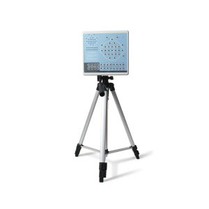 |  |  |  |  |  |
| SKU | SF1033560084-249 | SF1033560012-20 | SF1033560075-2 | SF1033560075-5 | SF1033560075-1 | SF1033560012-11 |
| Rating | ||||||
| Price | $1,320.00 | $4,620.00 | $6,542.00 | $1,166.00 | $4,202.00 |
|
| Stock | ||||||
| Availability | ||||||
| Add to cart | ||||||
| Description | Shipped From Abroad
KT88-3200 Digital Brain Electric Activity Mapping collects EEG signal with electrodes, via integrated amplification, A/D transformation, PC auto-analysis, FFT, to form an electroencephalogram that displays with color depth. The product is applicable for checking such diseases as epilepsy, intracranial inflammation, cerebrovascular diseases, and brain tumors.
| Shipped from Abroad SonoScape has developed a new probe and function for the E1 Exp. With these additions the E1 Exp will bring users a more efficient examination experience with satisfying image quality and a smooth workflow. Delivery & Availability: Typically 5-7 working days – excluding furniture and heavy/bulky equipment. Please contact us for further information. | Shipped from Abroad It is a system with professional tool dedicated to exercise and resting ECG examination. Treadmill has 12 lead ECG modules. With ECG Analyzing Software. Delivery & Availability: Typically 21 working days – excluding furniture and heavy/bulky equipment. Please contact us for further information. | Shipped from Abroad Electrocardiograph AsCARD Grey v.07.225 - is a 1, 3, 6, 12 channel ECG unit which enables to make electrocardiogram in full 12 leads. It is intended to conduct ECG examinations of adults and paediatric patients in all types of health care centres. ECG examination may be recorded in manual or automatic mode, with the possibility of analysis and interpretation. The device can be powered from 100 V ÷ 240 V mains supply or by an internal battery. Delivery & Availability: Typically 10 working days – excluding furniture and heavy/bulky equipment. Please contact us for further information. | Shipped from Abroad Ergometer CRG 200 is dedicated for Exercise Stress Tests System CardioTEST. The Ergometer has been designed according to modern technologies. It is controlled from PC equipped in CardioTEST software. Load level is controlled by a microprocessor, therefore it does not depend on speed, which in turn can be adjusted according to patient’s individual needs. Ergometer is equipped with ECG mode recording 12 standard leads. Delivery & Availability: Typically 21 working days – excluding furniture and heavy/bulky equipment. Please contact us for further information. | Shipped from Abroad Easily accomplish more with SonoScape’s new P50 ultrasound system. Incorporating single crystal clarity, automatic corrections and calculation, and user defined flexibility promises a confident diagnostic experience as well as opening new doors of opportunity for ultrasound use. Delivery & Availability: Typically 7-14 working days – excluding furniture and heavy/bulky equipment. Please contact us for further information. |
| Content | KT88-3200 Digital Brain Electric Activity Mapping collects EEG signal with electrodes, via integrated amplification, A/D transformation, PC auto-analysis, FFT, to form electroencephalogram that displays with color depth. The product is applicable for checking such diseases as epilepsy, intracranial inflammation, cerebrovascular diseases and brain tumors.
Features:
Technical Specifications:
| DETAILS
Efficient Diagnosis
μ-Scan, Speckle Reduction & Edge Enhancement
Spatial Compound Imaging
PIH - Pure Inversion Harmonic
Wide Scan - Enlarged Image Area
Tissue-Specific Imaging
SR Flow
Ergonomic Designs
Up to 2 Transducer Ports
Light Weight and Compact
15.6 inch Anti-flickering HD LED Screen
Tilting Monitor Angle Adjustment
Backlit Keyboard and Intelligent Panel
Long-lasting Battery for 90 mins
Ease of Use
Quick Boot Up
Auto-Brightness Adjustment
Auto Image Optimization
Auto IMT
Auto Trace
Equipped Accessories
Wi-Fi and Bluetooth Available
DICOM
500GB Hard Disk
Height Adjustable Trolley
Durable, Carry-on Site Suitcase
Click Here To Download Catalogue | It is a system with professional tool dedicated to exercise and resting ECG examination. Treadmill has 12 lead ECG modules. With ECG Analyzing Software.
Technical Specification:
Click Here To Download Catalogue |
Electrocardiograph AsCARD Grey v.07.225 - is a 1, 3, 6, 12 channel ECG unit which enables to make electrocardiogram in full 12 leads. It is intended to conduct ECG examinations of adults and paediatric patients in all types of health care centres. ECG examination may be recorded in manual or automatic mode, with the possibility of analysis and interpretation. The device can be powered from 100 V ÷ 240 V mains supply or by an internal battery.
Technical Specification:1. Visualisation of 1, 3, 6 or 12 ECG waveforms, analysis results and interpretations, examinations stored in memory.
2. Recording of 12 standard leads.
3. Print out in 1, 3, 6 or 12 ECG waveforms mode. Printing of a selected group:
Click Here To Download Catalogue | Ergometer CRG 200 is dedicated for Exercise Stress Tests System CardioTEST. The Ergometer has been designed according to modern technologies. It is controlled from PC equipped in CardioTEST software. Load level is controlled by a microprocessor, therefore it does not depend on speed, which in turn can be adjusted according to patient’s individual needs. Ergometer is equipped with ECG mode recording 12 standard leads.
Technical Specifications:
Click Here To Download Catalogue | DETAILS
Powerful Compact Precision
Taking into consideration the evolving expectations and needs for ultrasound, the P50 is a slim and unobtrusive trolley system that is comfortable in tight, congested spaces with little room to work in. Providing everything you need for a comfortable examination in a small space for both you and your patient.
Single Crystal Transducer
Wideband single crystal probes greatly improve the signal ratio, acquire stunning images and provide superior sensitivity and resolution for both the near and far-fields.
μ-Scan+
The new generation μ-Scan imaging technologies give you better image quality by reducing noise, improving signal strength and improving visualization.
Dynamic Color
Dynamic colour improves upon already existing colour Doppler technologies for clear capture of colour flow and detail visualization of even tiny veins with lower velocities.
Solution for Radiology
P50, is a leading-edge ultrasound system that can meet the demands of any clinical setting. You can experience a superior performance in multi-dimensional imaging for a full range of clinical applications – abdominal, breast and cardiovascular.
C-xlasto Imaging
By understanding that tissue stiffness varies depending on the type of tissue, we can use C-xlasto Imaging to easily find abnormalities and tumours within soft tissue. The differences in tissue responses are detected and visualized in real-time by the elastography algorithms through different representations, which can be particularly helpful in analyzing breast, thyroid and musculoskeletal structures. Predominately used only in linear probes, SonoScape’s new transvaginal and bi-plane probe for gynaecology and urology are breaking the mould and expanding elastography applications.
Real-time Color Panoramic
With the combination of colour flow and real-time panoramic, visualizing the blood flow of an entire vein or artery is now an easy task. Accomplished in real-time for the convenience of the sonographers, any mistakes can also be easily backtracked and corrected without interrupting the scan.
Contrast Imaging
Contrast Imaging on P50 makes full use of the infra harmonic signal and second harmonic signal to improve the image resolution and deep penetration. What’s more, the Dynamic Acoustic Control technology effectively controls the acoustic pressure for the contrast agent, decreasing the required agent dose and assures uniform image quality, guaranteeing longer contrast agent duration and better lesion perfusion of delayed phase observation.
Solution for OB/GYN
P50 has superior image quality, automated measurement tools, and a variety of volume technologies to provide ideal solutions for clinical examinations such as pregnancy examinations, and gynecologic disease diagnosis. With a new 4D transvaginal probe, P50 helps you to see and detect fetal abnormalities and significantly improves your diagnostic confidence during your examinations.
S-Live Silhouette
A unique transparent 3D anatomical image of the fetus for improved initial anatomical review. By using this new application, the system can create completely different fetal images from conventional ultrasound images, which can depict the fetal's intracorporeal anatomical structure.
Pelvic Floor 4D
Working in conjunction with SonoScape’s latest transvaginal probes, trans-perineal 4D pelvic floor ultrasound provides a useful clinical assessment of the impact of vaginal delivery on the female anterior compartment. Allowing doctors to judge whether the pelvic organs prolapsed or not, the extent of prolapse, and determining whether the pelvic muscles tore correctly.
S-Guide
S-Guide gives the user an extensive list of example obstetric ultrasound images as reference guides and a convenient checklist system to keep track of their progress during their obstetrics examination.
Auto Face
Automatically removes masking layers in front of the fetus’s face for a clearer vision of the fetus’s face.
AVC Follicle
AVC Follicle automatically identifies how many follicles are present and calculates their individual volumes.
Solution for Cardiology
P50 provides clear 2D clinical images and Doppler sensitivity to assess critical cardiac performance. Compatible with SonoScape’s single crystal probes, the P50 can provide images with better resolution and penetration in Cardiac diagnosis.
Tissue Doppler Imaging
Tissue Doppler Imaging allows clinical doctors to quantitatively evaluate local myocardial movements and functions, facilitating them with the ability to analyze and compare the motions of the different parts of the patient’s heart.
Stress Echo
Stress echocardiography is the combination of 2D echocardiography with physical, pharmacological or electrical stress of the patient. It also then provides users with report management tools such as configurable template editor, multiple loops to select one for storage, wall motion scoring, stress echo report, etc
Auto IMT
Auto IMT is used when determining the level of vascular sclerosis present in the patient by automatically tracing and calculating the thickness of the carotid vessels. What distinguishes the P50 is that it provides an instant and accurate Mean and Max index at the touch of a single button.
Auto EF
Automated 2D Cardiac Quantification is a fully intelligent trace function for endocardium with 19 easily-adjustable points providing rapid access to proven 2D EF and volumes.
Click Here To Download Catalogue |
| Weight | N/A | N/A | N/A | N/A | N/A | N/A |
| Dimensions | N/A | N/A | N/A | N/A | N/A | N/A |
| Additional information |


Mbah Prince
Cost of KT88-3200