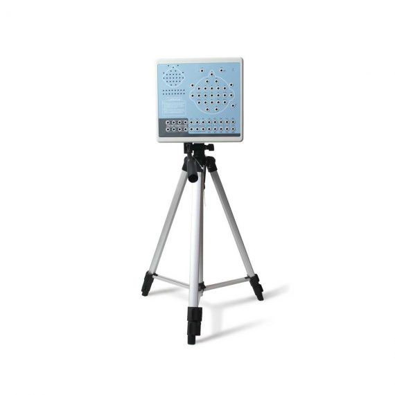
Contec KT88-3200 Electroencephalogram (EEG) Machine
$1,320.00
Shipped From Abroad
KT88-3200 Digital Brain Electric Activity Mapping collects EEG signal with electrodes, via integrated amplification, A/D transformation, PC auto-analysis, FFT, to form an electroencephalogram that displays with color depth. The product is applicable for checking such diseases as epilepsy, intracranial inflammation, cerebrovascular diseases, and brain tumors.
Delivery & Availability:
Typically 5-7 working days – excluding furniture and heavy/bulky equipment. Please contact us for further information.
Description
KT88-3200 Digital Brain Electric Activity Mapping collects EEG signal with electrodes, via integrated amplification, A/D transformation, PC auto-analysis, FFT, to form electroencephalogram that displays with color depth. The product is applicable for checking such diseases as epilepsy, intracranial inflammation, cerebrovascular diseases and brain tumors.
Features:
- Method of 10/20 electrodes placement under international standard system, leads can be changed during replaying. Support different types of combined leads during sampling.
- Adopt bioelectricity amplifier for distilling brainwave, continuous recording time can be up to 24 hours, integrated full-automatic calibration system.
- Powerful playback function: amplitude and display speed are adjustable. The special subdividing time line divides the waveform in one second into 5 parts, which is easy for doctors to look over the waveform.
- Digital filter system can be set as required, providing different window types
- EEG signal clipping function, analyze and store any section of EEG wave, and select several waveform segments for automatically analyzing and distilling to different parameters.
- Electronic frequency ruler, convenient to measure the basic information of any appointed EEG waveform. With partial enlarging window, accurate measurement of EEG period, amplitude and frequency, which can be adjusted according to personnel judgment.
- Mark EEG wave under the events of opening eyes, closing eyes and flashing with different colors, and user-defined events can be added, waveform color for evoked event can also be set freely, ensures that the waveform in corresponding time can be rapidly found by event name during case playback.
- Powerful automatic analysis function, can carry through the power spectrum analysis and pathologic wave detection for appointed waveform. Many graphs can be displayed in the same screen, including kinds of BEAM, numerical BEAM, compressed spectrum graph, trend graph, and so on.
- Professional isolation transformer, dual power supply isolation system and optoelectronic data transmission to ensure security. Use USB interface to transmit data which just need to be inserted.
- Multifunctional flash stimulator of USB interface, and flashing can be controlled manually or automatically. A flash stimulation scheme can be set and performed in the process of sampling.
- Perfect case management function, provides many means for research and quick statistic information; convenient case export and import function, and stores with MO or CD-RW disk, which is easy for data research.
- Integrative image and character report, report can be edited in mode and switched to Word document.
- Case files can be transformed into EDF and BDF data format, convenient for data exchange, academic exchange and further analysis.
- System parameters and display modes can be set as required, which meets different user’s requirement.
- Add marks and annotations to the waveform designated, which can rapidly find the waveform in that time by marks.
- Optional video function: USB camera is easy to install, convenient to use and exact to record. With flexible playback function, which can browse the waveform of any time along with the corresponding isochronous sampled image.
- SpO2 function is optional.
Technical Specifications:
- 32 channels of EEG
- Sampling rate: 200 dots/s
- Accuracy: 12bit
- Input impedance: ≥10MΩ
- Patient leak current: < 10µA
- Noise level: ≤5µVp-p
- CMRR: ≥90dB
- Magnification multiple: 10000
- Filter constant: all digital and free enactment
- Display speed(paper speed): 5, 10, 15, 30, 60, 120 mm/s
- Amplitude: 1, 1.5, 2, 3, 5, 7.5, 10, 12, 15, 20, 30, 50 mm/50µV
- Playback speed:1 time, 2 times, 3 times, 10 times, 20 times, 40 times, 60 times
- 50Hz interference suppression: ≥30dB
- Safety type:Class II, type BF applied part
Accesories:
- EEG lead
- EEG electrode
- Data line
- Headgear
- Strobe light
- Strobe light power supply
- PC software CD
- User Manual
- Earth wire
- EEG bracket
- Aluminum block
Review(1)
Quick Comparison
| Contec KT88-3200 Electroencephalogram (EEG) Machine remove | Sonoscape P20 Ultrasound Machine remove | ASPEL Stress ECG with Treadmill and Software remove | Topaz Digital X-ray Machine remove | Sonoscape S8 Exp Portable Ultrasound remove | ASPEL Ambulatory BP Machine remove | |
|---|---|---|---|---|---|---|
| Name | Contec KT88-3200 Electroencephalogram (EEG) Machine remove | Sonoscape P20 Ultrasound Machine remove | ASPEL Stress ECG with Treadmill and Software remove | Topaz Digital X-ray Machine remove | Sonoscape S8 Exp Portable Ultrasound remove | ASPEL Ambulatory BP Machine remove |
| Image | 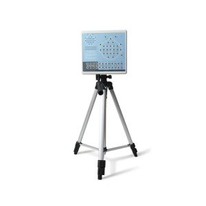 |  |  |  |  | 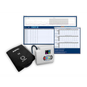 |
| SKU | SF1033560084-249 | SF1033560012-9 | SF1033560075-2 | SF1033560074-1 | SF1033560012-15 | SF1033560075-13 |
| Rating | ||||||
| Price | $1,320.00 |
| $6,542.00 |
| $9,350.00 | $920.00 |
| Stock | ||||||
| Availability | ||||||
| Add to cart | ||||||
| Description | Shipped From Abroad
KT88-3200 Digital Brain Electric Activity Mapping collects EEG signal with electrodes, via integrated amplification, A/D transformation, PC auto-analysis, FFT, to form an electroencephalogram that displays with color depth. The product is applicable for checking such diseases as epilepsy, intracranial inflammation, cerebrovascular diseases, and brain tumors.
| Shipped from Abroad Incorporating innovative technologies, P20’s user-friendly design with a simple operation panel, intuitive user interface and a variety of intelligent auxiliary scanning tools, will significantly improve your daily examination experience. Besides general imaging applications, P20 has entitled with diagnostic 4D technology which has an extraordinary performance in obstetrics and gynecology applications. Delivery & Availability: Typically 5-7 working days – excluding furniture and heavy/bulky equipment. Please contact us for further information. | Shipped from Abroad It is a system with professional tool dedicated to exercise and resting ECG examination. Treadmill has 12 lead ECG modules. With ECG Analyzing Software. Delivery & Availability: Typically 21 working days – excluding furniture and heavy/bulky equipment. Please contact us for further information. | In Stock DRGEM’s TOPAZ X-ray machine is a state-of-the-art mobile digital radiography system, designed with maximum comfort for patients and users in mind. From its user-friendly software to smooth movements, TOPAZ is made to improve your workflow and provide you with high-quality images. Delivery & Availability: Typically 21 working days – excluding furniture and heavy/bulky equipment. Please contact us for further information. | Shipped from Abroad With ultra-modern innovative design and the clinically-proven technologies, S8 Exp is portable ultrasound scanner well equipped as a low-physical-effort and enhanced-image-quality ultrasound scanner, which not only provides optimized solutions for versatile applications, but does help to improve the user-experience for both routine and non-traditional challenges. Delivery & Availability: Typically 5-7 working days – excluding furniture and heavy/bulky equipment. Please contact us for further information. | Shipped from Abroad ASPEL Ambulatory BP Machine - is a recorder of long-term records of non-invasive measurement of blood pressure intended for use in clinics, hospitals, outpatient centers and specialist surgeries. The recorder enables the assessment of blood pressure by the oscillometric method in adult patients, pregnant women, including preeclampsia and pediatric patients (from 3 years of age). Blood pressure is assessed by using an inflatable cuff, an accurate pressure transducer, and a deflation valve. Delivery & Availability: Typically 10 working days – excluding furniture and heavy/bulky equipment. Please contact us for further information. |
| Content | KT88-3200 Digital Brain Electric Activity Mapping collects EEG signal with electrodes, via integrated amplification, A/D transformation, PC auto-analysis, FFT, to form electroencephalogram that displays with color depth. The product is applicable for checking such diseases as epilepsy, intracranial inflammation, cerebrovascular diseases and brain tumors.
Features:
Technical Specifications:
| DETAILS
Upgraded Images with More Clarity
SonoScape never stops making progress in improving the image quality of its ultrasound products to enhance the confidence of diagnosis for doctors. With extraordinary images provided by P20, the anatomy structures are clearer than ever.
C-Xlasto Imaging
With C-xlasto Imaging, P20 enables comprehensive quantitative elastic analysis. Meanwhile, C-xlasto on P20 is supported by linear, convex and transvaginal probes, to ensure good reproducibility and highly consistent quantitative elastic results.
S-Live
S-Live allows for detailed visualization of subtle anatomical features, thereby enabling intuitive diagnosis with real-time 3D images and enriching patient communication.
Pelvic Floor 4D
Transperineal 4D pelvic floor ultrasound can provide useful clinical values in assessing the vaginal delivery impact on the female anterior compartment, judging whether the pelvic organs are prolapsed or not and the extent, determining if the pelvic muscles were torn accurately.
Anatomic M Mode
Anatomic M Mode helps you observe the myocardial motion at different phases by freely placing sample lines. It accurately measures the myocardial thickness and the heart size of even difficult patients and supports the myocardial function and LV wall-motion assessment.
Tissue Doppler Imaging
P20 is endowed with Tissue Doppler Imaging which provides velocities and other clinical information on myocardial functions, facilitating clinical doctors with the ability to analyze and compare the motions of different parts of the patient's heart.
Click Here To Download Catalogue | It is a system with professional tool dedicated to exercise and resting ECG examination. Treadmill has 12 lead ECG modules. With ECG Analyzing Software.
Technical Specification:
Click Here To Download Catalogue | TOPAZ X-ray machine is among the high end X-ray machine manufactured by DRGEM, a digital X-ray system that provides quality images with little or no effort.
It begins with Advanced Technology
Integrating high technology and over a decade of experience in conventional and digital radiography systems, DRGEM’s TOPAZ X-ray machine is a state-of-the-art mobile digital radiography system, designed with maximum comfort for patients and users. From its user-friendly software to smooth movements, TOPAZ X-ray machine is made to improve your workflow and provide you with high-quality images.
Full Featured Imaging Software & Excellent Digital Image Processing
With a high-performance, built-in touchscreen, TOPAZ X-ray machine offers a user-friendly interface and powerful software for easy operation and increased workflow. The anatomical view-based digital image processing, automatically optimizes and enhances the quality of the image. it also comes with automatic image storage and print with DICOM 3.0 networking capability. additionally, the system offers increasing exam throughput while decreasing examination time.
Click Here To Download Catalogue | Sonoscape S8 Exp Portable Ultrasound scannerDETAILS Agile and Versatile With ultra-modern innovative design and the clinically-proven technologies, S8 Exp Portable Ultrasound scanner is well equipped as a low-physical-effort and enhanced-image-quality ultrasound scanner, which not only provides optimized solutions for versatile applications but does help to improve the user experience for both routine and non-traditional challenges. Working with S8 Exp, it will trigger your unlimited reverie and endow you with endless charm. Carrying forward the classical design of SonoScape's portable ultrasound products, S8 Exp successfully combines the best ergonomics, attractive design and ease of use. This charismatic identity is also enhanced by a sophisticated color palette—with sedate grey as its interior paint color and pearl white as exterior cover, S8 Exp reveals a style of aristocrat and strong character among SonoScape's ultrasound systems. Workflow The S8 Exp is a portable ultrasound scanner that adapts to your workflow, whether you are in the consulting room, at the bedside, or at a remote location. With easy-to-use new platform designed for sonographers' needs and full connection interfaces for easy connectivity and data sharing, S8 Exp leads to improved user comfort and clinical outcome as well as patient throughput and working efficiency. Powerful Platform Embedded with SonoScape's core imaging technologies such as μ-scan, PHI and Spatial Compound, S8 Exp boasts exceptional 2D image, sensitive spectral, Color and Power Doppler, displaying well-defined anatomy and pathology and facilitating a highly optimized diagnostic user environment for conclusive diagnoses. Besides, S8 Exp offers a comprehensive selection of electronic probes to maximally extend its capabilities to meet a wide range of applications including the abdomen, pediatric, OB/GYN, cardiovascular, musculoskeletal, etc. The advanced probe technologies also effectively enhance the image quality and confidence in reaching clinical diagnoses even in difficult patients.Click Here To Download Catalogue | ASPEL Ambulatory BP Machine - is a recorder of long-term records of non-invasive measurement of blood pressure intended for use in clinics, hospitals, outpatient centers and specialist surgeries. The recorder enables the assessment of blood pressure by the oscillometric method in adult patients, pregnant women, including preeclampsia and pediatric patients (from 3 years of age). Blood pressure is assessed by using an inflatable cuff, an accurate pressure transducer, and a deflation valve.
Features:
Save-2-Safe: Double security system
Thanks to the use of two independent measuring systems with an additional valve, it meets the highest standards and takes care of patient safety even better.
Start-Easy: Quick start in two moves
The quick launch function allows you to use the device instantly, easily allows you to start recording in holter mode.
Memo-Care: Cuff pressure memory
Recorder remembers the pressure in the cuff. Thanks to the use of Intelligent Solutions, it adapts individually to the patient.
Power-Usb: USB connection
The device can work without batteries: by connecting to a computer via a USB cable.
Technical Specification:
Click Here To Download Catalogue |
| Weight | N/A | N/A | N/A | N/A | N/A | N/A |
| Dimensions | N/A | N/A | N/A | N/A | N/A | N/A |
| Additional information |


Mbah Prince
Cost of KT88-3200