CROSSLINKING CERATHOS
$0.00
Shipped From Abroad
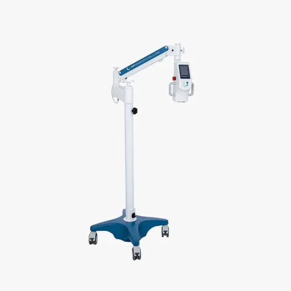
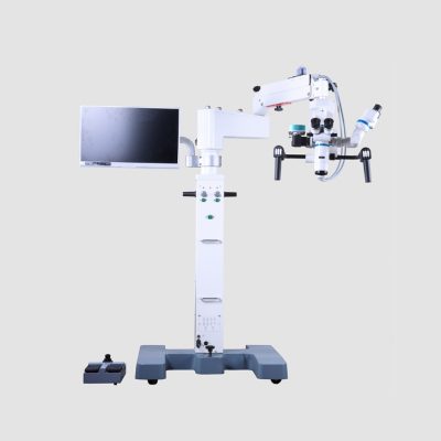
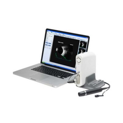
DFV Cerathos was designed with state-of-the-art technology to provide safe and effective treatment. Its optical system was developed to provide a safe dosage of energy homogeneously over the cornea to be treated. It has a dedicated microprocessor system that controls all functions and monitors the amount of energy emitted by the equipment. It’s versatile and accurate!
Delivery & Availability:
Typically 10-21 working days – excluding furniture and heavy/bulky equipment. Please contact us for further information.
Typically 10-21 working days – excluding furniture and heavy/bulky equipment. Please contact us for further information.
Quick Comparison
| CROSSLINKING CERATHOS remove | Portable Fundus Camera remove | Pantoscopic Ophthalmoscope remove | Applanation Tonometer remove | Portable Keratometer remove | Retinoscope remove | |||||||||||||||||||||||||||||||||||||||||||||||||||||||||||||||||
|---|---|---|---|---|---|---|---|---|---|---|---|---|---|---|---|---|---|---|---|---|---|---|---|---|---|---|---|---|---|---|---|---|---|---|---|---|---|---|---|---|---|---|---|---|---|---|---|---|---|---|---|---|---|---|---|---|---|---|---|---|---|---|---|---|---|---|---|---|---|---|
| Name | CROSSLINKING CERATHOS remove | Portable Fundus Camera remove | Pantoscopic Ophthalmoscope remove | Applanation Tonometer remove | Portable Keratometer remove | Retinoscope remove | ||||||||||||||||||||||||||||||||||||||||||||||||||||||||||||||||
| Image | 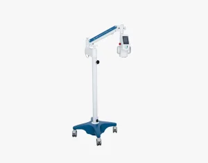 | 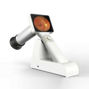 | 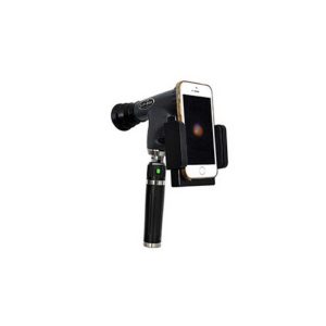 | 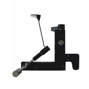 | 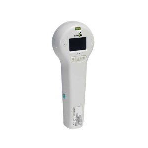 | 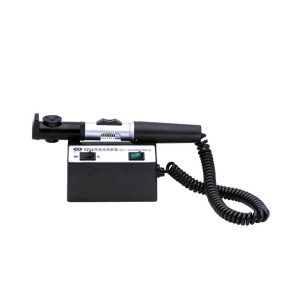 | ||||||||||||||||||||||||||||||||||||||||||||||||||||||||||||||||
| SKU | SF103356013091-3 | SF1033560107-23 | SF1033560107-3 | SF1033560107-1 | SF1033560107-16 | SF1033560107-12 | ||||||||||||||||||||||||||||||||||||||||||||||||||||||||||||||||
| Rating | ||||||||||||||||||||||||||||||||||||||||||||||||||||||||||||||||||||||
| Price |
| $2,310.00 |
|
|
| $165.00 | ||||||||||||||||||||||||||||||||||||||||||||||||||||||||||||||||
| Stock | ||||||||||||||||||||||||||||||||||||||||||||||||||||||||||||||||||||||
| Availability | ||||||||||||||||||||||||||||||||||||||||||||||||||||||||||||||||||||||
| Add to cart | ||||||||||||||||||||||||||||||||||||||||||||||||||||||||||||||||||||||
| Description | Shipped From Abroad
DFV Cerathos was designed with state-of-the-art technology to provide safe and effective treatment. Its optical system was developed to provide a safe dosage of energy homogeneously over the cornea to be treated. It has a dedicated microprocessor system that controls all functions and monitors the amount of energy emitted by the equipment. It's versatile and accurate!
Delivery & Availability:
Typically 10-21 working days – excluding furniture and heavy/bulky equipment. Please contact us for further information.
| 83Shipped from abroad
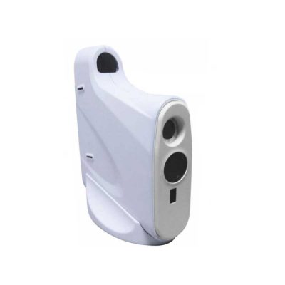
This is a portable medical camera for fundus imaging, diagnosis, and especially for fundus disease screening. It's compact, easy to obtain high definition fundus image. It can be conveniently applied to rapid screening, out diagnosis, bedside diagnosis and remote medical treatment, etc.
| Shipped from abroad
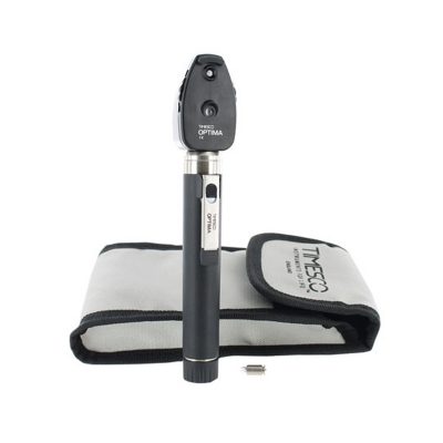
The brand-new Pantoscopic Ophthalmoscope is a portable digital imaging device which makes it possible to view and take pictures of the eyes.
| Shipped from abroad
The product is designed on the principle basis of Goldman tonometer. It can be connected with slit lamp(Carl Zeiss type).
| Shipped from abroad
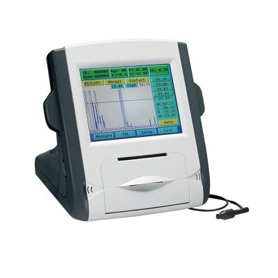
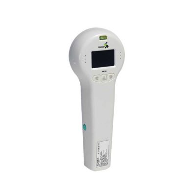
Ultra-small corneal curvature tester, is mainly used to measure the corneal curvature radius and diopter, wireless output print data.
| Shipped from abroad
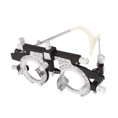
The product can quickly and precisely measure the astigmatism axis and is one of the necessary instruments in optometry inspection.
| ||||||||||||||||||||||||||||||||||||||||||||||||||||||||||||||||
| Content | Portable Fundus Camera is a portable medical camera for fundus imaging, diagnosis, and especially for fundus disease screening. It's compact, easy to obtain high definition fundus image. It can be conveniently applied to rapid screening, out diagnosis, bedside diagnosis and remote medical treatment, etc.
Features of Portable Fundus Camera:
| The brand-new Pantoscopic Ophthalmoscope is a portable digital imaging device which makes it possible to view and take pictures of the eyes. The optical access of the Pantoscopic Ophthalmoscope is aligned to the visual axis of the smartphone camera by the adaptor which allows to you take pictures of the fundus and retinal nerve in high resolution. You could save pictures for each patient or email and print as needed. The Pantoscopic Ophthalmoscope provides a 5X larger view of the fundus compared with the standard ophthalmoscope. It has a wider view field of 230. Without dilating the pupil, the fundus imagines could be captured at any time and places.
Features:
| Applanation Tonometer is designed on the principle basis of Goldman tonometer. It can be connected with slit lamp(Carl Zeiss type).
Features of Applanation Tonometer:
| Portable Keratometer Features:
Ultra-small corneal curvature tester, is mainly used to measure the corneal curvature radius and diopter, wireless output print data.
Technical Specifications:
| The product can quickly and precisely measure the astigmatism axis and is one of the necessary instruments in optometry inspection.
Features:
| |||||||||||||||||||||||||||||||||||||||||||||||||||||||||||||||||
| Weight | N/A | N/A | N/A | N/A | N/A | N/A | ||||||||||||||||||||||||||||||||||||||||||||||||||||||||||||||||
| Dimensions | N/A | N/A | N/A | N/A | N/A | N/A | ||||||||||||||||||||||||||||||||||||||||||||||||||||||||||||||||
| Additional information | ||||||||||||||||||||||||||||||||||||||||||||||||||||||||||||||||||||||


Reviews
There are no reviews yet.