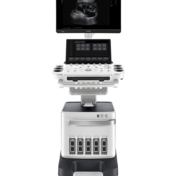
The CV45/CV40 (also known as CU40) is a compact, fully digital B‑mode and spectral Doppler ultrasound system offering high-resolution imaging, intuitive workflow, and versatile clinical presets ideal for general diagnostics in human and veterinary practice.
Delivery & Availability: Typically 10-21 working days – excluding furniture and heavy/bulky equipment. Please contact us for further information.
Shipped From AbroadThe CV45/CV40 (also known as CU40) is a compact, fully digital B‑mode and spectral Doppler ultrasound system offering high-resolution imaging, intuitive workflow, and versatile clinical presets ideal for general diagnostics in human and veterinary practice.
Delivery & Availability:Typically 10-21 working days – excluding furniture and heavy/bulky equipment. Please contact us for further information.
Shipped from Abroad
OPENMARK 5000 is 0.51T MRI. It's approved by FDA and has CE mark. It adopts two-pillar magnet design with 280 degree openness and equipped with powerful
RF and gradient system, together with advanced imaging technology, making it as a high-end system which is comparable to high-field MRI.
Delivery & Availability:
Typically 90 working days – excluding furniture and heavy/bulky equipment. Please contact us for further information.
In StockGXR Analogue X-ray system matches with a radiographic room which perfectly fits your workow and can be easily upgraded to DR system with the help of DR interface and PC interface in GXR generator as well as Bucky suitable to Flat Panel Detector. GXR X-ray system is equipped with a high frequency X-ray generator which consistently produces high quality radiograph in favor of high quality X-ray output with a very small kV ripple and accurate mA and mAs. GXR X-ray system is designed to provide convenience to operator and comfort to patient.Delivery & Availability:
Typically 21 working days – excluding furniture and heavy/bulky equipment. Please contact us for further information.
Shipped from Abroad
This Machine gives a possibility to perform computed tomography without any problems and on high quality level. This device is used to conduct exams of internal organs and their functioning. With its help, a physician has a possibility to assess the condition of the human body as a whole.
Delivery & Availability:
Typically 90 working days – excluding furniture and heavy/bulky equipment. Please contact us for further information.
In stock
Length: 355 mm
Height: 560 mm
Diameter: 150 mm
Power: 30 w
Scattering diffusion: Greater than 0.9
Delivery & Availability:
Typically 5-7 working days – excluding furniture and heavy/bulky equipment. Please contact us for further information.
In Stock
DRGEM’s TOPAZ X-ray machine is a state-of-the-art mobile digital radiography system, designed with maximum comfort for patients and users in mind. From its user-friendly software to smooth movements, TOPAZ is made to improve your workflow and provide you with high-quality images.
Delivery & Availability:
Typically 21 working days – excluding furniture and heavy/bulky equipment. Please contact us for further information.
Content
Key Features
High‑Resolution LCD (10″ – 12″ adjustable angle display) – clear visuals with anti‑glare and enhanced brightness
Digital Beamforming + Tissue Harmonic Imaging (THI) – for sharper contrast and deeper penetration
Back‑lit Control Panel & User‑Defined Keys – ergonomic and adaptable for varied environments
Portable Power & Connectivity – built‑in battery, DVI/USB/DICOM support, image/video/patient data storage & export
Extensive Clinical Presets – packages for general, OB/GYN, cardiac, MSK, urology, small parts, orthopedics
Specifications
Specification
CV40 / CU40
Display
10″–12″ non‑interlaced, adjustable angle
Imaging Technology
Digital beamforming, THI, compounding
Modes
B‑mode, M‑mode, Doppler (spectral, color optional in C6)
Probe Connectors
2
TGC Control
8 segments
Storage
Internal HDD + image/video export formats
Connectivity
USB, DVI video, wireless DICOM optional
Power
Integrated long-life battery
Control Panel
Back-lit keys, user-defined soft keys
Ultrasound Presets
General, OB/GYN, Cardiac, Urology, MSK, Small parts, Orthopedics
Interventional Aids
Biopsy and lithotripsy guide overlays
Video Capture
Cine loop with thumbnail clip viewer
OPENMARK 5000 is 0.51T MRI. It's approved by FDA and has CE mark. It adopts two-pillar magnet design with 280 degree openness and equipped with powerful
RF and gradient system, together with advanced imaging technology, making it as a high-end system which is comparable to high-field MRI.
Features:
With the highest system stability and the highest homogeneity of the
magnet field in permanent MRI
Screens on both sides facilitate positioning; 280 degree two-pillar magnet
design ensures stable magnet structure and facilitates interventional
treatment.
Active and passive shimming calibrate technology ensures the magnetic
field uniformity
Motor-driven patient couch makes it easier for patients to access and for
positioning
Powerful hardware and software platforms ensure the scan speed, image
quality and make it possible for advanced imaging functions
Fast scan speed eliminates motion artifact
Rich scan sequences, advanced imaging technology and powerful postprocessing
technology ensure image quality, extend more applications,
which can fully satisfy the clinical needs
Intelligent user-friendly operating system ensures you easy operation
Technical Specifications:
No.
Technique Description
Parameter
1
Magnet System
1.1
Magnet Type
Permanent Magnet
Automatic constant temperature
system
DrGem GXR Floor Mounted Analogue X-ray system matches with a radiographic room which perfectly fits your workflow and can be easily upgraded to DR system with the help of DR interface and PC interface in GXR generator as well as Bucky suitable to Flat Panel Detector. GXR (Analogue X-ray)system is equipped with a high frequency X-ray generator which consistently produces high quality radiograph in favor of high quality X-ray output with a very small kV ripple and accurate mA and mAs. GXR (Analogue X-ray) system is designed to provide convenience to operator and comfort to patient.Features of DrGem GXR Floor Mounted Analogue X-ray:
4 way Tabletop Patient Table (PBT-4)
A large tabletop with extended travel enables all radiography studies with minimal patient movement, and supporting patient weight up to 300kg. Fully at tabletop without a frame on the edge makes cleanliness and odors free
Floor Mount Tube Stand (TS-FM6)
Floor Rail type tube stand provides all radiography studies with smooth movement on the rail.
Wall Bucky Stand (WBS)
Elegant design, durable and easy-to-use Wall Bucky Stand provides full satisfaction.
Technical Specifications of DrGem GXR Floor Mounted Analogue X-ray:
This Machine gives a possibility to perform computed tomography without any problems and on high quality level. This device is used to conduct exams of internal organs and their functioning. With its help, a physician has a possibility to assess the condition of the human body as a whole.
Features:
It is easy to use;
Convenience;
Multi functionality;
Obtained images are of high definition;
High-definition 3D images of the area under study;
The procedure is pain-free;
The data is processed fast;
The image can be stored in the computer memory;
The diagnostics does not take a lot of time;
Acceptable radiation dose.
Technical Specifications:
No.
Technical Features
Descriptions
1
Gantry
1.01
Gantry type
Low voltage slip-ring
1.02
Gantry driven type
Strap-driven
1.03
Patient opening
70cm
1.04
Gantry tilt mode
Digital gantry tilt
1.05
Digital tilt capability
±50°
1.06
Detector type
OptiWave rare-earth ceramic detector
1.07
Numbers of detector rows
16
1.08
Width of Z-axle detector
20mm
1.09
Detector columns of channels per row
848
1.10
Numbers of detector columns
13568
1.11
Data-transfer type
RF, optical fiber communication
1.12
Distance of focus-ISO-center
53cm
1.13
Distance of focus-detector
94cm
1.14
3D laser orientation
Provided
1.15
13" integrated display panel
Provided
1.16
Adose automatic exposure control (mA
Modulation)
Provided
1.17
Auto-voice manager
Breath Graphical Display
Hold Message (Record/Playback)
Breath Message (Record/Playback)
1.18
AccuSaving energy conservation management
Provided
2
HVPS and X-ray tube
2.01
Maximum continuous output of HVgenerator
42kW
2.02
Tube kV selections
70kV, 80kV, 100 kV, 120 kV, 140 kV
2.03
Tube mA range
10~350mA
2.04
Tube anode heat capacity
3.5MHU
2.05
Max. anode cooling rate
735kHU/min
2.06
Type of cooling
Oil cooling + Air cooling
2.07
Tube focus
Large: 1.2mm×1.4mm
Small: 0.7mm×0.8mm
2.08
Collimator width selection
4-level election
2.09
Focus spot tracking technology
Provided
3
Patient table
3.01
Maximum horizontal-movable range
1850mm
3.02
Table horizontal-scannablerange
1800mm
3.03
Table horizontal-position repeatability
±0.25mm
3.04
Minimum height above floor
430mm
3.05
Maximum vertical-movable range
500mm
3.06
Maximum speed of vertical movement
35mm
3.07
Maximum speed of horizontal movement
150mm/s
3.08
Maximum patient weight
205kg
3.09
Foot pedal of patient table control
Provided
4
Computer
4.01
CPU
3.5GHz
4.02
Memory
32GB
4.03
Storage of hard-disk
1TB×2
4.04
Monitor
24’’ LCD Monitor
4.05
Resolution of monitor
1920×1200
4.06
Image-data external storage type
CD/DVD/USB
4.07
Time of image reconstruction (512×512)
33.3ms/image
4.08
Speed of image reconstruction (512×12)
30fps
4.09
DICOM 3.0 interface
Provided
4.10
Printer DICOM 3.0 interface
Provided
4.11
Auto filming
Provided
4.12
Worklist function
Provided
5
Scan parameters
5.01
Shortest 360 degree rotation time
0.75s
5.02
Allowed rotation times
0.75s, 1.0s, 1.5s, 2.0s, 3.0s, 4.0s
5.03
Maximum slice numbers per rotation
32
5.04
Minimum slice thickness of scan
1.25mm
5.05
Minimum slice thickness of reconstruction
0.625mm
5.06
Maximum slice thickness of scan
20mm
5.07
Nominal reconstruction slice thickness
0.625mm, 1.25mm, 2.5mm, 5.0mm, 7.5mm,
10mm, 20mm
5.08
Speed of image reconstruction (512×512)
30 frames/s
5.09
Scan FOV
50cm
5.10
Image reconstruction matrix
512×512, 1024×1024 (Optional)
5.11
Image reconstruction matrix
512×512, 1024×1024 (Optional)
5.12
Image display matrix
512×512, 1024×1024 (Optional)
5.13
Maximum continuous scan duration
120s
5.14
Maximum continuous scan length
180cm
5.15
Direction of TOPO
Front-back, Left-right
5.16
Max. length of TOPO
180cm
5.17
Range of pitch
0.5~1.5
5.18
Scan mode
Scout scan
Axial scan
Helical scan
Cine scan
6
Image Quality
6.01
High contrast resolution
21lp/cm@0%MTF
6.02
Low contrast resolution
2.0mm@0.30%
6.03
Isotropic imaging resolution
0.24mm
6.04
Range of CT numbers
-32767~32768
6.05
Image noise
≤0.29@28mGy
7
Advanced application
7.01
Multi-Planar Reconstruction (MPR)
Provided
7.02
Curve Multi-Planar Reconstruction (CPR)
Provided
7.03
Surface Shaded Display (SSD)
Provided
7.04
Volume Rendering (VR)
Provided
7.05
Maximum Intensity Projection (MIP)
Provided
7.06
Minimum Intensity Projection (MinIP)
Provided
7.07
Virtual Endoscopy (VE)
Provided
7.08
CT angiography (CTA)
Provided
7.09
Tissue segmentation
Provided
7.10
One click bone remove
Provided
7.11
One click patient table remove
Provided
7.12
Bolus-tracking Technology
Provided
7.13
Spiral auto start
Provided
7.14
Cine display
Provided
7.15
AbastTM bone artifact suppression technology
Provided
7.16
AmastTM metal artifact suppression technology
Provided
7.17
Admir3D all-domain iterative reconstruction
Provided
7.18
Low-dose pediatric scan technology
Provided
7.19
Low-dose lung scan technology
Provided
7.20
AccuHead grey-white matter enhanced
technology
Provided
7.21
AccuOrgan lung high resolution scan technology
Provided
7.22
AccuOrgan inner-ear high resolution scan
technology
Provided
7.23
AccuOrgan body high resolution scan technology
Provided
7.24
AccuOrgan bone high resolution scan technology
Provided
7.25
AccuMatter dual-energy with Admir3D for new
application
TOPAZ X-ray machine is among the high end X-ray machine manufactured by DRGEM, a digital X-ray system that provides quality images with little or no effort.
It begins with Advanced Technology
Integrating high technology and over a decade of experience in conventional and digital radiography systems, DRGEM’s TOPAZ X-ray machine is a state-of-the-art mobile digital radiography system, designed with maximum comfort for patients and users. From its user-friendly software to smooth movements, TOPAZ X-ray machine is made to improve your workflow and provide you with high-quality images.
Full Featured Imaging Software & Excellent Digital Image Processing
With a high-performance, built-in touchscreen, TOPAZ X-ray machine offers a user-friendly interface and powerful software for easy operation and increased workflow. The anatomical view-based digital image processing, automatically optimizes and enhances the quality of the image. it also comes with automatic image storage and print with DICOM 3.0 networking capability. additionally, the system offers increasing exam throughput while decreasing examination time.
Provides convenient user interface and easy operation
Anatomical view-based digital image processing
Radiographic stand and automatic collimator control function
DICOM 3.0 networking interface features include: work-list, print, store, and query for
integration with any PACS or RIS.
Features of Topaz X-ray Machine:
Outstanding image quality by optimized digital image processing
Easy driving and maneuverable with ergonomic and compact design
Convenient and enough space for detector, battery and other necessary stuff
Swift mobility with 5km/h speed allows you to save time, cost and satisfy your patient with quick processing
Accurate positioning and precise movement provided with 4 direction buttons on this control panel
Longer arm stroke and high column provide wider coverage and patient-friendly operation service
A safety function with front safety bumper & brake, spring loaded front wheel and status LED indicator
Provide best satisfaction and convenience for your patient and operator. It will prevent any unexpected and secondary accident


Reviews
There are no reviews yet.