DECIUS ADVANCED MICROSCOPE
$0.00
Shipped From Abroad
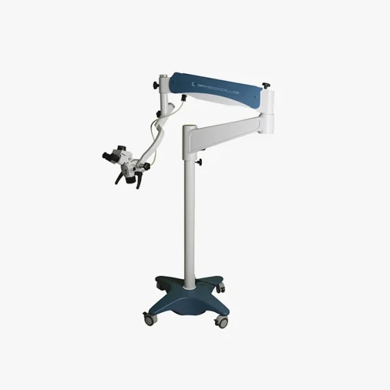
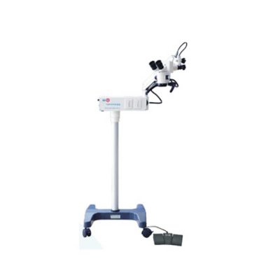
The Decius Advanced Microscope meets the most demanding requirements of dentistry and its specialties. Its optical system allows you to view the smallest details, further improving the quality of your exams and treatments. It is highly versatile, as it has a flexible articulation system, making it adaptable to all your needs.Its modern structure has a design that provides more comfortable ergonomics, reducing tension and fatigue, maintaining adequate posture regardless of the patient’s position. The DECIUS microscope is excellence in your hands.
With the automation of functions through a multi-tasking Joystick, it allows the professional to have full control of focus, alternation between filter colors, increase and decrease of lighting and on/off button, during the procedure in an easy way.
Typically 10-21 working days – excluding furniture and heavy/bulky equipment. Please contact us for further information.
Quick Comparison
| DECIUS ADVANCED MICROSCOPE remove | Tonometer remove | Ophthalmic Ultrasound Pachymeter remove | Portable Keratometer remove | Slit Lamp remove | Portable Fundus Camera remove | |||||||||||||||||||||||||||||||||||||||||||||||||||||||||||||||||||||||||||||||||||||||||||||||
|---|---|---|---|---|---|---|---|---|---|---|---|---|---|---|---|---|---|---|---|---|---|---|---|---|---|---|---|---|---|---|---|---|---|---|---|---|---|---|---|---|---|---|---|---|---|---|---|---|---|---|---|---|---|---|---|---|---|---|---|---|---|---|---|---|---|---|---|---|---|---|---|---|---|---|---|---|---|---|---|---|---|---|---|---|---|---|---|---|---|---|---|---|---|---|---|---|---|---|---|---|
| Name | DECIUS ADVANCED MICROSCOPE remove | Tonometer remove | Ophthalmic Ultrasound Pachymeter remove | Portable Keratometer remove | Slit Lamp remove | Portable Fundus Camera remove | ||||||||||||||||||||||||||||||||||||||||||||||||||||||||||||||||||||||||||||||||||||||||||||||
| Image |  | 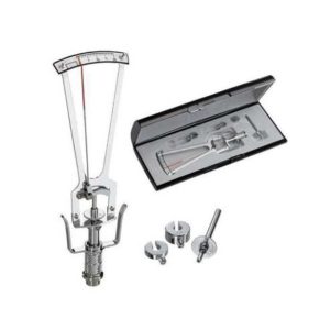 | 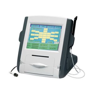 | 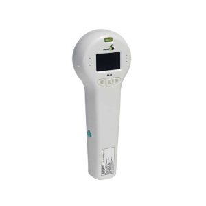 | 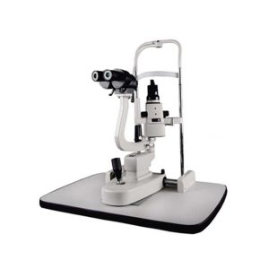 | 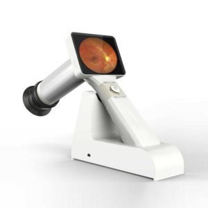 | ||||||||||||||||||||||||||||||||||||||||||||||||||||||||||||||||||||||||||||||||||||||||||||||
| SKU | SF103356013091-5 | SF1033560107-9 | SF1033560107-18 | SF1033560107-16 | SF1033560107-15 | SF1033560107-23 | ||||||||||||||||||||||||||||||||||||||||||||||||||||||||||||||||||||||||||||||||||||||||||||||
| Rating | ||||||||||||||||||||||||||||||||||||||||||||||||||||||||||||||||||||||||||||||||||||||||||||||||||||
| Price |
| $220.00 | $2,365.00 |
| $1,375.00 | $2,310.00 | ||||||||||||||||||||||||||||||||||||||||||||||||||||||||||||||||||||||||||||||||||||||||||||||
| Stock | ||||||||||||||||||||||||||||||||||||||||||||||||||||||||||||||||||||||||||||||||||||||||||||||||||||
| Availability | ||||||||||||||||||||||||||||||||||||||||||||||||||||||||||||||||||||||||||||||||||||||||||||||||||||
| Add to cart | ||||||||||||||||||||||||||||||||||||||||||||||||||||||||||||||||||||||||||||||||||||||||||||||||||||
| Description | Shipped From Abroad
The Decius Advanced Microscope meets the most demanding requirements of dentistry and its specialties. Its optical system allows you to view the smallest details, further improving the quality of your exams and treatments. It is highly versatile, as it has a flexible articulation system, making it adaptable to all your needs.Its modern structure has a design that provides more comfortable ergonomics, reducing tension and fatigue, maintaining adequate posture regardless of the patient's position. The DECIUS microscope is excellence in your hands.
With the automation of functions through a multi-tasking Joystick, it allows the professional to have full control of focus, alternation between filter colors, increase and decrease of lighting and on/off button, during the procedure in an easy way.
Delivery & Availability:
Typically 10-21 working days – excluding furniture and heavy/bulky equipment. Please contact us for further information. | Shipped from abroad
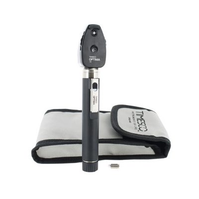
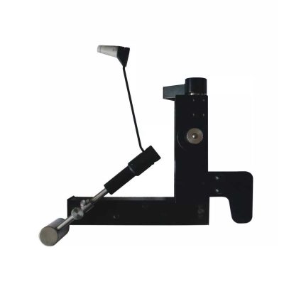
This product is used to measure the intraocular pressure (IOP) by measuring the depth produced on the surface of the cornea by a load of a known weight. Each division on the scale corresponds to 1/20mm corneal depth.
| Shipped from abroad
| Shipped from abroad
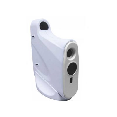
Ultra-small corneal curvature tester, is mainly used to measure the corneal curvature radius and diopter, wireless output print data.
| Shipped from abroad
| 83Shipped from abroad
This is a portable medical camera for fundus imaging, diagnosis, and especially for fundus disease screening. It's compact, easy to obtain high definition fundus image. It can be conveniently applied to rapid screening, out diagnosis, bedside diagnosis and remote medical treatment, etc.
| ||||||||||||||||||||||||||||||||||||||||||||||||||||||||||||||||||||||||||||||||||||||||||||||
| Content | This product is used to measure the intraocular pressure (IOP) by measuring the depth produced on the surface of the cornea by a load of a known weight. Each division on the scale corresponds to 1/20mm corneal depth.
Features:
| Features:
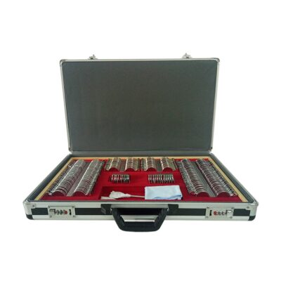
| Portable Keratometer Features:
Ultra-small corneal curvature tester, is mainly used to measure the corneal curvature radius and diopter, wireless output print data.
Technical Specifications:
| Slit Lamp Features:
| Portable Fundus Camera is a portable medical camera for fundus imaging, diagnosis, and especially for fundus disease screening. It's compact, easy to obtain high definition fundus image. It can be conveniently applied to rapid screening, out diagnosis, bedside diagnosis and remote medical treatment, etc.
Features of Portable Fundus Camera:
| |||||||||||||||||||||||||||||||||||||||||||||||||||||||||||||||||||||||||||||||||||||||||||||||
| Weight | N/A | N/A | N/A | N/A | N/A | N/A | ||||||||||||||||||||||||||||||||||||||||||||||||||||||||||||||||||||||||||||||||||||||||||||||
| Dimensions | N/A | N/A | N/A | N/A | N/A | N/A | ||||||||||||||||||||||||||||||||||||||||||||||||||||||||||||||||||||||||||||||||||||||||||||||
| Additional information | ||||||||||||||||||||||||||||||||||||||||||||||||||||||||||||||||||||||||||||||||||||||||||||||||||||


Reviews
There are no reviews yet.