Elmiko Digitrack Electroencephalogram (EEG) Machine
Ask for Price$0.00
Shipped From Abroad
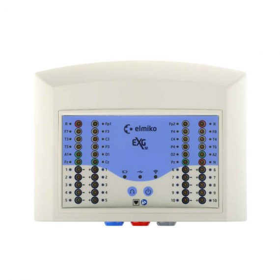
Basic EEG device for private clinics. Available in stationary or mobile version.This device is similar in terms of software functionality to the more expensive EEGDigiTrack simpleEEG_32 device. It stands out, however, with an extremely low price, because the set does not include a trolley, printer, uninterruptible power supply and a tripod to the head. It is a very efficient, user-friendly tool for routine clinical EEG. The newest and most advanced 32-channel EEG amplifier and many additional modules allows to make a full range of neurological diagnostics. The EEGDigiTrack basicEEG_32 amplifier (equipped with wide range of sensors) can be also used for polysomnography.
Delivery & Availability:
Typically 5-7 working days – excluding furniture and heavy/bulky equipment. Please contact us for further information.
Description
Basic EEG device for private clinics. Available in stationary or mobile version.This device is similar in terms of software functionality to the more expensive EEGDigiTrack simpleEEG_32 device. It stands out, however, with an extremely low price, because the set does not include a trolley, printer, uninterruptible power supply and a tripod to the head. It is a very efficient, user-friendly tool for routine clinical EEG. The newest and most advanced 32-channel EEG amplifier and many additional modules allows to make a full range of neurological diagnostics. The EEGDigiTrack basicEEG_32 amplifier (equipped with wide range of sensors) can be also used for polysomnography.
Features:
- NEW! 32-channels ExG-32 headbox with wireless communication (with Sp02 module and a patient’s button),
- a new, touch screen-adapted, exceptionally user-friendly user interface,
- completely new networked, dispersed, multi-station EEGDigiTrack Manager database automatically synchronises examinations, descriptions and other data with any computer, eliminating the need for a data server,
- ability to conduct EEG analysis on mobile devices (tablets, smartphones) with Windows OS.,
- optional multi-camera videometry with optical and digital zoom to monitor your patient on several feeds at once
- ability to turn video recording on and off independently from EEG,
- extensive, user-editable, medical events manager, with a database of over 400 pre-defined options,
- ability to create interactive notes and inserting markers during EEG recording,
- new, advanced CSA and DSA signals analyses,
- multi-dimensional FFT analysis,
- new 3D mapping algorithms,
- advanced electrocorticographic mapping to automatically recognize and register signals from all types of reference electrodes,
- ability to connect with a corticographic stimulator,
- automatic artefact detection,
- automatic spike detection,
- independent component analysis (ICA),
- new function – back averaging,
- LORETA analysis integration,
- new, optional polisomnography module with automatic analyses of artefacts and sleep spindles as well as automatic hypnogram,
- a new module for Visual Evoked Potentials (VEP),
- an optional module for EEG (aEEG) signal trend analysis for long-term neurological and cardiological monitoring,
- HL7 standard compatible, with ability to create own plug-ins,
- extraordinary configuration flexibility and easy way to translate the software to any language with a specially designed application.
Parameters:
- 33-channel headbox
- WIFI or USB connection
- 22 reference channels
- 10 bipolar channels with seperate reference for each channel!
- POX channel
- ONLINE impedance (from headbox and software)
- Impedance saved to EEG file
- Automatic calibration
- LED impedance indicator
- 24-bits A/D converter
- Flashlamp with build in battery
- Possibility of connecting Event Marker and Fotodetector
Reviews(2)
Quick Comparison
| Settings | Elmiko Digitrack Electroencephalogram (EEG) Machine remove | DrGem Diamond All-In-One Digital X-ray Machine remove | Sonoscape E1 Ultrasound Machine With Two Probes remove | Sonoscape P20 Ultrasound Machine remove | Sonoscape P15 Ultrasound Machine With Four Probes remove | Sonoscape P50 Ultrasound Machine remove |
|---|---|---|---|---|---|---|
| Name | Elmiko Digitrack Electroencephalogram (EEG) Machine remove | DrGem Diamond All-In-One Digital X-ray Machine remove | Sonoscape E1 Ultrasound Machine With Two Probes remove | Sonoscape P20 Ultrasound Machine remove | Sonoscape P15 Ultrasound Machine With Four Probes remove | Sonoscape P50 Ultrasound Machine remove |
| Image |  |  |  |  |  |  |
| SKU | SF1033560084-250 | SF1033560074-3 | SF1033560012-20 | SF1033560012-9 | SF1033560012-8 | SF1033560012-11 |
| Rating | ||||||
| Price | Ask for Price | Ask for Price | Ask for Price | Ask for Price | $13,900.00 | Ask for Price |
| Stock | ||||||
| Availability | ||||||
| Add to cart | ||||||
| Description | Shipped From Abroad
Basic EEG device for private clinics. Available in stationary or mobile version.This device is similar in terms of software functionality to the more expensive EEGDigiTrack simpleEEG_32 device. It stands out, however, with an extremely low price, because the set does not include a trolley, printer, uninterruptible power supply and a tripod to the head. It is a very efficient, user-friendly tool for routine clinical EEG. The newest and most advanced 32-channel EEG amplifier and many additional modules allows to make a full range of neurological diagnostics. The EEGDigiTrack basicEEG_32 amplifier (equipped with wide range of sensors) can be also used for polysomnography.
| Shipped from Abroad DrGem Diamond All-In-One Digital X-ray Machine is a fully automatic digital radiography system providing state-of-the-art image quality, image processing and user interface. With a wide selection of anatomical studies on the imaging software, DIAMOND automatically sets up the x-ray generator’s preprogrammed exposure technique settings, motorized radiographic stand positioning, x-ray collimation and post-image processing for the selected study. Specifically designed to increase workflow, this fully digital system offers convenient auto-positioning and advanced image processing to achieve big performance with little effort. Delivery & Availability: Typically 21 working days – excluding furniture and heavy/bulky equipment. Please contact us for further information. | Shipped from Abroad SonoScape has developed a new probe and function for the E1 Exp. With these additions the E1 Exp will bring users a more efficient examination experience with satisfying image quality and a smooth workflow. Delivery & Availability: Typically 5-7 working days – excluding furniture and heavy/bulky equipment. Please contact us for further information. | Shipped from Abroad Incorporating innovative technologies, P20’s user-friendly design with a simple operation panel, intuitive user interface and a variety of intelligent auxiliary scanning tools, will significantly improve your daily examination experience. Besides general imaging applications, P20 has entitled with diagnostic 4D technology which has an extraordinary performance in obstetrics and gynecology applications. Delivery & Availability: Typically 5-7 working days – excluding furniture and heavy/bulky equipment. Please contact us for further information. | In Stock A feature-rich system inheriting the Wi-Sono high-end platform, the P15 uses an array of advanced tools to help enhance the image quality. It's a cost-effective, simplified console with an intuitive user interface and multiple intelligent functions. Delivery & Availability: Typically 2 working days – excluding furniture and heavy/bulky equipment. Please contact us for further information. | Shipped from Abroad Easily accomplish more with SonoScape’s new P50 ultrasound system. Incorporating single crystal clarity, automatic corrections and calculation, and user defined flexibility promises a confident diagnostic experience as well as opening new doors of opportunity for ultrasound use. Delivery & Availability: Typically 7-14 working days – excluding furniture and heavy/bulky equipment. Please contact us for further information. |
| Content | Basic EEG device for private clinics. Available in stationary or mobile version.This device is similar in terms of software functionality to the more expensive EEGDigiTrack simpleEEG_32 device. It stands out, however, with an extremely low price, because the set does not include a trolley, printer, uninterruptible power supply and a tripod to the head. It is a very efficient, user-friendly tool for routine clinical EEG. The newest and most advanced 32-channel EEG amplifier and many additional modules allows to make a full range of neurological diagnostics. The EEGDigiTrack basicEEG_32 amplifier (equipped with wide range of sensors) can be also used for polysomnography.
Features:
| DrGem Diamond All-In-One Digital X-ray Machine is a fully automatic digital radiography system providing state-of-the-art image quality, image processing and user interface. With a wide selection of anatomical studies on the imaging software, DIAMOND automatically sets up the x-ray generator’s pre-programmed exposure technique settings, motorized radiographic stand positioning, x-ray collimation and post-image processing for the selected study. Specifically designed to increase workflow, this fully digital system offers convenient auto-positioning and advanced image processing to achieve big performance with little effort.
Features of DrGem Diamond All-In-One Digital X-ray Machine:
Outstanding Image Quality -
Digital radiography via at panel detector improves your workflow, exam speed and comfort with efficiency. Digital at panel detector with Csl screen provides excellent spatial resolution, MTF, DQE and stability based on ne pixel pitch. A 3-field ion-chamber is provided for AEC function.
Automatic Collimation –
Automatic x-ray eld size control of the motorized collimator corresponds to dierent SIDs. Includes user adjustable lamp timer with on/oswitch.
Automatic Positioning –
Click Here To Download Catalogue | DETAILS
Efficient Diagnosis
μ-Scan, Speckle Reduction & Edge Enhancement
Spatial Compound Imaging
PIH - Pure Inversion Harmonic
Wide Scan - Enlarged Image Area
Tissue-Specific Imaging
SR Flow
Ergonomic Designs
Up to 2 Transducer Ports
Light Weight and Compact
15.6 inch Anti-flickering HD LED Screen
Tilting Monitor Angle Adjustment
Backlit Keyboard and Intelligent Panel
Long-lasting Battery for 90 mins
Ease of Use
Quick Boot Up
Auto-Brightness Adjustment
Auto Image Optimization
Auto IMT
Auto Trace
Equipped Accessories
Wi-Fi and Bluetooth Available
DICOM
500GB Hard Disk
Height Adjustable Trolley
Durable, Carry-on Site Suitcase
Click Here To Download Catalogue | DETAILS
Upgraded Images with More Clarity
SonoScape never stops making progress in improving the image quality of its ultrasound products to enhance the confidence of diagnosis for doctors. With extraordinary images provided by P20, the anatomy structures are clearer than ever.
C-Xlasto Imaging
With C-xlasto Imaging, P20 enables comprehensive quantitative elastic analysis. Meanwhile, C-xlasto on P20 is supported by linear, convex and transvaginal probes, to ensure good reproducibility and highly consistent quantitative elastic results.
S-Live
S-Live allows for detailed visualization of subtle anatomical features, thereby enabling intuitive diagnosis with real-time 3D images and enriching patient communication.
Pelvic Floor 4D
Transperineal 4D pelvic floor ultrasound can provide useful clinical values in assessing the vaginal delivery impact on the female anterior compartment, judging whether the pelvic organs are prolapsed or not and the extent, determining if the pelvic muscles were torn accurately.
Anatomic M Mode
Anatomic M Mode helps you observe the myocardial motion at different phases by freely placing sample lines. It accurately measures the myocardial thickness and the heart size of even difficult patients and supports the myocardial function and LV wall-motion assessment.
Tissue Doppler Imaging
P20 is endowed with Tissue Doppler Imaging which provides velocities and other clinical information on myocardial functions, facilitating clinical doctors with the ability to analyze and compare the motions of different parts of the patient's heart.
Click Here To Download Catalogue | DETAILS
Super Wide-bandwidth Platform
Inheriting Wi-sono's ultra-wide system platform and with the advanced probe technology, high-resolution and deep penetration images are provided for precision medicine.
Spatial Compound Imaging
Spatial Compound Imaging utilizes several lines of sight for optimal contrast resolution, speckle reduction and border detection, with which P15 is ideal for superficial and abdominal imaging with better clarity and improved continuity of structures.
μ-Scan+
The new generation μ-Scan imaging technology gives you better image quality by reducing noise, improving signal strength and improving visualization.
Dynamic Color
Dynamic color improves upon already existing color Doppler technologies for a clearer capture of color flow and detailed visualization of even tiny veins with lower velocities.
Real-time Panoramic
With real-time panoramic, you can acquire an extended field of view for large organs or long vessels for easy measurement and diagnostic efficiency. Accomplished in real-time for the convenience of the sonographers, any mistake can also be easily back tracked and corrected without interrupting the scan.
3D/4D
Outstanding volume performance with speed and convenience makes P15 outshine others on volume imaging.
Tissue Doppler Imaging
Tissue Doppler Imaging allows clinical doctors to quantitatively evaluate local myocardial movements and functions, facilitating them with the ability to analyze and compare the motions of the different parts of the patient's heart.
Auto IMT
Quick measurement of intra-media vessel thickness ensures good reproducibility and high diagnostic efficiency.
Click Here To Download Catalogue | DETAILS
Powerful Compact Precision
Taking into consideration the evolving expectations and needs for ultrasound, the P50 is a slim and unobtrusive trolley system that is comfortable in tight, congested spaces with little room to work in. Providing everything you need for a comfortable examination in a small space for both you and your patient.
Single Crystal Transducer
Wideband single crystal probes greatly improve the signal ratio, acquire stunning images and provide superior sensitivity and resolution for both the near and far-fields.
μ-Scan+
The new generation μ-Scan imaging technologies give you better image quality by reducing noise, improving signal strength and improving visualization.
Dynamic Color
Dynamic colour improves upon already existing colour Doppler technologies for clear capture of colour flow and detail visualization of even tiny veins with lower velocities.
Solution for Radiology
P50, is a leading-edge ultrasound system that can meet the demands of any clinical setting. You can experience a superior performance in multi-dimensional imaging for a full range of clinical applications – abdominal, breast and cardiovascular.
C-xlasto Imaging
By understanding that tissue stiffness varies depending on the type of tissue, we can use C-xlasto Imaging to easily find abnormalities and tumours within soft tissue. The differences in tissue responses are detected and visualized in real-time by the elastography algorithms through different representations, which can be particularly helpful in analyzing breast, thyroid and musculoskeletal structures. Predominately used only in linear probes, SonoScape’s new transvaginal and bi-plane probe for gynaecology and urology are breaking the mould and expanding elastography applications.
Real-time Color Panoramic
With the combination of colour flow and real-time panoramic, visualizing the blood flow of an entire vein or artery is now an easy task. Accomplished in real-time for the convenience of the sonographers, any mistakes can also be easily backtracked and corrected without interrupting the scan.
Contrast Imaging
Contrast Imaging on P50 makes full use of the infra harmonic signal and second harmonic signal to improve the image resolution and deep penetration. What’s more, the Dynamic Acoustic Control technology effectively controls the acoustic pressure for the contrast agent, decreasing the required agent dose and assures uniform image quality, guaranteeing longer contrast agent duration and better lesion perfusion of delayed phase observation.
Solution for OB/GYN
P50 has superior image quality, automated measurement tools, and a variety of volume technologies to provide ideal solutions for clinical examinations such as pregnancy examinations, and gynecologic disease diagnosis. With a new 4D transvaginal probe, P50 helps you to see and detect fetal abnormalities and significantly improves your diagnostic confidence during your examinations.
S-Live Silhouette
A unique transparent 3D anatomical image of the fetus for improved initial anatomical review. By using this new application, the system can create completely different fetal images from conventional ultrasound images, which can depict the fetal's intracorporeal anatomical structure.
Pelvic Floor 4D
Working in conjunction with SonoScape’s latest transvaginal probes, trans-perineal 4D pelvic floor ultrasound provides a useful clinical assessment of the impact of vaginal delivery on the female anterior compartment. Allowing doctors to judge whether the pelvic organs prolapsed or not, the extent of prolapse, and determining whether the pelvic muscles tore correctly.
S-Guide
S-Guide gives the user an extensive list of example obstetric ultrasound images as reference guides and a convenient checklist system to keep track of their progress during their obstetrics examination.
Auto Face
Automatically removes masking layers in front of the fetus’s face for a clearer vision of the fetus’s face.
AVC Follicle
AVC Follicle automatically identifies how many follicles are present and calculates their individual volumes.
Solution for Cardiology
P50 provides clear 2D clinical images and Doppler sensitivity to assess critical cardiac performance. Compatible with SonoScape’s single crystal probes, the P50 can provide images with better resolution and penetration in Cardiac diagnosis.
Tissue Doppler Imaging
Tissue Doppler Imaging allows clinical doctors to quantitatively evaluate local myocardial movements and functions, facilitating them with the ability to analyze and compare the motions of the different parts of the patient’s heart.
Stress Echo
Stress echocardiography is the combination of 2D echocardiography with physical, pharmacological or electrical stress of the patient. It also then provides users with report management tools such as configurable template editor, multiple loops to select one for storage, wall motion scoring, stress echo report, etc
Auto IMT
Auto IMT is used when determining the level of vascular sclerosis present in the patient by automatically tracing and calculating the thickness of the carotid vessels. What distinguishes the P50 is that it provides an instant and accurate Mean and Max index at the touch of a single button.
Auto EF
Automated 2D Cardiac Quantification is a fully intelligent trace function for endocardium with 19 easily-adjustable points providing rapid access to proven 2D EF and volumes.
Click Here To Download Catalogue |
| Weight | N/A | N/A | N/A | N/A | N/A | N/A |
| Dimensions | N/A | N/A | N/A | N/A | N/A | N/A |
| Additional information |


my blog latestbtcnews.com
Its like you read my mind! You seem to know so much about this, like you wrote the book in it or something. I think that you can do with some pics to drive the message home a little bit, but instead of that, this is fantastic blog. A fantastic read. I’ll certainly be back.
RJbB
163148 458245Hmm is anyone else experiencing issues with the images on this blog loading? Im trying to discover out if its a issue on my end or if its the blog. Any feed-back would be greatly appreciated. 20461