Eonis Color 8MP (MDRC‑8132)
$0.00
Shipped From Abroad
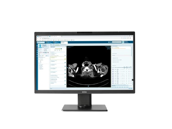
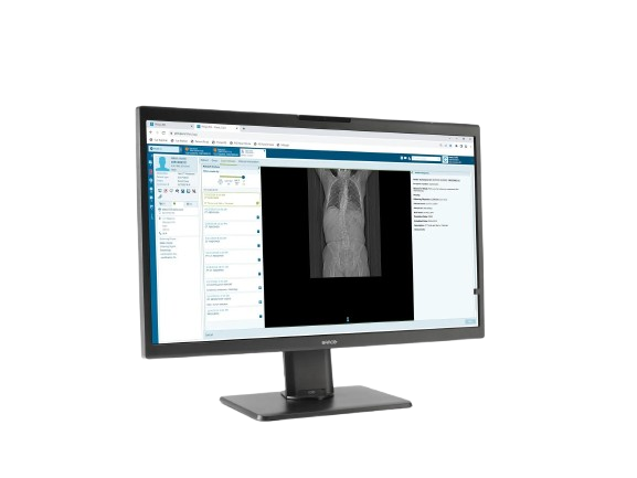
The Barco Eonis Color 8MP is a 32″ clinical review display offering extremely high resolution, built-in front sensor for consistent image quality, pathology mode, KVM, and remote QA management – ideal for advanced imaging and consultation workflows.
Typically 10-21 working days – excluding furniture and heavy/bulky equipment. Please contact us for further information.
Description
Eonis 8MP is a 32″ display built with healthcare specialists in mind. It combines consistent, high image quality and an attractive, versatile design with centralized quality assurance. It also includes built-in multimedia functionalities for easy virtual collaboration.
High image quality
Eonis 8MP offers you 8 megapixels of screen resolution, the highest in our Eonis range. This makes it an ideal choice for healthcare professionals seeking second opinions, emergency or trauma assessments, or referrals, which are not primary diagnoses. Moreover, the display incorporates an innovative front consistency sensor that seamlessly adjusts image quality whenever you power it on.
Additionally, Eonis 8MP has a pathology setting that triggers sRGB color calibration, resulting in excellent images with accurate color representation.
Virtual collaboration was never this easy
The Eonis 8MP extends its capabilities beyond its core functions by providing multimedia features. These streamline intra-hospital and virtual communication among healthcare practitioners, discussing a patient’s medical history, electronic medical records, and clinical images.
- Built-in camera, microphone, and speakers
- Consistent images and centralized quality assurance
- KVM for seamless switching between workstations
These features promote effective communication across locations and clinical disciplines, making Eonis 8MP an excellent tool for teleconsultations with patients as well.
Centralized quality assurance
Eonis monitors are bundled with Barco’s cloud-based QAWeb Enterprise software, an online platform for automated calibration, quality assurance, and asset management. Praised in hospitals around the world, QAWeb Enterprise allows healthcare IT and PACS administrators to centrally and remotely manage image quality across the healthcare organization, at the touch of a button.
A+ ecolabel for Eonis Color 8MP
The Eonis Color 8MP has been subjected to Barco’s ecoscoring protocol and has received an A+ rating. Some key factors that contributed to this rating are:
- Automatic standby mode when the device is not in use
- Use of plastics containing 30% recycled content >50% halogen-free cables & >65% halogen-free PCBs
- >90% recyclable packaging
- Unpainted housing to make recycling easier
Key Features
-
32″ flat panel with 8MP resolution (3840 × 2160)
-
True color + grayscale imaging (30-bit)
-
Front Consistency Sensor ensures image stability
-
Pathology mode with sRGB calibration
-
Integrated multimedia: camera, microphone, speakers
-
KVM switching between workstations
-
Remote QA and calibration via QAWeb
-
High luminance panel (500 cd/m² typ)
-
DICOM-calibrated luminance at 300 cd/m²
-
Wide viewing angles (178°)
-
RapidFrame refresh technology
-
Ambient light presets and reading room modes
-
Ergonomic stand, height/tilt range
-
Medical safety and imaging certifications
Technical Specifications
| Specification | Detail |
|---|---|
| Screen Technology | IPS panel |
| Diagonal Size | 812.80 mm (32.0″) |
| Active Area (H × V) | 708.48 × 398.52 mm |
| Aspect Ratio | 16:9 |
| Resolution | 8MP (3840 × 2160 @ 60 Hz) |
| Pixel Pitch | 0.1845 mm |
| Color & Gray Imaging | Yes |
| Bit Depth | 30 bit (true color + grayscale) |
| Viewing Angle | 178° horizontal / vertical |
| Max Luminance (Panel) | 500 cd/m² |
| DICOM Calibrated Luminance | 300 cd/m² |
| Contrast Ratio | 1000:1 (typical) |
| Response Time (Tr + Tf / 2) | 9.8 ms (typical) |
| Video Inputs | 2 × DisplayPort 1.4 |
| Video Output | 1 × DisplayPort (MST) |
| USB | 2 × USB-B upstream; 5 × USB-A downstream (1 charge port) |
| KVM | Yes |
| Power Supply | 24 V DC, 8.3 A |
| Power Consumption | ≈ 45.5 W (nominal); <0.35 W in hibernate/off |
| Dimensions with Stand | 743 × (518 to 618) × 238 mm |
| Dimensions without Stand | 743 × 459 × 63 mm |
| Net Weight | 13 kg (with stand), 8.4 kg (without stand) |
| Tilt | –5° to +25° |
| Swivel | –30° to +30° |
| Height Adjustment | 100 mm |
| Mounting | VESA 100 mm |
| Certifications & Compliance | Medical device safety standards (CE, FDA class I, IEC 60601-1, etc.) |
Quick Comparison
| Eonis Color 8MP (MDRC‑8132) remove | Bistos BT-770-12.1" Touchscreen Patient Monitor remove | Sonoscape P50 Ultrasound Machine remove | DrGem Diamond All-In-One Digital X-ray Machine remove | DrGem Ceiling Mounted Digital X-ray remove | DrGem GXR-SD 400mA Floor Mounted Digital X-ray remove | |||||||||||||||||||||||||||||||||||||||||||||||||||||||||
|---|---|---|---|---|---|---|---|---|---|---|---|---|---|---|---|---|---|---|---|---|---|---|---|---|---|---|---|---|---|---|---|---|---|---|---|---|---|---|---|---|---|---|---|---|---|---|---|---|---|---|---|---|---|---|---|---|---|---|---|---|---|---|
| Name | Eonis Color 8MP (MDRC‑8132) remove | Bistos BT-770-12.1" Touchscreen Patient Monitor remove | Sonoscape P50 Ultrasound Machine remove | DrGem Diamond All-In-One Digital X-ray Machine remove | DrGem Ceiling Mounted Digital X-ray remove | DrGem GXR-SD 400mA Floor Mounted Digital X-ray remove | ||||||||||||||||||||||||||||||||||||||||||||||||||||||||
| Image | 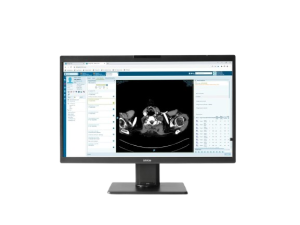 | 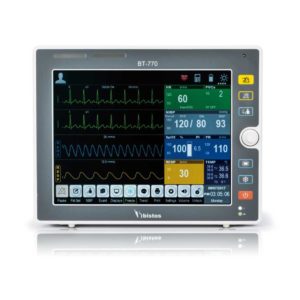 |  |  |  |  | ||||||||||||||||||||||||||||||||||||||||||||||||||||||||
| SKU | SF1033560059-1 | SF1033560012-11 | SF1033560074-3 | SF1033560074-4 | SF1033560074-5 | |||||||||||||||||||||||||||||||||||||||||||||||||||||||||
| Rating | ||||||||||||||||||||||||||||||||||||||||||||||||||||||||||||||
| Price |
| $902.00 |
|
|
|
| ||||||||||||||||||||||||||||||||||||||||||||||||||||||||
| Stock | ||||||||||||||||||||||||||||||||||||||||||||||||||||||||||||||
| Availability | ||||||||||||||||||||||||||||||||||||||||||||||||||||||||||||||
| Add to cart | ||||||||||||||||||||||||||||||||||||||||||||||||||||||||||||||
| Description | Shipped From Abroad
The Barco Eonis Color 8MP is a 32″ clinical review display offering extremely high resolution, built-in front sensor for consistent image quality, pathology mode, KVM, and remote QA management - ideal for advanced imaging and consultation workflows.
Delivery & Availability:
Typically 10-21 working days – excluding furniture and heavy/bulky equipment. Please contact us for further information.
| Shipped from Abroad The Bistos BT-770 patient monitor is equipped with a 12.1" touchscreen display, which allows for an easy operation and readability with a powerful rechargeable battery guaranteeing a continuous operation of 5 hours to monitor ECG, SpO2, NIBP, temperature and respiration Delivery & Availability: Typically 14 working days – excluding furniture and heavy/bulky equipment. Please contact us for further information. | Shipped from Abroad Easily accomplish more with SonoScape’s new P50 ultrasound system. Incorporating single crystal clarity, automatic corrections and calculation, and user defined flexibility promises a confident diagnostic experience as well as opening new doors of opportunity for ultrasound use. Delivery & Availability: Typically 7-14 working days – excluding furniture and heavy/bulky equipment. Please contact us for further information. | Shipped from Abroad DrGem Diamond All-In-One Digital X-ray Machine is a fully automatic digital radiography system providing state-of-the-art image quality, image processing and user interface. With a wide selection of anatomical studies on the imaging software, DIAMOND automatically sets up the x-ray generator’s preprogrammed exposure technique settings, motorized radiographic stand positioning, x-ray collimation and post-image processing for the selected study. Specifically designed to increase workflow, this fully digital system offers convenient auto-positioning and advanced image processing to achieve big performance with little effort. Delivery & Availability: Typically 21 working days – excluding furniture and heavy/bulky equipment. Please contact us for further information. | In Stock The GXR-SD is a diagnostic digital radiography system that provides reliable high quality digital radiographic images with a reduced dose. The GXR-SD DR systems offer comprehensive digital solutions to all radiography needs, featuring ACQUIDR digital imaging system with stationary or portable digital flat-panel detectors as well as reliable high-frequency x-ray generators that are known worldwide for their excellent performance, lifetime and stability. Patient tables and wall stands are also offered. Delivery & Availability: Typically 21 working days – excluding furniture and heavy/bulky equipment. Please contact us for further information. | In Stock The GXR-SD Digital X-ray is a diagnostic digital radiography system that provides reliable high quality digital radiographic images with a reduced dose. The GXR-SD DR systems offer comprehensive digital solutions to all radiography needs, featuring ACQUIDR digital imaging system with stationary or portable digital flat-panel detectors as well as reliable high-frequency x-ray generators that are known worldwide for their excellent performance, lifetime and stability. Patient tables and wall stands are also offered. Delivery & Availability: Typically 21 working days – excluding furniture and heavy/bulky equipment. Please contact us for further information. | ||||||||||||||||||||||||||||||||||||||||||||||||||||||||
| Content | Eonis 8MP is a 32" display built with healthcare specialists in mind. It combines consistent, high image quality and an attractive, versatile design with centralized quality assurance. It also includes built-in multimedia functionalities for easy virtual collaboration. High image qualityEonis 8MP offers you 8 megapixels of screen resolution, the highest in our Eonis range. This makes it an ideal choice for healthcare professionals seeking second opinions, emergency or trauma assessments, or referrals, which are not primary diagnoses. Moreover, the display incorporates an innovative front consistency sensor that seamlessly adjusts image quality whenever you power it on. Additionally, Eonis 8MP has a pathology setting that triggers sRGB color calibration, resulting in excellent images with accurate color representation.Virtual collaboration was never this easyThe Eonis 8MP extends its capabilities beyond its core functions by providing multimedia features. These streamline intra-hospital and virtual communication among healthcare practitioners, discussing a patient's medical history, electronic medical records, and clinical images.
Centralized quality assuranceEonis monitors are bundled with Barco's cloud-based QAWeb Enterprise software, an online platform for automated calibration, quality assurance, and asset management. Praised in hospitals around the world, QAWeb Enterprise allows healthcare IT and PACS administrators to centrally and remotely manage image quality across the healthcare organization, at the touch of a button.A+ ecolabel for Eonis Color 8MPThe Eonis Color 8MP has been subjected to Barco’s ecoscoring protocol and has received an A+ rating. Some key factors that contributed to this rating are:
Key Features
Technical Specifications
|
Bistos BT-770 is a 12.1" touchscreen patient monitor designed for easy operations.
SPECIFICATIONS
Click Here To Download Catalogue | DETAILS
Powerful Compact Precision
Taking into consideration the evolving expectations and needs for ultrasound, the P50 is a slim and unobtrusive trolley system that is comfortable in tight, congested spaces with little room to work in. Providing everything you need for a comfortable examination in a small space for both you and your patient.
Single Crystal Transducer
Wideband single crystal probes greatly improve the signal ratio, acquire stunning images and provide superior sensitivity and resolution for both the near and far-fields.
μ-Scan+
The new generation μ-Scan imaging technologies give you better image quality by reducing noise, improving signal strength and improving visualization.
Dynamic Color
Dynamic colour improves upon already existing colour Doppler technologies for clear capture of colour flow and detail visualization of even tiny veins with lower velocities.
Solution for Radiology
P50, is a leading-edge ultrasound system that can meet the demands of any clinical setting. You can experience a superior performance in multi-dimensional imaging for a full range of clinical applications – abdominal, breast and cardiovascular.
C-xlasto Imaging
By understanding that tissue stiffness varies depending on the type of tissue, we can use C-xlasto Imaging to easily find abnormalities and tumours within soft tissue. The differences in tissue responses are detected and visualized in real-time by the elastography algorithms through different representations, which can be particularly helpful in analyzing breast, thyroid and musculoskeletal structures. Predominately used only in linear probes, SonoScape’s new transvaginal and bi-plane probe for gynaecology and urology are breaking the mould and expanding elastography applications.
Real-time Color Panoramic
With the combination of colour flow and real-time panoramic, visualizing the blood flow of an entire vein or artery is now an easy task. Accomplished in real-time for the convenience of the sonographers, any mistakes can also be easily backtracked and corrected without interrupting the scan.
Contrast Imaging
Contrast Imaging on P50 makes full use of the infra harmonic signal and second harmonic signal to improve the image resolution and deep penetration. What’s more, the Dynamic Acoustic Control technology effectively controls the acoustic pressure for the contrast agent, decreasing the required agent dose and assures uniform image quality, guaranteeing longer contrast agent duration and better lesion perfusion of delayed phase observation.
Solution for OB/GYN
P50 has superior image quality, automated measurement tools, and a variety of volume technologies to provide ideal solutions for clinical examinations such as pregnancy examinations, and gynecologic disease diagnosis. With a new 4D transvaginal probe, P50 helps you to see and detect fetal abnormalities and significantly improves your diagnostic confidence during your examinations.
S-Live Silhouette
A unique transparent 3D anatomical image of the fetus for improved initial anatomical review. By using this new application, the system can create completely different fetal images from conventional ultrasound images, which can depict the fetal's intracorporeal anatomical structure.
Pelvic Floor 4D
Working in conjunction with SonoScape’s latest transvaginal probes, trans-perineal 4D pelvic floor ultrasound provides a useful clinical assessment of the impact of vaginal delivery on the female anterior compartment. Allowing doctors to judge whether the pelvic organs prolapsed or not, the extent of prolapse, and determining whether the pelvic muscles tore correctly.
S-Guide
S-Guide gives the user an extensive list of example obstetric ultrasound images as reference guides and a convenient checklist system to keep track of their progress during their obstetrics examination.
Auto Face
Automatically removes masking layers in front of the fetus’s face for a clearer vision of the fetus’s face.
AVC Follicle
AVC Follicle automatically identifies how many follicles are present and calculates their individual volumes.
Solution for Cardiology
P50 provides clear 2D clinical images and Doppler sensitivity to assess critical cardiac performance. Compatible with SonoScape’s single crystal probes, the P50 can provide images with better resolution and penetration in Cardiac diagnosis.
Tissue Doppler Imaging
Tissue Doppler Imaging allows clinical doctors to quantitatively evaluate local myocardial movements and functions, facilitating them with the ability to analyze and compare the motions of the different parts of the patient’s heart.
Stress Echo
Stress echocardiography is the combination of 2D echocardiography with physical, pharmacological or electrical stress of the patient. It also then provides users with report management tools such as configurable template editor, multiple loops to select one for storage, wall motion scoring, stress echo report, etc
Auto IMT
Auto IMT is used when determining the level of vascular sclerosis present in the patient by automatically tracing and calculating the thickness of the carotid vessels. What distinguishes the P50 is that it provides an instant and accurate Mean and Max index at the touch of a single button.
Auto EF
Automated 2D Cardiac Quantification is a fully intelligent trace function for endocardium with 19 easily-adjustable points providing rapid access to proven 2D EF and volumes.
Click Here To Download Catalogue | DrGem Diamond All-In-One Digital X-ray Machine is a fully automatic digital radiography system providing state-of-the-art image quality, image processing and user interface. With a wide selection of anatomical studies on the imaging software, DIAMOND automatically sets up the x-ray generator’s pre-programmed exposure technique settings, motorized radiographic stand positioning, x-ray collimation and post-image processing for the selected study. Specifically designed to increase workflow, this fully digital system offers convenient auto-positioning and advanced image processing to achieve big performance with little effort.
Features of DrGem Diamond All-In-One Digital X-ray Machine:
Outstanding Image Quality -
Digital radiography via at panel detector improves your workflow, exam speed and comfort with efficiency. Digital at panel detector with Csl screen provides excellent spatial resolution, MTF, DQE and stability based on ne pixel pitch. A 3-field ion-chamber is provided for AEC function.
Automatic Collimation –
Automatic x-ray eld size control of the motorized collimator corresponds to dierent SIDs. Includes user adjustable lamp timer with on/oswitch.
Automatic Positioning –
Click Here To Download Catalogue | DrGem Ceiling Mounted Digital X-ray is a diagnostic digital radiography system that provides reliable high quality digital radiographic images with a reduced dose. The GXR-SD DR systems offer comprehensive digital solutions to all radiography needs, featuring ACQUIDR digital imaging system with stationary or portable digital flat-panel detectors as well as reliable high-frequency x-ray generators that are known worldwide for their excellent performance, lifetime and stability. Patient tables and wall stands are also offered.
Features:
Click Here To Download Catalogue | DrGem GXR-SD 400mA Floor Mounted Digital X-ray system matches with a radiographic room which perfectly fits your workow and can be easily upgraded to DR system with the help of DR interface and PC interface in GXR generator as well as Bucky suitable to Flat Panel Detector. GXR X-ray system is equipped with a high frequency X-ray generator which consistently produces high quality radiograph in favor of high quality X-ray output with a very small kV ripple and accurate mA and mAs. GXR X-ray system is designed to provide convenience to operator and comfort to patient
Features of DrGem GXR-SD 400mA Floor Mounted Digital X-ray:
Click Here To Download Catalogue | ||||||||||||||||||||||||||||||||||||||||||||||||||||||||
| Weight | N/A | N/A | N/A | N/A | N/A | N/A | ||||||||||||||||||||||||||||||||||||||||||||||||||||||||
| Dimensions | N/A | N/A | N/A | N/A | N/A | N/A | ||||||||||||||||||||||||||||||||||||||||||||||||||||||||
| Additional information |


Reviews
There are no reviews yet.