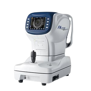
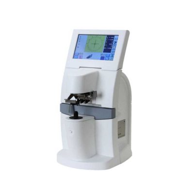
FA-6100B Auto Refractometer Auto Kerato-Refractometer
$0.00
Shipped From Abroad
The FA-6100B Auto Refractometer / Auto Kerato-Refractometer delivers fast, accurate, and reliable measurement of refractive errors and corneal curvature. With advanced optics, ergonomic design, and seamless operation, it enhances efficiency in clinics and hospitals. Compact, user-friendly, and precise, it is ideal for modern eye care professionals.
Typically 10-21 working days – excluding furniture and heavy/bulky equipment. Please contact us for further information.
Description
The FA-6100B Auto Refractometer / Auto Kerato-Refractometer is a high-precision ophthalmic diagnostic device that combines refraction and keratometry in one compact system. Designed to provide accurate, quick, and repeatable results, it measures refractive errors such as myopia, hyperopia, and astigmatism, along with corneal curvature and pupil distance. Built with advanced optical technology, it reduces testing errors and ensures patient comfort during examinations. Its ergonomic chin rest and head support improve stability, while the intuitive LCD interface allows easy operation by practitioners. The FA-6100B is equipped with a built-in thermal printer for instant report generation and supports data transfer for integration with external systems. Compact and reliable, this dual-function device enhances efficiency in optical centers, eye hospitals, and ophthalmology practices. By streamlining workflow and improving diagnostic accuracy, the FA-6100B is an essential tool for modern vision assessment, prescription verification, and pre-surgical evaluations.
Features
- Faster and more accurate
- Easy to operate,one-hand control
- Convenient user menu setting
- Keratometry and refractometry mode selection
- Auto/manual focus function
- Auto PD measurement
- A broad measurement range of Min. pupil size to Φ2.0mm
Specifications
|
Refractometer |
|
| Vertex distance | 0mm, 12mm, 13.75mm |
| Spherical | -20~+20m-1(VD=12) 0.12/0.25m-1Step |
| Cylinder | -8~+8m-1 0.12/0.25m-1 Step |
| Axis | 0~180° 1°Step |
| Cylinder form | -, + , ± |
| Pupil distance | 45~88mm, 1mm Step |
| Min. pupil size | 2.0mm |
| Keratometry | |
| Radius of curvature | 5.0~10mm(increment:1mm) |
| Corneal power | 33.75~67.50m-1 (when corner equivalent refractive index is 1.337) |
| (increment selectable from 0.12, 0.25m-1) | |
| Corneal astigmatism | 0.0~8.00m (increment selectable from 0.12,0.25m-1) |
| Axis | 1~180°(increment:1°) |
| Corneal diameter | 2.0~14.0mm(increment: 0.1mm) |
| Others | |
| Chart | Auto fog |
| Memory of data | 10 measure value for each right and left eye |
| Display | 5.6″LCD/TFT |
| Thermal printer | |
| Power supply | 100~240V 50/60Hz |
| Dimension | 490mm×280mm×470mm |
| Weight | ~15.5kg |
Quick Comparison
| FA-6100B Auto Refractometer Auto Kerato-Refractometer remove | Portable Fundus Camera remove | Ophthalmic AB Scan Machine remove | Pantoscopic Ophthalmoscope remove | Timesco Ophthalmoscope remove | Portable Slit Lamp remove | ||||||||||||||||||||||||||||||||||||||||||||||||||||||||||||||||||||||||||||||||||||
|---|---|---|---|---|---|---|---|---|---|---|---|---|---|---|---|---|---|---|---|---|---|---|---|---|---|---|---|---|---|---|---|---|---|---|---|---|---|---|---|---|---|---|---|---|---|---|---|---|---|---|---|---|---|---|---|---|---|---|---|---|---|---|---|---|---|---|---|---|---|---|---|---|---|---|---|---|---|---|---|---|---|---|---|---|---|---|---|---|---|
| Name | FA-6100B Auto Refractometer Auto Kerato-Refractometer remove | Portable Fundus Camera remove | Ophthalmic AB Scan Machine remove | Pantoscopic Ophthalmoscope remove | Timesco Ophthalmoscope remove | Portable Slit Lamp remove | |||||||||||||||||||||||||||||||||||||||||||||||||||||||||||||||||||||||||||||||||||
| Image | | 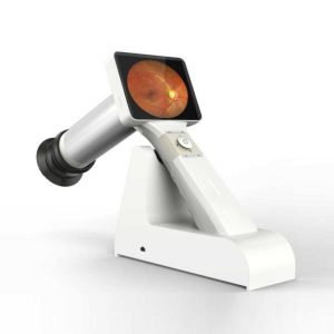 | 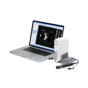 | 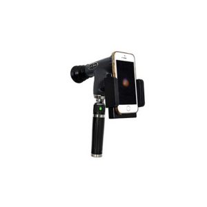 | 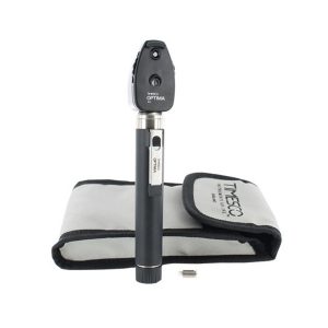 | 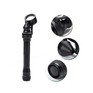 | |||||||||||||||||||||||||||||||||||||||||||||||||||||||||||||||||||||||||||||||||||
| SKU | SF1033560107-23 | SF1033560107-8 | SF1033560107-3 | SF1033560084-282 | SF1033560107-6 | ||||||||||||||||||||||||||||||||||||||||||||||||||||||||||||||||||||||||||||||||||||
| Rating | |||||||||||||||||||||||||||||||||||||||||||||||||||||||||||||||||||||||||||||||||||||||||
| Price |
| $2,310.00 | $4,895.00 |
| $140.00 |
| |||||||||||||||||||||||||||||||||||||||||||||||||||||||||||||||||||||||||||||||||||
| Stock | |||||||||||||||||||||||||||||||||||||||||||||||||||||||||||||||||||||||||||||||||||||||||
| Availability | |||||||||||||||||||||||||||||||||||||||||||||||||||||||||||||||||||||||||||||||||||||||||
| Add to cart | |||||||||||||||||||||||||||||||||||||||||||||||||||||||||||||||||||||||||||||||||||||||||
| Description | Shipped From Abroad
The FA-6100B Auto Refractometer / Auto Kerato-Refractometer delivers fast, accurate, and reliable measurement of refractive errors and corneal curvature. With advanced optics, ergonomic design, and seamless operation, it enhances efficiency in clinics and hospitals. Compact, user-friendly, and precise, it is ideal for modern eye care professionals.
Delivery & Availability:
Typically 10-21 working days – excluding furniture and heavy/bulky equipment. Please contact us for further information.
| 83Shipped from abroad
This is a portable medical camera for fundus imaging, diagnosis, and especially for fundus disease screening. It's compact, easy to obtain high definition fundus image. It can be conveniently applied to rapid screening, out diagnosis, bedside diagnosis and remote medical treatment, etc.
| Shipped from abroad
| Shipped from abroad
The brand-new Pantoscopic Ophthalmoscope is a portable digital imaging device which makes it possible to view and take pictures of the eyes.
| In Stock
| Shipped from abroad
This ultra-portable is an excellent diagnostic instrument for the examination of anterior segment structures and ocular abnormalities.
| |||||||||||||||||||||||||||||||||||||||||||||||||||||||||||||||||||||||||||||||||||
| Content | The FA-6100B Auto Refractometer / Auto Kerato-Refractometer is a high-precision ophthalmic diagnostic device that combines refraction and keratometry in one compact system. Designed to provide accurate, quick, and repeatable results, it measures refractive errors such as myopia, hyperopia, and astigmatism, along with corneal curvature and pupil distance. Built with advanced optical technology, it reduces testing errors and ensures patient comfort during examinations. Its ergonomic chin rest and head support improve stability, while the intuitive LCD interface allows easy operation by practitioners. The FA-6100B is equipped with a built-in thermal printer for instant report generation and supports data transfer for integration with external systems. Compact and reliable, this dual-function device enhances efficiency in optical centers, eye hospitals, and ophthalmology practices. By streamlining workflow and improving diagnostic accuracy, the FA-6100B is an essential tool for modern vision assessment, prescription verification, and pre-surgical evaluations.
Features
Specifications
| Portable Fundus Camera is a portable medical camera for fundus imaging, diagnosis, and especially for fundus disease screening. It's compact, easy to obtain high definition fundus image. It can be conveniently applied to rapid screening, out diagnosis, bedside diagnosis and remote medical treatment, etc.
Features of Portable Fundus Camera:
| Functions of Ophthalmic AB Scan Machine:
| The brand-new Pantoscopic Ophthalmoscope is a portable digital imaging device which makes it possible to view and take pictures of the eyes. The optical access of the Pantoscopic Ophthalmoscope is aligned to the visual axis of the smartphone camera by the adaptor which allows to you take pictures of the fundus and retinal nerve in high resolution. You could save pictures for each patient or email and print as needed. The Pantoscopic Ophthalmoscope provides a 5X larger view of the fundus compared with the standard ophthalmoscope. It has a wider view field of 230. Without dilating the pupil, the fundus imagines could be captured at any time and places.
Features:
| Timesco Ophthalmoscope features a head made from lightweight hermetically sealed durable plastic, precision optics and a latex free rubber eyebrow rest. A bright white light from long life standard bulbs provides crystal clear illumination in ophthalmic diagnostic procedures.
Click Here To Download Catalogue | Features:
| |||||||||||||||||||||||||||||||||||||||||||||||||||||||||||||||||||||||||||||||||||
| Weight | N/A | N/A | N/A | N/A | N/A | N/A | |||||||||||||||||||||||||||||||||||||||||||||||||||||||||||||||||||||||||||||||||||
| Dimensions | N/A | N/A | N/A | N/A | N/A | N/A | |||||||||||||||||||||||||||||||||||||||||||||||||||||||||||||||||||||||||||||||||||
| Additional information | |||||||||||||||||||||||||||||||||||||||||||||||||||||||||||||||||||||||||||||||||||||||||


Reviews
There are no reviews yet.