| Description | Shipped From Abroad
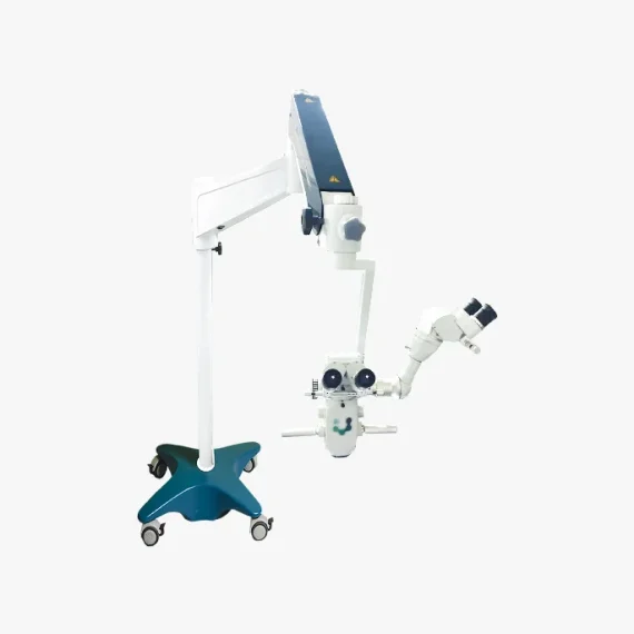
The FOCUS series is a line of equipment specially designed in detail to meet the most demanding standards of anterior and posterior segment surgical procedures.The optics developed for FOCUS line microscopes enable external transmission of LED light, generating excellent clarity and image depth of the observed field.
Ideal for Cataract procedures.
Delivery & Availability:
Typically 10-21 working days – excluding furniture and heavy/bulky equipment. Please contact us for further information.
| Shipped from abroad
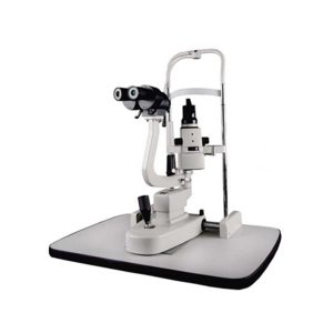
- Top Optical System.
- High eyepoint, comfort.
- Five steps drum zoom design, easier to use 6x, 10x, 16x, 25x, 40x magnification.
- High Definition Eyepieces, More Comfortable for Viewing.
Delivery & Availability:
Typically 14 working days – excluding furniture and heavy/bulky equipment. Please contact us for further information.
| 83Shipped from abroad
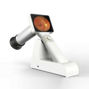
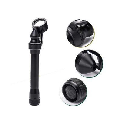
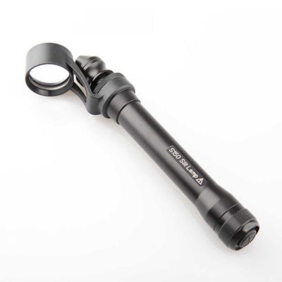
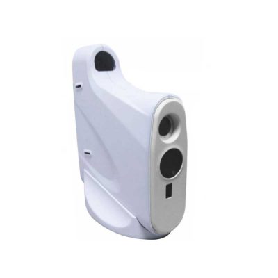
This is a portable medical camera for fundus imaging, diagnosis, and especially for fundus disease screening. It's compact, easy to obtain high definition fundus image. It can be conveniently applied to rapid screening, out diagnosis, bedside diagnosis and remote medical treatment, etc.
Delivery & Availability:
Typically 14 working days – excluding furniture and heavy/bulky equipment. Please contact us for further information.
| Shipped from abroad
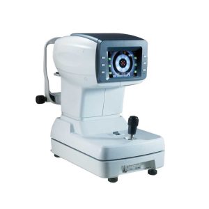
- Light rack with integral casting, CNC machining, the measuring system is more stable, good consistency.
- Color LCD display, 5.7 inches man-machine interface is more comfortable.
- PD automatic measurement function, automatically PD value.
Delivery & Availability:
Typically 14 working days – excluding furniture and heavy/bulky equipment. Please contact us for further information.
| Shipped from abroad
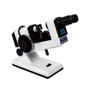
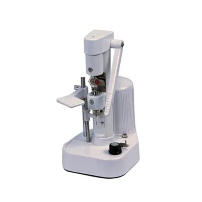
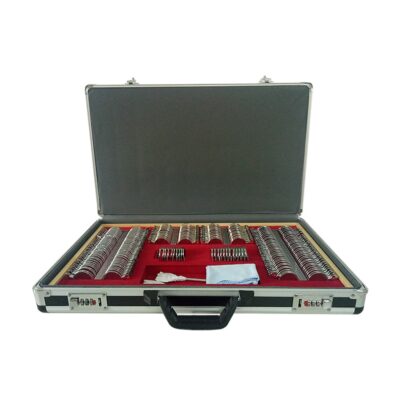
The NJC-4 Lensmeter is used to measure the diopters of spherical lens and cylinder lens, the axis of cylinder lens, the strength and the baseline direction of shuttle lens, it can also stamp the optical center of lens, the axis of cylinder len and the base direction of shuttle lens.
Delivery & Availability:
Typically 14 working days – excluding furniture and heavy/bulky equipment. Please contact us for further information.
| Ship from abroad
- Easy installation and maintenance: It is quite easy to replace light bulks, and unnecessary to adjust its focus and position.
- Customer Programmable: Icons can be displayed in predefined sequence by programming.
- Display Single Icon: Display single icon by multi-function mask plate.
- Tilted Placement: Perfect image position can be obtained by adjusting the horizontal axis of projector.
- Standards parts: Metal plate with polarized coating (410x 410mm)
Delivery & Availability:
Typically 14 working days – excluding furniture and heavy/bulky equipment. Please contact us for further information.
|
| Content | Description
Versatility in multiple configuration possibilities
Focus Microscopes stand out in the market for presenting multidisciplinary characteristics and high optical quality. Additionally, they allow you to add options and accessories according to the need for use, including binoculars, double iris, image inverter, among others.
The Focus* line of microscopes has a pedal-operated motorized microfocusing and zooming system, aiming for greater precision and comfort for the user during surgical procedures.
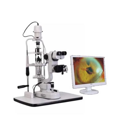
| Slit Lamp Features:
- Top Optical System.
- High eyepoint, comfort.
- Five steps drum zoom design, easier to use 6x, 10x, 16x, 25x, 40x magnification.
- High Definition Eyepieces, More Comfortable for Viewing.
Technical Specifications:
| Microscope Type |
Galileo Parallel |
| Optics |
Super Optic System |
| Magnification Change Way |
Drum Rotation |
| Eyepiece Magnification |
12.5x |
| Total Magnifications |
6x 10x, 16x, 25x, 40x |
| Diopter Adjustment |
-5D ~+5D |
| Slit Width |
0-14MM Continuous |
| Slit Height |
1-14MM Continuous |
| Slit Angle |
0°- 180° Adjustable |
| Light Source |
LED |
| Light Spot Diameter |
0.2mm, 2mm, 3mm, 5mm, 10mm, 14mm |
| Filter |
Heat Absorption; Grey; Redfree; Cobalt Blue |
| Fixation |
Red LED |
| Electrical |
|
| Illumination Bulb |
LED |
| Input Voltage |
110V/220V (±10%) |
| Applanation Tonometer Interface |
Included |
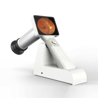
| Portable Fundus Camera is a portable medical camera for fundus imaging, diagnosis, and especially for fundus disease screening. It's compact, easy to obtain high definition fundus image. It can be conveniently applied to rapid screening, out diagnosis, bedside diagnosis and remote medical treatment, etc.
Features of Portable Fundus Camera:
- Images real-time display, one-button magnifying
- Non-mydriatic and high definition imaging.
- Micro SD memory card up to 32GB for 80,000 images
- USB and WiFi connection
- Low weight 450g, movable and portable
- A rechargeable battery provides up to more than 4 hours of continuous operation.
- Optional imaging modules for examination: posterior ophthalmoscope, anterior segment ophthalmoscope, otoscope, rhinoscope, laryngoscope and dermatoscope
- High definition and stable image
- Easy one-handed operation and excellent portability
- Large data service and a variety of image acquisition mode
Technical Specifications of Portable Fundus Camera:
| Model |
MC-600 |
| FOV |
45° |
| Minimum pupil |
2.5mm |
| Refractive compensation |
-20D~+20D |
| Light source |
White LED/IR |
| Image resolution |
1920×1080 |
| Focus Mode |
Manual |
| Screen |
3.5" color |
| Power |
≤6VA |
| Storage |
8GB Micro SD card |
| Power supply |
3.7VLithium Battery |
| Interface |
Mini USB/Wifi |
| N. Weight |
450g(Typical) |
| G. Weight |
2.5kg |
| Size |
160mm*90mm*190mm |
| Packing size |
360mm*310mm*160mm |
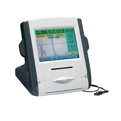
| Features:
- Using the ARM processor and the latest domestic image processing, the system is fast and the image is clear.
- Japan’s mature optical path system, humanized automatic mist measurement process, to reduce the error caused by adjustment, more precise measurements.
- Light rack with integral casting, CNC machining, the measuring system is more stable, good consistency.
- Color LCD display, 5.7 inches man-machine interface is more comfortable.
- PD automatic measurement function, automatically PD value.
- One key lock function, quickly locking mobile platforms.
- Innovative designs, according to the structure of human body engineering, to bring customers good aesthetic feeling and comfortable.
- Data transmission with CV7200, improved efficiency of online 5x magnification options, respond to different viewing needs.
Technical Specifications:
Measurement Range
Vertex distance.......................................0.0,12.0,13.75,15mm
Sphere.......................................-20D~+20D(0.12/0.25D step)
Cylinder...........................................0~±10D(0.12/0.25D step)
Axis...............................................................1~180°(1° step)
PD distance............................................................30~85mm
PD..........................................................................10~86mm
Minimum pupil diameter..............................................Ø2.0mm
Target................................................automatic fogging target
Hardware specifications
Monitor.............................................................5.7" color LCD
Printer......................................................Fast thermal printer
Power saving function....................OFF, 5/15 mins (selectable)
Power supply................................AC110~220V, 60/50Hz, 50W
Dimension/weight..............................288x500x480mm(14kgs) | The NJC-4 Lensmeter is used to measure the diopters of the spherical lens and cylinder lens, the axis of cylinder lens, the strength and the baseline direction of shuttle lens, it can also stamp the optical center of the lens, the axis of cylinder lens, and the base direction of shuttle lens. This instrument (Both Ac and Dc are permitted Two cells When Dc) has clear readings and graduations as well as high objective precision and reliabilities except that it can be operated easily and conveniently, AU the lenses and the made glasses can be measured by it, therefore, it is a required and ideal measuring instrument for glasses manufactures, glasses stores and ophthalmological hospitals.
Technical Specifications:
- Eyepiece focusing range: ±5D
- The top point diopter measuring range: 0—±20D
- The minimum measuring square value: 0.125D
- The shuttle lens measuring range: 5A
- The shuttle lens square value: 1A
- The cylinder lens axial range: 0—180°
- The cylinder lens axial square value: 5°
- The maximum diameter of the measured lenses l00m/m
- Power supply: AC 110V-6V DC 3V
- Note: This item is AC 110V; The battery is not included!
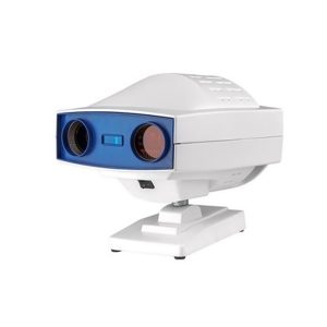
| Digital Chart Projector-Features:
- Easy installation and maintenance: It is quite easy to replace light bulks, and unnecessary to adjust its focus and position.
- Customer Programmable: Icons can be displayed in predefined sequence by programming.
- Display Single Icon: Display single icon by multi-function mask plate.
- Tilted Placement: Perfect image position can be obtained by adjusting the horizontal axis of projector.
- Standards parts: Metal plate with polarized coating (410x 410mm)
Technical Specifications of Digital Chart Projector:
|
Projection Distance |
1.5m~6m |
|
Projection Magnification |
30x (at 5m) |
|
Projection Size |
330x270mm (at 5 m) |
|
Chart Switching Speed |
One chart per 0.03s |
|
Mask Switching Speed |
1 open, 5 horizontal different lines, 8 vertical lines, 21 single letters, 1 red/green |
|
Program |
2 sets programs, each program contains up to 30 steps |
|
Speed of mask conversion |
One mask per 0.03s |
|
Lamp |
LED lamp |
|
Auto-off function |
After 10 minutes idle time |
|
Power Source |
AC 220V, 50Hz or 110V, 60Hz |
|
Power consumption |
40W |
|
Accessories |
Remote control, polarized metal screen, halogen lamp, polarized glasses, fuses (2), batteries (2) |
|


Reviews
There are no reviews yet.