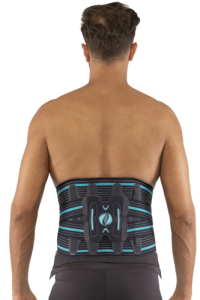
Lumbomove Basic Duo

$0.00

SF103356013086-22
Brands:: Vizor

Description

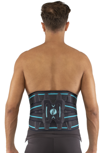
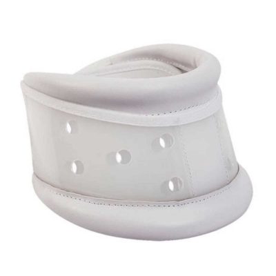
  • It is made of flexible, breathable and sweat-proof elastic fabric.
  • There are 4 steel underwires at the back, suitable for the anatomical structure.
  • Steel underwires can be tilted manually according to lumbar lordosis.
  • The front is in two parts. The wide lower part supports the lower part of the abdomen.
  • The narrow piece is worn diagonally in the front.
  • It reduces pain by alleviating the load on the discs in the lumbar region.

Reviews

There are no reviews yet.

Be the first to review “Lumbomove Basic Duo”

Your email address will not be published. Required fields are marked *

Quick Comparison

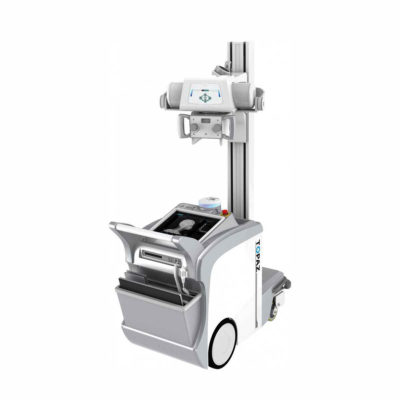
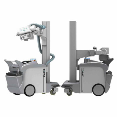
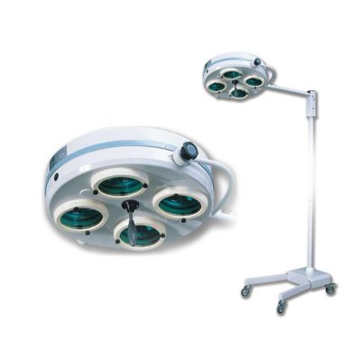
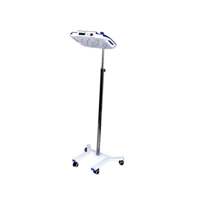
SettingsLumbomove Basic Duo removeDrGem Floor Mounted Analogue X-ray removeDrGem Ceiling Mounted Digital X-ray removeIBIS Neeo R9 Digital Surgical C-Arm removeKeewell FT-2800 Blood & Infusion Warmer removeDRGEM DR System remove
NameLumbomove Basic Duo removeDrGem Floor Mounted Analogue X-ray removeDrGem Ceiling Mounted Digital X-ray removeIBIS Neeo R9 Digital Surgical C-Arm removeKeewell FT-2800 Blood & Infusion Warmer removeDRGEM DR System remove
ImageDrGem GXR-S 400mA Floor Mounted Analogue X-ray MachineDrGem GXR-SD 400mA Ceiling Mounted Digital X-ray MachineIBIS Neeo R9 Digital Surgical C-ArmDrGem Digital Radiography (DR) System
SKUSF103356013086-22SF1033560074-6SF1033560074-4SF1033560011-1SF1033560075SF1033560074-8
Rating
Price

$0.00

$26,924.00

$68,468.00

$0.00

$0.00

$0.00

Stock
Availability
Add to cart

DescriptionIn Stock GXR Analogue X-ray system matches with a radiographic room which perfectly fits your workow and can be easily upgraded to DR system with the help of DR interface and PC interface in GXR generator as well as Bucky suitable to Flat Panel Detector. GXR X-ray system is equipped with a high frequency X-ray generator which consistently produces high quality radiograph in favor of high quality X-ray output with a very small kV ripple and accurate mA and mAs. GXR X-ray system is designed to provide convenience to operator and comfort to patient. Delivery & Availability: Typically 21 working days – excluding furniture and heavy/bulky equipment. Please contact us for further information.In Stock The GXR-SD is a diagnostic digital radiography system that provides reliable high quality digital radiographic images with a reduced dose. The GXR-SD DR systems offer comprehensive digital solutions to all radiography needs, featuring ACQUIDR digital imaging system with stationary or portable digital flat-panel detectors as well as reliable high-frequency x-ray generators that are known worldwide for their excellent performance, lifetime and stability. Patient tables and wall stands are also offered. Delivery & Availability: Typically 21 working days – excluding furniture and heavy/bulky equipment. Please contact us for further information.Shipped from Abroad Our Neeo “C” arms are easy to place, use and are specifically designed to be used in orthopedics, traumatology, abdominal surgery, urology, cardiology and operating rooms. Delivery & Availability: Typically 21 working days – excluding furniture and heavy/bulky equipment. Please contact us for further information.Shipped From Abroad
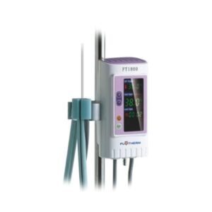
Technology of control: Microprocessor integrated with temperature sensors provides high precise control Safety system: Permanent running self-tests, 24 hours continuous operating Double independent over-heating protections and automatic cut off Visual and acoustic alarm for high temperature / low temperature/sensor fault Dual channels: Each channel can work independently with own temp. setting and temp control Two channels can be combined to enlarge the warming capacity Heating up to the patient: IV tube is completely wrapped in, no heat loss Delivery & Availability: Typically 7-14 working days – excluding furniture and heavy/bulky equipment. Please contact us for further information.
Ship from abroad ACQUIDR is the digital imaging system composed of a Flat Panel Detector(FPD) and an imaging workstation with software. The digital FPD and full-feature imaging software with excellent digital image processing, designed for DRGEM X-ray machine. Delivery & Availability: Typically 21 working days – excluding furniture and heavy/bulky equipment. Please contact us for further information.
Content
  • It is made of flexible, breathable and sweat-proof elastic fabric.
  • There are 4 steel underwires at the back, suitable for the anatomical structure.
  • Steel underwires can be tilted manually according to lumbar lordosis.
  • The front is in two parts. The wide lower part supports the lower part of the abdomen.
  • The narrow piece is worn diagonally in the front.
  • It reduces pain by alleviating the load on the discs in the lumbar region.
DrGem GXR Floor Mounted Analogue X-ray system matches with a radiographic room which perfectly fits your workflow and can be easily upgraded to DR system with the help of DR interface and PC interface in GXR generator as well as Bucky suitable to Flat Panel Detector. GXR (Analogue X-ray)system is equipped with a high frequency X-ray generator which consistently produces high quality radiograph in favor of high quality X-ray output with a very small kV ripple and accurate mA and mAs. GXR (Analogue X-ray) system is designed to provide convenience to operator and comfort to patient. Features of DrGem GXR Floor Mounted Analogue X-ray:
  • 4 way Tabletop Patient Table (PBT-4) A large tabletop with extended travel enables all radiography studies with minimal patient movement, and supporting patient weight up to 300kg. Fully at tabletop without a frame on the edge makes cleanliness and odors free
  • Floor Mount Tube Stand (TS-FM6) Floor Rail type tube stand provides all radiography studies with smooth movement on the rail.
  • Wall Bucky Stand (WBS) Elegant design, durable and easy-to-use Wall Bucky Stand provides full satisfaction.
Technical Specifications of DrGem GXR Floor Mounted Analogue X-ray:
  • Power Rating - 32KW
  • Generator - GXR-32S
  • Rotor - Dual Speed Starter(DSS)
  • Input Power - 400/480VAC, Three phase
  • Line Frequency - 50/60Hz
  • X-ray tube - DXT-12M, (0.6/1.2mm, 300kHU)
  • Tube Voltage - 40 to 150kV, 1kV Step
  • Tube Current – 10 to 640mA
  • Output - 640mA@81kV,  500mA@104kV, 400mA@130kV, 320mA@150kV
  • Time Range - 1ms to 10s
  • mAs Range - 0.1 to 800mAs
  • Reproducibility - Coe­cient of Variation : kV < 0.005, Time < 0.005,mAs < 0.01
  • Accuracy - kV < ±(1%+1kV), mA < ±(3%+1mA), Time <±(1%+0.5ms), mAs < ±(3%+0.1mAs)
  • Linearity - Coe­cient of Linearity < 0.01 : CL = (X1-X2)/(X1+X2), where X is mR/mAs
 
Click Here To Download Catalogue
DrGem Ceiling Mounted Digital X-ray is a diagnostic digital radiography system that provides reliable high quality digital radiographic images with a reduced dose. The GXR-SD DR systems offer comprehensive digital solutions to all radiography needs, featuring ACQUIDR digital imaging system with stationary or portable digital flat-panel detectors as well as reliable high-frequency x-ray generators that are known worldwide for their excellent performance, lifetime and stability. Patient tables and wall stands are also offered. Features:
  • TS-CSA-A (Vertical movement, 1.6m stroke, rail length 3x4meter) including HV cable 15m
  • WBS-TA: Vertical movement
  1. V Stroke:1,450mm in Uprigh Bucky Position,
  2. 1,526mm in Horizontal Bucky position.
  • PBT-4 is a 4 way Floating Tabletop. A large tabletop with extended travel enables all radiography studies with minimal patient movement. Fully fat tabletop without a frame on the edge makes cleanliness and odors free
  • Digital Flat Panel Detector (FPD) – Wireless 17X14 (Csl, 4336W) with Auto Exposure Detection (AED) function, there is no DR trigger cable between detector and generator.
  • Full Featured Imaging Software & Excellent Digital Image Processing:
  1. Provides convenient user interface and easy operation
  2. Anatomical view-based digital image processing automatically optimizes and enhances the quality of the captured image for the pictured anatomy.
  3. Radiographic stand & automatic collimator control function
  4. DICOM 3.0 networking interface includes Worklist, Print, Store, Query for integration with any PACS or RIS
  • Included – Software, HP Laptop Computer
  1. CPU≥3.2GHz
  2. Memory capacity:≥4GB
  3. Hard drive capacity :≥500 GB
  4. Resolution: 1280 x 1024
  5. Display size: 21 inch color LCD screen
  6. 64 bit Windows 10 operation system
  7. Core: i5
  Technical Specification:
  • Power Rating - 32KW
  • Generator - GXR-32S
  • Rotor - Dual Speed Starter(DSS)
  • Input Power - 400/480VAC, Three phase
  • Line Frequency - 50/60Hz
  • X-ray tube - DXT-12M, (0.6/1.2mm, 300kHU)
  • Tube Voltage - 40 to 150kV, 1kV Step
  • Tube Current – 10 to 640mA
  • Output - 640mA@81kV,  500mA@104kV, 400mA@130kV, 320mA@150kV
  • Time Range - 1ms to 10s
  • mAs Range - 0.1 to 800mAs
  • Reproducibility - Coe­cient of Variation : kV < 0.005, Time < 0.005,mAs < 0.01
  • Accuracy - kV < ±(1%+1kV), mA < ±(3%+1mA), Time <±(1%+0.5ms), mAs < ±(3%+0.1mAs)
  • Linearity - Coe­cient of Linearity < 0.01 : CL = (X1-X2)/(X1+X2), where X is mR/mAs
  • Mechanical Parts:
-TS-CSA-A (Vertical movement, 1.6m, stroke rail length 3x4meter) including HV cable 15m - PBT-4: 4 way Floating Tabletop. - WBS-TA: a. Vertical    movement
  1. V Stroke:1,450mm in Upright Bucky
  2. Position, 1,526mm in Horizontal Bucky position.
- HVC-15: 15M HV cable - Auto Collimator  
Click Here To Download Catalogue
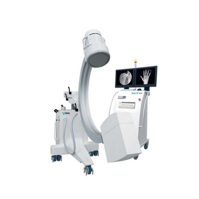
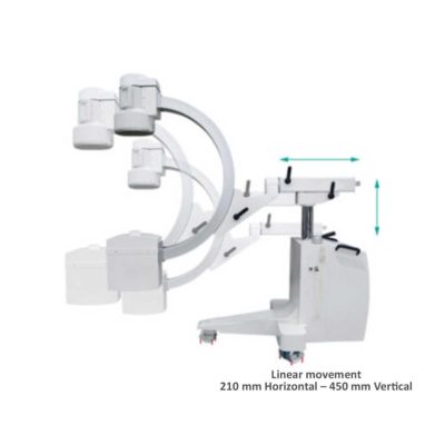
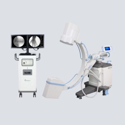
Our Neeo “C” arms are easy to place, use and are specifically designed to be used in orthopedics, traumatology, abdominal surgery, urology, cardiology and operating rooms. Using Neeo with the RTP (Real Time Processing) option it is possible to perform vascular, urological and cardiological diagnostics. One of the main functions, digital image subtraction, allows to see, as an example, the passage of contrast liquids in a tissue or in a venous or arterial duct; thanks to the possibility of looping, the acquired video can be reproduced several times to monitor more accurately the passage of the fluid within the area in question. Angiographic measurement is another useful function in the vascular field (QA Quantitative Angiography) that allows the measurement of stenoses. Finally, fluoroscopy allows the correct positioning of stents or expanders. Neeo is used in various interventional and diagnostic procedures in traumatology and orthopedics wards and operating rooms as well. Thanks to low-dose fluoroscopy, it is possible to use the device for positioning bone or subcutaneous grafts, inserting K-wire (Kirschner wire) for stabilization of bone fragments or for the correct positioning of prostheses. The low dose emitted ensures safe use for both the patient and the surgeon or doctor on the operating field. On the control panel there is a large touch screen display that allows to adjust the basic functions of the equipment. From this display it is possible to select and adjust the fluoroscopic data for the examination, activate or deactivate the laser pointer, select between pulsed, one shot or standard fluoroscopy, rotate the image and perform all operations on collimator. The four side buttons on the display offer the possibility to move the bow vertically thanks to an extremely silent motor. Neeo has two 19 “medical grade monitors that can be positioned according to the needs of the medical practitioner. Work monitors and feedback monitors are separated to be managed independently. The possible movements are: rotation, revolution, tilting and possibility of height adjustment. Features:
  • Continuous fluoroscopy
  • Pulsed fluorography
  • One Shot fluoroscopy
  • Low dose fluoroscopy
  • Radiography with cassette (up to 5KW)
  • 110,000 images of memory (Standard)
  • 19 ″ Medical Flat Monitor (X2)
  • Touch control panel
  • Image Processing – 1K X 1K, 16 bit
  • DAP-Dose Area Meter
  • DSA Road Mapping
  • Laser Marker
Technical Specification:
  • Power: 5KW
  • kV Range (kV): 40 - 120
  • mAs Range (mAs): 1 - 250
  • SID (mm): 1000
  • Free Space (mm): 730
DICOM Feature:
  • RTP-SW DSA
  • RTP-SW QA
  • RTP-SW Worklist
  • RTP-SW Print
  • RTP-SW CD DVD Saving
  • RTP-SW MPPS
  • RTP-SW Query/Retrieve
  • RTP-SW HCF
  • RTP-SW BASE
  • RTP-SW STCOMMIT
Configuration: C-Arm Structure, Trolley for Monitor, Collimator, Monoblock, Toshiba 12” Image Intensifier, 19″ Philips Medical Flat Monitor (X2), Camera CD1030CA 1KX1K, Sony Printer UP-X898MD, Cassette Holder
Click Here To Download Catalogue
Features:
Technology of control: Microprocessor integrated with temperature sensors provides high precise control
Safety system:
Permanent running self-tests, 24 hours continuous operating Double independent over-heating protections and automatic cut off Visual and acoustic alarm for high temperature / low temperature/sensor fault
Dual channels:
Each channel can work independently with own temp. setting and temp control Two channels can be combined to enlarge the warming capacity Heating up to the patient: IV tube is completely wrapped in, no heat loss
User friendly interface:
Big LED screen showing set temp., actual temp., heating time and fault situation Easy and quick to set up, ready to use within minutes. Open system: Accepts standard IV tube, no special disposables needed The most economical warming solution without extra consumable costs TECHNICAL SPECIFICATIONS Model: FT2800 Temperature setting: 33 - 41˚C Power supply: a.c.100~240V/50~60Hz Power consumption: Max. 180VA Type of protection against electric shock: Class I Degree of protection against electric shock: BF Applied part; Defibrillation-protected Degree of protection against ingress of liquids: IPX2 Temperature accuracy: ±1.0˚C Overheat protection: 42˚C /43˚C Low temperature alarm: 32˚C Warming up time: From 20˚C to 36˚C approx.2 min. Operating mode: Continuous Dimension (W*D*H): 200×130×250mm Net Weight: 2.3kg Warming profile:  - length 1400mm - compatible IV tube 3.5-5.0mm O.D.  
Click Here To Download Catalogue 
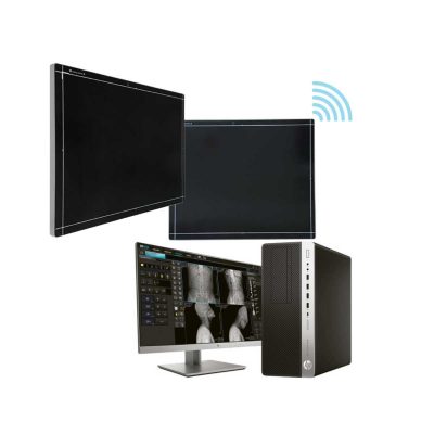
DRGEM ACQUIDR (DRGEM DR System) is the digital imaging system composed of a Flat Panel Detector(FPD) and an imaging workstation with software. The digital FPD and full-feature imaging software with excellent digital image processing will meet all your needs in the diagnostic digital radiographic field. Features of DRGEM DR System:
  • Adaptable with most existing analog x-ray systems
  • AED(Auto Exposure Detection) function available
  • Simple and convenient installation
  • Intuitive graphic user interface
  • Over 1300 pre-loaded exam profiles with DRGEM’s generator
  • Configurable automatic study
  • PMS / EMR /HIS / RIS work list support
  • Multiple printing formats
  • DICOM 3.0 compliant
  • Brightness and contrast adjustment
  • Rotation
  • Horizontal and vertical flip
  • Zoom and pan
  • Left / right makers
  • Optimized work flow
  • Automatic shutters
  • Support DAP interface
  • Exposure index (EI)
  • DR upgrade solution by retrofit.
  • Portable and Wireless FPD DR solution for maximum flexibility in virtually all general radiographic specialties.
  • Portable and wireless detector fits into most existing analogue x-ray system.
  • Faster workflow after DR upgrade.
  • With AED(Auto Exposure Detection) function, there is no DR trigger cable between detector and generator providing very easy DR upgrade.
  • Easy to interface with any kind of x-ray generator.
  • Radmax acquisition workstation
  • Turns any analogue X-Ray system into a fully DR system
  • Optional image stitching program
  • Vet software available
  • Detector format: 17×17” / 17×14", wired/wireless
WeightN/AN/AN/AN/AN/AN/A
DimensionsN/AN/AN/AN/AN/AN/A
Additional information
Select the fields to be shown. Others will be hidden. Drag and drop to rearrange the order.
  • Image
  • Rating
  • Price
  • Description
Click outside to hide the comparison bar
Compare