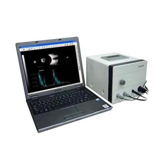
Ophthalmic AB Scan Machine
Ask for Price$0.00
Shipped from abroad
Features:
1. Capable of capturing both still images and video clips, provides excellent image resolution with full set of features
2. Easy to use measurement calipers for distance and angle measurements
3. Frame-by-frame review of video clips provided for selection of optimal image
4. Patient database and image library is provided with printing and download capabilities
Delivery & Availability:
Typically 14 working days – excluding furniture and heavy/bulky equipment. Please contact us for further information.
Description
1. Capable of capturing both still images and video clips, provides excellent image resolution with full set of features
2. Easy to use measurement calipers for distance and angle measurements
3. Frame-by-frame review of video clips provided for selection of optimal image
4. Patient database and image library is provided with printing and download capabilities
Technical Specifications:
A probe Detecting
1.Probe Frequency: 10MHZ Error±0.5 MHZ
2.Precision: 0.05mm
3.Measurement parameters: ACD depth, lens, axial length, and its average
4.IOL Calculation: SRK/II, SRK/T, Holladay, SCDK, Hoffer-Q
5.Data Processing: IOL table
6.Operation: Automatic, Manual
7.Information storage: Mass memory Build-in Case data
8.Images reports: External high definition ink-jet printer
B probe Detecting
1.B probe mode: Mechanic Sector scans
2.Scan angle: 53°
3.Detect Depth: 50mm
4.Probe frequency: 10 MHZ Error±0.5 MHZ
5.Resolution: Vertical≤0.5mm, Lateral≤1.0mm
6.Gray Scale: 256
7.Display Mode: B; B+B; B+A
8.Storage: 10 Frames
9.Image Post Processing: Brightness, Contrast, Smooth, Sharp, stretch, Equalize, etc.
Quick Comparison
| Settings | Ophthalmic AB Scan Machine remove | Sonoscape P15 Ultrasound Machine With Four Probes remove | DrGem Ceiling Mounted Digital X-ray remove | Sonoscape S11 Ultrasound Machine remove | Sonoscape P20 Ultrasound Machine remove | ASPEL AsPEKT 712 Holter Monitor and Software remove |
|---|---|---|---|---|---|---|
| Name | Ophthalmic AB Scan Machine remove | Sonoscape P15 Ultrasound Machine With Four Probes remove | DrGem Ceiling Mounted Digital X-ray remove | Sonoscape S11 Ultrasound Machine remove | Sonoscape P20 Ultrasound Machine remove | ASPEL AsPEKT 712 Holter Monitor and Software remove |
| Image |  |  |  |  |  |  |
| SKU | SF1033560107-5 | SF1033560012-8 | SF1033560074-4 | SF1033560012-1 | SF1033560012-9 | SF1033560075-4 |
| Rating | ||||||
| Price | Ask for Price | $13,900.00 | $68,468.00 | $6,950.00 | Ask for Price | Ask for Price |
| Stock | ||||||
| Availability | ||||||
| Add to cart | ||||||
| Description | Shipped from abroad
Features:
1. Capable of capturing both still images and video clips, provides excellent image resolution with full set of features
2. Easy to use measurement calipers for distance and angle measurements
3. Frame-by-frame review of video clips provided for selection of optimal image
4. Patient database and image library is provided with printing and download capabilities
| In Stock A feature-rich system inheriting the Wi-Sono high-end platform, the P15 uses an array of advanced tools to help enhance the image quality. It's a cost-effective, simplified console with an intuitive user interface and multiple intelligent functions. Delivery & Availability: Typically 2 working days – excluding furniture and heavy/bulky equipment. Please contact us for further information. | In Stock The GXR-SD is a diagnostic digital radiography system that provides reliable high quality digital radiographic images with a reduced dose. The GXR-SD DR systems offer comprehensive digital solutions to all radiography needs, featuring ACQUIDR digital imaging system with stationary or portable digital flat-panel detectors as well as reliable high-frequency x-ray generators that are known worldwide for their excellent performance, lifetime and stability. Patient tables and wall stands are also offered. Delivery & Availability: Typically 21 working days – excluding furniture and heavy/bulky equipment. Please contact us for further information. | In Stock A Value Choice beyond Your Expectation. SonoScape’s trolley color Doppler system S11 redefines price and performance with practical design. The S11 will go beyond your expectations but not your budget. Delivery & Availability: Typically 2 working days – excluding furniture and heavy/bulky equipment. Please contact us for further information. | Shipped from Abroad Incorporating innovative technologies, P20’s user-friendly design with a simple operation panel, intuitive user interface and a variety of intelligent auxiliary scanning tools, will significantly improve your daily examination experience. Besides general imaging applications, P20 has entitled with diagnostic 4D technology which has an extraordinary performance in obstetrics and gynecology applications. Delivery & Availability: Typically 5-7 working days – excluding furniture and heavy/bulky equipment. Please contact us for further information. | Shipped from Abroad The Holta Monitor allows quick analysis of ECG examination and detection, reviewing and editing capability in the qualitative assessment of VE, VT, Single SVE, PSVT, Pauses, Irregular Rhythm, VT, IVR, Brady - and Tachycardia, Couplets, ST-segment elevation and depression, Maximum, Minimum and averaged Heart Rates, artifacts Delivery & Availability: Typically 10 working days – excluding furniture and heavy/bulky equipment. Please contact us for further information. |
| Content | Features:
1. Capable of capturing both still images and video clips, provides excellent image resolution with full set of features
2. Easy to use measurement calipers for distance and angle measurements
3. Frame-by-frame review of video clips provided for selection of optimal image
4. Patient database and image library is provided with printing and download capabilities
Technical Specifications:
A probe Detecting
1.Probe Frequency: 10MHZ Error±0.5 MHZ
2.Precision: 0.05mm
3.Measurement parameters: ACD depth, lens, axial length, and its average
4.IOL Calculation: SRK/II, SRK/T, Holladay, SCDK, Hoffer-Q
5.Data Processing: IOL table
6.Operation: Automatic, Manual
7.Information storage: Mass memory Build-in Case data
8.Images reports: External high definition ink-jet printer
B probe Detecting
1.B probe mode: Mechanic Sector scans
2.Scan angle: 53°
3.Detect Depth: 50mm
4.Probe frequency: 10 MHZ Error±0.5 MHZ
5.Resolution: Vertical≤0.5mm, Lateral≤1.0mm
6.Gray Scale: 256
7.Display Mode: B; B+B; B+A
8.Storage: 10 Frames
9.Image Post Processing: Brightness, Contrast, Smooth, Sharp, stretch, Equalize, etc. | DETAILS
Super Wide-bandwidth Platform
Inheriting Wi-sono's ultra-wide system platform and with the advanced probe technology, high-resolution and deep penetration images are provided for precision medicine.
Spatial Compound Imaging
Spatial Compound Imaging utilizes several lines of sight for optimal contrast resolution, speckle reduction and border detection, with which P15 is ideal for superficial and abdominal imaging with better clarity and improved continuity of structures.
μ-Scan+
The new generation μ-Scan imaging technology gives you better image quality by reducing noise, improving signal strength and improving visualization.
Dynamic Color
Dynamic color improves upon already existing color Doppler technologies for a clearer capture of color flow and detailed visualization of even tiny veins with lower velocities.
Real-time Panoramic
With real-time panoramic, you can acquire an extended field of view for large organs or long vessels for easy measurement and diagnostic efficiency. Accomplished in real-time for the convenience of the sonographers, any mistake can also be easily back tracked and corrected without interrupting the scan.
3D/4D
Outstanding volume performance with speed and convenience makes P15 outshine others on volume imaging.
Tissue Doppler Imaging
Tissue Doppler Imaging allows clinical doctors to quantitatively evaluate local myocardial movements and functions, facilitating them with the ability to analyze and compare the motions of the different parts of the patient's heart.
Auto IMT
Quick measurement of intra-media vessel thickness ensures good reproducibility and high diagnostic efficiency.
Click Here To Download Catalogue | DrGem Ceiling Mounted Digital X-ray is a diagnostic digital radiography system that provides reliable high quality digital radiographic images with a reduced dose. The GXR-SD DR systems offer comprehensive digital solutions to all radiography needs, featuring ACQUIDR digital imaging system with stationary or portable digital flat-panel detectors as well as reliable high-frequency x-ray generators that are known worldwide for their excellent performance, lifetime and stability. Patient tables and wall stands are also offered.
Features:
Click Here To Download Catalogue | DETAILS
SonoScape’s trolley colour Doppler system S11 redefines price and performance with practical design. The S11 will go beyond your expectations but not your budget. As an easy-to-use ultrasound system, the S11 is integrated with a new software platform, especially optimized for a smooth workflow and convenient operation. The system speeds up the exam process and makes file management easier.
SPECIFICATION
- 15-inch high definition LCD monitor with articulating arm
- Compact and agile trolley design
- 3 active transducer sockets available for a wide range of applications
- Duplex, Color Doppler, DPI, PW Doppler, tissue harmonic imaging, μ-scan speckle reduction imaging, compound imaging, trapezoidal imaging
- Customized settings based on your own working style
- Full patient database and image management solutions
Click Here To Download Catalogue | DETAILS
Upgraded Images with More Clarity
SonoScape never stops making progress in improving the image quality of its ultrasound products to enhance the confidence of diagnosis for doctors. With extraordinary images provided by P20, the anatomy structures are clearer than ever.
C-Xlasto Imaging
With C-xlasto Imaging, P20 enables comprehensive quantitative elastic analysis. Meanwhile, C-xlasto on P20 is supported by linear, convex and transvaginal probes, to ensure good reproducibility and highly consistent quantitative elastic results.
S-Live
S-Live allows for detailed visualization of subtle anatomical features, thereby enabling intuitive diagnosis with real-time 3D images and enriching patient communication.
Pelvic Floor 4D
Transperineal 4D pelvic floor ultrasound can provide useful clinical values in assessing the vaginal delivery impact on the female anterior compartment, judging whether the pelvic organs are prolapsed or not and the extent, determining if the pelvic muscles were torn accurately.
Anatomic M Mode
Anatomic M Mode helps you observe the myocardial motion at different phases by freely placing sample lines. It accurately measures the myocardial thickness and the heart size of even difficult patients and supports the myocardial function and LV wall-motion assessment.
Tissue Doppler Imaging
P20 is endowed with Tissue Doppler Imaging which provides velocities and other clinical information on myocardial functions, facilitating clinical doctors with the ability to analyze and compare the motions of different parts of the patient's heart.
Click Here To Download Catalogue | The Holter Monitor allows quick analysis of ECG examination (arrhythmias and ST segment).
Technical specifications:
HolCARD 24W Software:
Click Here To Download Catalogue |
| Weight | N/A | N/A | N/A | N/A | N/A | N/A |
| Dimensions | N/A | N/A | N/A | N/A | N/A | N/A |
| Additional information |


Reviews
There are no reviews yet.