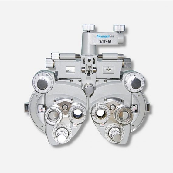
View Tester (Manual Phoropter)
Ask for Price$0.00
Ship from abroad
- Equipped with comprehensive measuring functions, it provides SPH, CYL, AXIS and pupil distance optometry
- Durable and easy to operate
- Easily and intuitively read the sphere focal scale value
- High eco-friendly materials
- Design fitting the face curve and no stimulation
Delivery & Availability:
Typically 14 working days – excluding furniture and heavy/bulky equipment. Please contact us for further information.
Description
Features:
- Equipped with comprehensive measuring functions, it provides SPH, CYL, AXIS and pupil distance optometry
- Durable and easy to operate
- Easily and intuitively read the sphere focal scale value
- High eco-friendly materials
- Design fitting the face curve and no stimulation
- Easy to take and clean
- Free switch between the cross-cylindrical lens and the rotary prism
- When the rotating risk is turning by the sphere, it can make sphere power adjust 3.00D for big scope.
- It is designed expediently and smartly for a particular cross cylinder. Supporting supplementary lens could increase scope of measurement.
Technical Specifications:
|
Sphere
|
Range:-19.00~+16.75m-1 Step: 0.25m-1, 3.00m-1
|
||
|
Cylinder
|
Range: 0.00~-6.00m-1(Measuring Range With Accessories0.00~-8.00m-1) Step: 0.25m-1
|
||
|
Cylinder Axis
|
Range: 0~180°, Step:5°
|
||
|
Distance of Optical center (also known as Pupil)
|
Range: 50~75mmStep: 1mm
|
||
|
Sight Switch
|
Range:∞~380mm (distance of Optical center is64mm)
|
||
|
Front Chin Test
|
Range: 0~16mm
|
||
|
Distance (from cornea vertex to the lens surface)
|
16mm
|
||
|
Standard Accessories Lens
|
two pieces of Auxiliary Cylinder -2.00m-1 and -0.12m-1 respectively
|
||
|
Standard Accessories
|
one piece of M2 Hexagon wrench , one piece of a Myopia Standard Card, two piece of Myopia Standard Card , one piece of standard card holder , a dust cover
|
||
|
Auxiliary Lens
|
“O”:Open aperture “R”:Retinoscope lens “R”:Retinoscope lens “R”:Retinoscope lens *Lens of +1.50m-1 ,It is suit for the distance of 67 centimeters “P”:Polaroid * it is used for examining the dioptric balance of eyes , Implicit strabismus and stereo vision “RMV”:Red Vertical maddox *Be used to examine Implicit strabismus “RMH”:Red horizontal maddox *Be used to examine Implicit strabismus “WMV”:Plane Vertical Maddox *Be used to examine Implicit strabismus “WMH”:Plane horizontal maddox *Be used to examine Implicit strabismus “RL”:Red lens *Be used to examine eye function, Blending function and Implicit strabismus “GL”:Green lens *Be used to examine eye function, Blending function and Implicit strabismus “+”:Test mark of optical center adjustment “+.12”:Dioptric of the Spherical Lens is +0.12m-1 *Be used for the semi-adjustment of sphere lens, 0.25m-1 “PH”:1mmPinhole lens *Be used to exclude visual non-refractive errors of the tested eye “6ΔU”:6ΔBottom-up prism *Be used to examine the rotating prism with the detection of nearly horizontal squint “10ΔI”:10ΔBottom-up prism *Be used to examine the rotating prism with the detection of nearly horizontal squint “±0.50”:Cross-cylindrical lens *Be used to examine the corrected dioptric of the Presbyopia and spherical lens “OC”:Black lens |
||
|
size
|
338(L)×99(W)×292(H)mm
|
||
|
NW
|
about 5kg
|
||
Quick Comparison
| Settings | View Tester (Manual Phoropter) remove | Sonoscape P20 Ultrasound Machine remove | Sonoscape P15 Ultrasound Machine With Four Probes remove | DrGem Ceiling Mounted Digital X-ray remove | Sonoscape S8 Exp Portable Ultrasound remove | Sonoscape S11 Ultrasound Machine remove | ||||||||||||||||||||||||||||||||||||||||||||||||
|---|---|---|---|---|---|---|---|---|---|---|---|---|---|---|---|---|---|---|---|---|---|---|---|---|---|---|---|---|---|---|---|---|---|---|---|---|---|---|---|---|---|---|---|---|---|---|---|---|---|---|---|---|---|---|
| Name | View Tester (Manual Phoropter) remove | Sonoscape P20 Ultrasound Machine remove | Sonoscape P15 Ultrasound Machine With Four Probes remove | DrGem Ceiling Mounted Digital X-ray remove | Sonoscape S8 Exp Portable Ultrasound remove | Sonoscape S11 Ultrasound Machine remove | ||||||||||||||||||||||||||||||||||||||||||||||||
| Image | 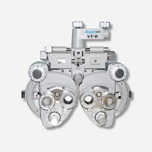 |  |  |  |  |  | ||||||||||||||||||||||||||||||||||||||||||||||||
| SKU | SF1033560107-26 | SF1033560012-9 | SF1033560012-8 | SF1033560074-4 | SF1033560012-15 | SF1033560012-1 | ||||||||||||||||||||||||||||||||||||||||||||||||
| Rating | ||||||||||||||||||||||||||||||||||||||||||||||||||||||
| Price | Ask for Price | Ask for Price | $13,900.00 | $68,468.00 | $7,700.00 | $6,950.00 | ||||||||||||||||||||||||||||||||||||||||||||||||
| Stock | ||||||||||||||||||||||||||||||||||||||||||||||||||||||
| Availability | ||||||||||||||||||||||||||||||||||||||||||||||||||||||
| Add to cart | ||||||||||||||||||||||||||||||||||||||||||||||||||||||
| Description | Ship from abroad
| Shipped from Abroad Incorporating innovative technologies, P20’s user-friendly design with a simple operation panel, intuitive user interface and a variety of intelligent auxiliary scanning tools, will significantly improve your daily examination experience. Besides general imaging applications, P20 has entitled with diagnostic 4D technology which has an extraordinary performance in obstetrics and gynecology applications. Delivery & Availability: Typically 5-7 working days – excluding furniture and heavy/bulky equipment. Please contact us for further information. | In Stock A feature-rich system inheriting the Wi-Sono high-end platform, the P15 uses an array of advanced tools to help enhance the image quality. It's a cost-effective, simplified console with an intuitive user interface and multiple intelligent functions. Delivery & Availability: Typically 2 working days – excluding furniture and heavy/bulky equipment. Please contact us for further information. | In Stock The GXR-SD is a diagnostic digital radiography system that provides reliable high quality digital radiographic images with a reduced dose. The GXR-SD DR systems offer comprehensive digital solutions to all radiography needs, featuring ACQUIDR digital imaging system with stationary or portable digital flat-panel detectors as well as reliable high-frequency x-ray generators that are known worldwide for their excellent performance, lifetime and stability. Patient tables and wall stands are also offered. Delivery & Availability: Typically 21 working days – excluding furniture and heavy/bulky equipment. Please contact us for further information. | Shipped from Abroad With ultra-modern innovative design and the clinically-proven technologies, S8 Exp is portable ultrasound scanner well equipped as a low-physical-effort and enhanced-image-quality ultrasound scanner, which not only provides optimized solutions for versatile applications, but does help to improve the user-experience for both routine and non-traditional challenges. Delivery & Availability: Typically 5-7 working days – excluding furniture and heavy/bulky equipment. Please contact us for further information. | In Stock A Value Choice beyond Your Expectation. SonoScape’s trolley color Doppler system S11 redefines price and performance with practical design. The S11 will go beyond your expectations but not your budget. Delivery & Availability: Typically 2 working days – excluding furniture and heavy/bulky equipment. Please contact us for further information. | ||||||||||||||||||||||||||||||||||||||||||||||||
| Content | Features:
| DETAILS
Upgraded Images with More Clarity
SonoScape never stops making progress in improving the image quality of its ultrasound products to enhance the confidence of diagnosis for doctors. With extraordinary images provided by P20, the anatomy structures are clearer than ever.
C-Xlasto Imaging
With C-xlasto Imaging, P20 enables comprehensive quantitative elastic analysis. Meanwhile, C-xlasto on P20 is supported by linear, convex and transvaginal probes, to ensure good reproducibility and highly consistent quantitative elastic results.
S-Live
S-Live allows for detailed visualization of subtle anatomical features, thereby enabling intuitive diagnosis with real-time 3D images and enriching patient communication.
Pelvic Floor 4D
Transperineal 4D pelvic floor ultrasound can provide useful clinical values in assessing the vaginal delivery impact on the female anterior compartment, judging whether the pelvic organs are prolapsed or not and the extent, determining if the pelvic muscles were torn accurately.
Anatomic M Mode
Anatomic M Mode helps you observe the myocardial motion at different phases by freely placing sample lines. It accurately measures the myocardial thickness and the heart size of even difficult patients and supports the myocardial function and LV wall-motion assessment.
Tissue Doppler Imaging
P20 is endowed with Tissue Doppler Imaging which provides velocities and other clinical information on myocardial functions, facilitating clinical doctors with the ability to analyze and compare the motions of different parts of the patient's heart.
Click Here To Download Catalogue | DETAILS
Super Wide-bandwidth Platform
Inheriting Wi-sono's ultra-wide system platform and with the advanced probe technology, high-resolution and deep penetration images are provided for precision medicine.
Spatial Compound Imaging
Spatial Compound Imaging utilizes several lines of sight for optimal contrast resolution, speckle reduction and border detection, with which P15 is ideal for superficial and abdominal imaging with better clarity and improved continuity of structures.
μ-Scan+
The new generation μ-Scan imaging technology gives you better image quality by reducing noise, improving signal strength and improving visualization.
Dynamic Color
Dynamic color improves upon already existing color Doppler technologies for a clearer capture of color flow and detailed visualization of even tiny veins with lower velocities.
Real-time Panoramic
With real-time panoramic, you can acquire an extended field of view for large organs or long vessels for easy measurement and diagnostic efficiency. Accomplished in real-time for the convenience of the sonographers, any mistake can also be easily back tracked and corrected without interrupting the scan.
3D/4D
Outstanding volume performance with speed and convenience makes P15 outshine others on volume imaging.
Tissue Doppler Imaging
Tissue Doppler Imaging allows clinical doctors to quantitatively evaluate local myocardial movements and functions, facilitating them with the ability to analyze and compare the motions of the different parts of the patient's heart.
Auto IMT
Quick measurement of intra-media vessel thickness ensures good reproducibility and high diagnostic efficiency.
Click Here To Download Catalogue | DrGem Ceiling Mounted Digital X-ray is a diagnostic digital radiography system that provides reliable high quality digital radiographic images with a reduced dose. The GXR-SD DR systems offer comprehensive digital solutions to all radiography needs, featuring ACQUIDR digital imaging system with stationary or portable digital flat-panel detectors as well as reliable high-frequency x-ray generators that are known worldwide for their excellent performance, lifetime and stability. Patient tables and wall stands are also offered.
Features:
Click Here To Download Catalogue | Sonoscape S8 Exp Portable Ultrasound scannerDETAILS Agile and Versatile With ultra-modern innovative design and the clinically-proven technologies, S8 Exp Portable Ultrasound scanner is well equipped as a low-physical-effort and enhanced-image-quality ultrasound scanner, which not only provides optimized solutions for versatile applications but does help to improve the user experience for both routine and non-traditional challenges. Working with S8 Exp, it will trigger your unlimited reverie and endow you with endless charm. Carrying forward the classical design of SonoScape's portable ultrasound products, S8 Exp successfully combines the best ergonomics, attractive design and ease of use. This charismatic identity is also enhanced by a sophisticated color palette—with sedate grey as its interior paint color and pearl white as exterior cover, S8 Exp reveals a style of aristocrat and strong character among SonoScape's ultrasound systems. Workflow The S8 Exp is a portable ultrasound scanner that adapts to your workflow, whether you are in the consulting room, at the bedside, or at a remote location. With easy-to-use new platform designed for sonographers' needs and full connection interfaces for easy connectivity and data sharing, S8 Exp leads to improved user comfort and clinical outcome as well as patient throughput and working efficiency. Powerful Platform Embedded with SonoScape's core imaging technologies such as μ-scan, PHI and Spatial Compound, S8 Exp boasts exceptional 2D image, sensitive spectral, Color and Power Doppler, displaying well-defined anatomy and pathology and facilitating a highly optimized diagnostic user environment for conclusive diagnoses. Besides, S8 Exp offers a comprehensive selection of electronic probes to maximally extend its capabilities to meet a wide range of applications including the abdomen, pediatric, OB/GYN, cardiovascular, musculoskeletal, etc. The advanced probe technologies also effectively enhance the image quality and confidence in reaching clinical diagnoses even in difficult patients.Click Here To Download Catalogue | DETAILS
SonoScape’s trolley colour Doppler system S11 redefines price and performance with practical design. The S11 will go beyond your expectations but not your budget. As an easy-to-use ultrasound system, the S11 is integrated with a new software platform, especially optimized for a smooth workflow and convenient operation. The system speeds up the exam process and makes file management easier.
SPECIFICATION
- 15-inch high definition LCD monitor with articulating arm
- Compact and agile trolley design
- 3 active transducer sockets available for a wide range of applications
- Duplex, Color Doppler, DPI, PW Doppler, tissue harmonic imaging, μ-scan speckle reduction imaging, compound imaging, trapezoidal imaging
- Customized settings based on your own working style
- Full patient database and image management solutions
Click Here To Download Catalogue | ||||||||||||||||||||||||||||||||||||||||||||||||
| Weight | N/A | N/A | N/A | N/A | N/A | N/A | ||||||||||||||||||||||||||||||||||||||||||||||||
| Dimensions | N/A | N/A | N/A | N/A | N/A | N/A | ||||||||||||||||||||||||||||||||||||||||||||||||
| Additional information | ||||||||||||||||||||||||||||||||||||||||||||||||||||||


Reviews
There are no reviews yet.