
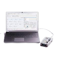
Neuro MEP Micro-Portable EMG and NCS System
$0.00
Shipped From Abroad
In coloproctology centers, EMG of external sphincter and pelvic floor muscles is often required. Portable EMG and NCS system Neuro-MEP-Micro packed with the necessary techniques is the right solution for these needs.
Typically 10-21 working days – excluding furniture and heavy/bulky equipment. Please contact us for further information.
Description
Features
Specific techniques for neurophysiological assessment of external sphincter and other pelvic floor neuromuscular structures
The system is used to perform various neurophysiological examinations of pelvic floor for diagnostic and scientific purposes, i.e.:
- bulbocavernosus and other sacral reflex tests with electrical stimulation of pudendal nerve to assess the conduction of reflex arcs (S2-S4);
- surface EMG of external sphincter and pelvic floor muscles to assess the level of tonic contraction;
- needle EMG with quantitative motor unit potential (MUP) analysis coupled with sacral reflex test to detect sacral denervation;
- stimulation EMG used with a disposable St. Mark’s electrode to stimulate the distal part of pudendal nerve;
- pudendal nerve somatosensory evoked potentials (pSEPs) especially in patients with preserved sacral reflexes and hypoesthesia of perineum;
- sympathetic skin responses (SSR) recorded from perineal area to assess the conduction velocity of sympathetic unmyelinated fibers and myelinated sensory nerve fibers;
- analysis of motor evoked potentials (MEP) in perineal muscles during the cortical and sacral magnetic stimulation (the assessment of corticospinal tract conduction with recording of pelvic floor motor evoked potentials (if the magnetic stimulator is available)).
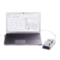
Portable design
Neuro-MEP-Micro requires very little space and is connected to a computer via the USB cable which ensures data uploading and power supply of the device. If not powered from mains, the device can operate from the notebook battery.
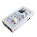
Electrical stimulator with two outputs
Two software switchable outputs to plug in the electrical stimulator allow a specialist to place two pairs of stimulating electrodes on a patient and connect them to the device. Thus, there is no need to switch the electrodes as the stimulating electrode is software-defined.
Quick Comparison
| Neuro MEP Micro-Portable EMG and NCS System remove | Sonoscape P50 Ultrasound Machine remove | Lab/Ward Coat remove | Ecleris HD Digital Video Colposcope remove | CryoIQ Pro with LED Light for 16 Gram Gas, & Gas cartridge 25 Gram No2 with Valve remove | Sonoscape S22 Ultrasound Machine remove | |||||||||||||||||||||||||||||||||||
|---|---|---|---|---|---|---|---|---|---|---|---|---|---|---|---|---|---|---|---|---|---|---|---|---|---|---|---|---|---|---|---|---|---|---|---|---|---|---|---|---|
| Name | Neuro MEP Micro-Portable EMG and NCS System remove | Sonoscape P50 Ultrasound Machine remove | Lab/Ward Coat remove | Ecleris HD Digital Video Colposcope remove | CryoIQ Pro with LED Light for 16 Gram Gas, & Gas cartridge 25 Gram No2 with Valve remove | Sonoscape S22 Ultrasound Machine remove | ||||||||||||||||||||||||||||||||||
| Image | 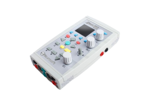 |  |  | 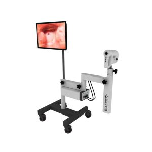 | 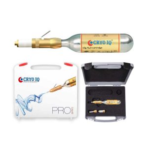 |  | ||||||||||||||||||||||||||||||||||
| SKU | SF1033560130187-16 | SF1033560012-11 | SF1033560084-222 | SF1033560087-2 | SF1033560096-6 | SF1033560012-3 | ||||||||||||||||||||||||||||||||||
| Rating | ||||||||||||||||||||||||||||||||||||||||
| Price |
|
| $11.00 | $3,564.00 |
| $9,350.00 | ||||||||||||||||||||||||||||||||||
| Stock | ||||||||||||||||||||||||||||||||||||||||
| Availability | ||||||||||||||||||||||||||||||||||||||||
| Add to cart | ||||||||||||||||||||||||||||||||||||||||
| Description | Shipped From Abroad
In coloproctology centers, EMG of external sphincter and pelvic floor muscles is often required. Portable EMG and NCS system Neuro-MEP-Micro packed with the necessary techniques is the right solution for these needs.
Delivery & Availability:
Typically 10-21 working days – excluding furniture and heavy/bulky equipment. Please contact us for further information.
| Shipped from Abroad Easily accomplish more with SonoScape’s new P50 ultrasound system. Incorporating single crystal clarity, automatic corrections and calculation, and user defined flexibility promises a confident diagnostic experience as well as opening new doors of opportunity for ultrasound use. Delivery & Availability: Typically 7-14 working days – excluding furniture and heavy/bulky equipment. Please contact us for further information. | In stock
| Shipped from Abroad Content Includes: Floor Mounted with Base with 4 Castors, Video Head, Positioning Arm, LED Light Included on Video Head, 110/220V Power Cable and User´s Guide, Stand For LCD Monitor and Printer, Digital Capturing System for Images, Videos and Sounds, USB 2.0. Includes Main Unit Processor with three Camera Inputs, USB Cable, Software, Hands Free Microphone and Footswitch. Delivery & Availability: Typically 10 working days – excluding furniture and heavy/bulky equipment. Please contact us for further information. | Shipped from Abroad
CryoIQ® PRO Contact is based on our contact freezing system (closed gold plated metal contact tips with diameters of 1, 3, 5, or 7 mm). It provides precise treatment of the affected skin areas only in the size of the contact applicator without damage to the surrounding healthy tissue.
| Shipped from Abroad As SonoScape steps forward to add value and efficiency to ultrasound, the latest S22 was designed in a user-friendly platform to address current and future demanding needs. It represents an excellent mix in performance and price. Delivery & Availability: Typically 5-7 working days – excluding furniture and heavy/bulky equipment. Please contact us for further information. | ||||||||||||||||||||||||||||||||||
| Content | FeaturesSpecific techniques for neurophysiological assessment of external sphincter and other pelvic floor neuromuscular structures The system is used to perform various neurophysiological examinations of pelvic floor for diagnostic and scientific purposes, i.e.:
| DETAILS
Powerful Compact Precision
Taking into consideration the evolving expectations and needs for ultrasound, the P50 is a slim and unobtrusive trolley system that is comfortable in tight, congested spaces with little room to work in. Providing everything you need for a comfortable examination in a small space for both you and your patient.
Single Crystal Transducer
Wideband single crystal probes greatly improve the signal ratio, acquire stunning images and provide superior sensitivity and resolution for both the near and far-fields.
μ-Scan+
The new generation μ-Scan imaging technologies give you better image quality by reducing noise, improving signal strength and improving visualization.
Dynamic Color
Dynamic colour improves upon already existing colour Doppler technologies for clear capture of colour flow and detail visualization of even tiny veins with lower velocities.
Solution for Radiology
P50, is a leading-edge ultrasound system that can meet the demands of any clinical setting. You can experience a superior performance in multi-dimensional imaging for a full range of clinical applications – abdominal, breast and cardiovascular.
C-xlasto Imaging
By understanding that tissue stiffness varies depending on the type of tissue, we can use C-xlasto Imaging to easily find abnormalities and tumours within soft tissue. The differences in tissue responses are detected and visualized in real-time by the elastography algorithms through different representations, which can be particularly helpful in analyzing breast, thyroid and musculoskeletal structures. Predominately used only in linear probes, SonoScape’s new transvaginal and bi-plane probe for gynaecology and urology are breaking the mould and expanding elastography applications.
Real-time Color Panoramic
With the combination of colour flow and real-time panoramic, visualizing the blood flow of an entire vein or artery is now an easy task. Accomplished in real-time for the convenience of the sonographers, any mistakes can also be easily backtracked and corrected without interrupting the scan.
Contrast Imaging
Contrast Imaging on P50 makes full use of the infra harmonic signal and second harmonic signal to improve the image resolution and deep penetration. What’s more, the Dynamic Acoustic Control technology effectively controls the acoustic pressure for the contrast agent, decreasing the required agent dose and assures uniform image quality, guaranteeing longer contrast agent duration and better lesion perfusion of delayed phase observation.
Solution for OB/GYN
P50 has superior image quality, automated measurement tools, and a variety of volume technologies to provide ideal solutions for clinical examinations such as pregnancy examinations, and gynecologic disease diagnosis. With a new 4D transvaginal probe, P50 helps you to see and detect fetal abnormalities and significantly improves your diagnostic confidence during your examinations.
S-Live Silhouette
A unique transparent 3D anatomical image of the fetus for improved initial anatomical review. By using this new application, the system can create completely different fetal images from conventional ultrasound images, which can depict the fetal's intracorporeal anatomical structure.
Pelvic Floor 4D
Working in conjunction with SonoScape’s latest transvaginal probes, trans-perineal 4D pelvic floor ultrasound provides a useful clinical assessment of the impact of vaginal delivery on the female anterior compartment. Allowing doctors to judge whether the pelvic organs prolapsed or not, the extent of prolapse, and determining whether the pelvic muscles tore correctly.
S-Guide
S-Guide gives the user an extensive list of example obstetric ultrasound images as reference guides and a convenient checklist system to keep track of their progress during their obstetrics examination.
Auto Face
Automatically removes masking layers in front of the fetus’s face for a clearer vision of the fetus’s face.
AVC Follicle
AVC Follicle automatically identifies how many follicles are present and calculates their individual volumes.
Solution for Cardiology
P50 provides clear 2D clinical images and Doppler sensitivity to assess critical cardiac performance. Compatible with SonoScape’s single crystal probes, the P50 can provide images with better resolution and penetration in Cardiac diagnosis.
Tissue Doppler Imaging
Tissue Doppler Imaging allows clinical doctors to quantitatively evaluate local myocardial movements and functions, facilitating them with the ability to analyze and compare the motions of the different parts of the patient’s heart.
Stress Echo
Stress echocardiography is the combination of 2D echocardiography with physical, pharmacological or electrical stress of the patient. It also then provides users with report management tools such as configurable template editor, multiple loops to select one for storage, wall motion scoring, stress echo report, etc
Auto IMT
Auto IMT is used when determining the level of vascular sclerosis present in the patient by automatically tracing and calculating the thickness of the carotid vessels. What distinguishes the P50 is that it provides an instant and accurate Mean and Max index at the touch of a single button.
Auto EF
Automated 2D Cardiac Quantification is a fully intelligent trace function for endocardium with 19 easily-adjustable points providing rapid access to proven 2D EF and volumes.
Click Here To Download Catalogue |
| The Ecleris ColpoHD Digital Video Colposcope offers clinicians an illuminated, high definition magnified view of the cervix and tissues of the vagina and vulva. As a screening tool for sexual assault victims or for the early detection of precancerous lesions, the ColpoHD provides insight into relevant pathology changes of tissue shape and color with magnification of up to 50x and a digital zoom up to 128x. The integrated electronic green filter delivers exceptional pathology visualization, with clear, true color imaging and superb depth of field. The swing arm mounted high-definition (HD) camera can be easily maneuvered into place. Optional integrated image capture is available for documentation along with archiving and printing of images.
A powerful tool for cervical cancer screening
The ColpoHD is precise and easy to handle in all examination situations. High quality optics along bright LED illumination and high-tech CMOS image sensors guarantee fantastic quality HD images are displayed for every patient.
The ColpoHD is also ideally suited for the examination of sexual assault victims who may be uncomfortable with a traditional Colposcope examination with the physician working through the standard microscope binoculars. The ColpoHD offers distance and a level of privacy for these patients who may be in a vulnerable frame of mind. The ColpoHD also offers optional image archiving and documentation of the examination in high definition image quality which is vital for sexual assault cases.
Features at a Glance
Click Here To Download Catalogue | CryoIQ® PRO Contact is based on our contact freezing system (closed gold plated metal contact tips with diameters of 1, 3, 5, or 7 mm). It provides precise treatment of the affected skin areas only in the size of the contact applicator without damage to the surrounding healthy tissue. The most versatile model for contact freezing treatment of warts and common skin lesions. Its unique construction with a built-in non-return valve prevents gas leakage, thus giving maximum use of the gas content. With the new PRO model, you now have the possibility to change the tip for different areas of application, such as dermatology, dentistry, podiatry, gynecology/urology, and veterinary.
Features:
| DETAILS
As SonoScape steps forward to add value and efficiency to ultrasound, the latest S22 was designed in a user-friendly platform to address current and future demanding needs. It represents an excellent mix in performance and price.
S22, is a shared service ultrasound system with a slim and elegant package that has combined mobility with utility to fit in specific clinical situations including emergency department, ICU, operating room and so on. Furthermore, its ergonomic design, easy operating and flexible data management will give you a memorable experience.
SPECIFICATION
• Large high-resolution widescreen LED
• Sensitive touch screen
• Four transducer sockets plus one socket for pencil probe
• A comprehensive selection of probes: linear, Convex, Micro-convex, Volumetric, Endocavity, Bi-plane, Phased Array, TEE, Intraoperative, Pencil
• Premium application technology: 4D, μ-scan speckle reduction, compound imaging, Pulse Inversion Harmonic Imaging, Color M-Mode, Steer M-Mode, PDI, TDI, Real-time Panoramic Imaging, Trapezoid Imaging, Auto-IMT…
• Full patient database and image management solutions: DICOM 3.0, AVI/JPG, USB 2.0, HDD, DVD, PDF report
• Multi-Language Input Keyboard
• Built-in battery
Click Here To Download Catalogue | ||||||||||||||||||||||||||||||||||
| Weight | N/A | N/A | N/A | N/A | N/A | N/A | ||||||||||||||||||||||||||||||||||
| Dimensions | N/A | N/A | N/A | N/A | N/A | N/A | ||||||||||||||||||||||||||||||||||
| Additional information |


Reviews
There are no reviews yet.