
Neuro MEP Micro-Portable EMG and NCS System
$0.00
Shipped From Abroad
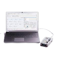
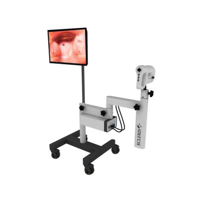
In coloproctology centers, EMG of external sphincter and pelvic floor muscles is often required. Portable EMG and NCS system Neuro-MEP-Micro packed with the necessary techniques is the right solution for these needs.
Typically 10-21 working days – excluding furniture and heavy/bulky equipment. Please contact us for further information.
Description
Features
Specific techniques for neurophysiological assessment of external sphincter and other pelvic floor neuromuscular structures
The system is used to perform various neurophysiological examinations of pelvic floor for diagnostic and scientific purposes, i.e.:
- bulbocavernosus and other sacral reflex tests with electrical stimulation of pudendal nerve to assess the conduction of reflex arcs (S2-S4);
- surface EMG of external sphincter and pelvic floor muscles to assess the level of tonic contraction;
- needle EMG with quantitative motor unit potential (MUP) analysis coupled with sacral reflex test to detect sacral denervation;
- stimulation EMG used with a disposable St. Mark’s electrode to stimulate the distal part of pudendal nerve;
- pudendal nerve somatosensory evoked potentials (pSEPs) especially in patients with preserved sacral reflexes and hypoesthesia of perineum;
- sympathetic skin responses (SSR) recorded from perineal area to assess the conduction velocity of sympathetic unmyelinated fibers and myelinated sensory nerve fibers;
- analysis of motor evoked potentials (MEP) in perineal muscles during the cortical and sacral magnetic stimulation (the assessment of corticospinal tract conduction with recording of pelvic floor motor evoked potentials (if the magnetic stimulator is available)).
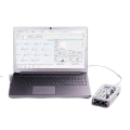
Portable design
Neuro-MEP-Micro requires very little space and is connected to a computer via the USB cable which ensures data uploading and power supply of the device. If not powered from mains, the device can operate from the notebook battery.
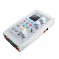
Electrical stimulator with two outputs
Two software switchable outputs to plug in the electrical stimulator allow a specialist to place two pairs of stimulating electrodes on a patient and connect them to the device. Thus, there is no need to switch the electrodes as the stimulating electrode is software-defined.
Quick Comparison
| Neuro MEP Micro-Portable EMG and NCS System remove | Female Urinal remove | Vigeo VTek Reusable Biopsy Device remove | CryoIQ CIQ-DP-L- Cryo DERM Plus Liquid Device with LED Light (CiQ-A-LED) remove | COMBI 2 Urine Test Kit remove | Sonoscape P15 Ultrasound Machine With Four Probes remove | |
|---|---|---|---|---|---|---|
| Name | Neuro MEP Micro-Portable EMG and NCS System remove | Female Urinal remove | Vigeo VTek Reusable Biopsy Device remove | CryoIQ CIQ-DP-L- Cryo DERM Plus Liquid Device with LED Light (CiQ-A-LED) remove | COMBI 2 Urine Test Kit remove | Sonoscape P15 Ultrasound Machine With Four Probes remove |
| Image | 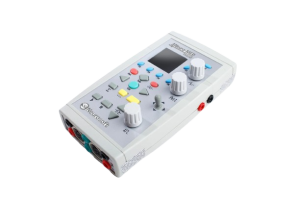 | 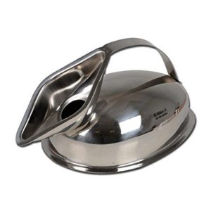 | 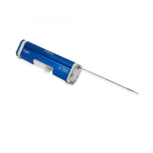 | 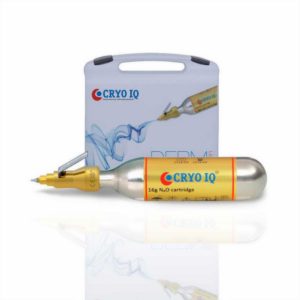 |  |  |
| SKU | SF1033560130187-16 | SF1033560084-82 | SF10335601282 | SF1033560096-4 | SF1033560084-231 | SF1033560012-8 |
| Rating | ||||||
| Price |
| $6.80 |
|
|
| $13,900.00 |
| Stock | ||||||
| Availability | ||||||
| Add to cart | ||||||
| Description | Shipped From Abroad
In coloproctology centers, EMG of external sphincter and pelvic floor muscles is often required. Portable EMG and NCS system Neuro-MEP-Micro packed with the necessary techniques is the right solution for these needs.
Delivery & Availability:
Typically 10-21 working days – excluding furniture and heavy/bulky equipment. Please contact us for further information.
| In stock Delivery & Availability: Typically 5-7 working days – excluding furniture and heavy/bulky equipment. Please contact us for further information. | Shipped From Abroad
Features:
| Shipped from Abroad
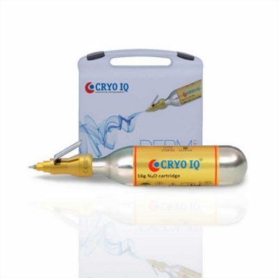
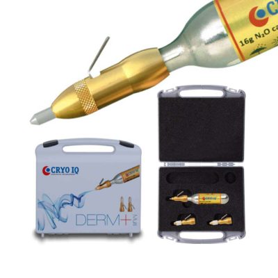
CryoIQ device is perfectly fitted for treatment of warts and other common skin lesions. Its unique construction with a built-in non-return valve prevents gas leakage thus giving a maximum use of the gas content. Areas of application are general practice, dermatology, pediatrics, podiatry, aesthetics, veterinary and gynecology/urology.
| In stock
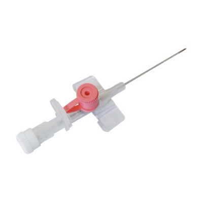
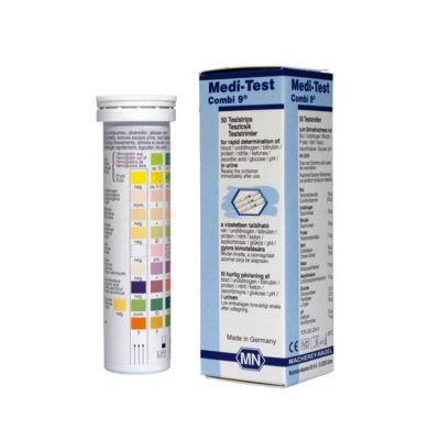
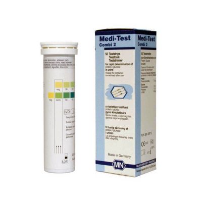
Medi-Test Combi 2 urine test strips from Macherey-Nagel are meant for use in urinalysis to quickly and easily detect the presence of glucose and protein in urine. These strips are very flexible, so the test can be carried out even with small samples. Thanks to the high resistance to interferences caused by ascorbic acid, there is usually no need to perform the test again.
| In Stock A feature-rich system inheriting the Wi-Sono high-end platform, the P15 uses an array of advanced tools to help enhance the image quality. It's a cost-effective, simplified console with an intuitive user interface and multiple intelligent functions. Delivery & Availability: Typically 2 working days – excluding furniture and heavy/bulky equipment. Please contact us for further information. |
| Content | FeaturesSpecific techniques for neurophysiological assessment of external sphincter and other pelvic floor neuromuscular structures The system is used to perform various neurophysiological examinations of pelvic floor for diagnostic and scientific purposes, i.e.:
| Vigeo VTek Reusable Biopsy Device Features:
Click Here To Download Catalogue | CryoIQ device is perfectly fitted for treatment of warts and other common skin lesions. Its unique construction with a built-in non-return valve prevents gas leakage thus giving a maximum use of the gas content. Areas of application are general practice, dermatology, pediatrics, podiatry, aesthetics, veterinary and gynecology/urology. The device uses a state-of-the-art cooling technology that does not require handling of potentially dangerous gases and liquids meaning that the gas cartridge is safely replaced when empty. The device features an ergonomic design where the cooling medium is evenly, accurately and economically distributed by depressing the metering lever.
Features:
| Medi-Test Combi 2 urine test strips from Macherey-Nagel are meant for use in urinalysis to quickly and easily detect the presence of glucose and protein in urine. These strips are very flexible, so the test can be carried out even with small samples. Thanks to the high resistance to interferences caused by ascorbic acid, there is usually no need to perform the test again.
Product Details for Medi-Test Combi 2
| DETAILS
Super Wide-bandwidth Platform
Inheriting Wi-sono's ultra-wide system platform and with the advanced probe technology, high-resolution and deep penetration images are provided for precision medicine.
Spatial Compound Imaging
Spatial Compound Imaging utilizes several lines of sight for optimal contrast resolution, speckle reduction and border detection, with which P15 is ideal for superficial and abdominal imaging with better clarity and improved continuity of structures.
μ-Scan+
The new generation μ-Scan imaging technology gives you better image quality by reducing noise, improving signal strength and improving visualization.
Dynamic Color
Dynamic color improves upon already existing color Doppler technologies for a clearer capture of color flow and detailed visualization of even tiny veins with lower velocities.
Real-time Panoramic
With real-time panoramic, you can acquire an extended field of view for large organs or long vessels for easy measurement and diagnostic efficiency. Accomplished in real-time for the convenience of the sonographers, any mistake can also be easily back tracked and corrected without interrupting the scan.
3D/4D
Outstanding volume performance with speed and convenience makes P15 outshine others on volume imaging.
Tissue Doppler Imaging
Tissue Doppler Imaging allows clinical doctors to quantitatively evaluate local myocardial movements and functions, facilitating them with the ability to analyze and compare the motions of the different parts of the patient's heart.
Auto IMT
Quick measurement of intra-media vessel thickness ensures good reproducibility and high diagnostic efficiency.
Click Here To Download Catalogue | |
| Weight | N/A | N/A | N/A | N/A | N/A | N/A |
| Dimensions | N/A | N/A | N/A | N/A | N/A | N/A |
| Additional information |


Reviews
There are no reviews yet.