
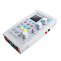
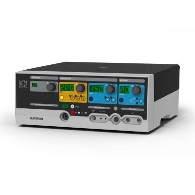
Neuro MEP Micro-Portable EMG and NCS System
$0.00
Shipped From Abroad
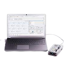
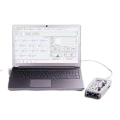
In coloproctology centers, EMG of external sphincter and pelvic floor muscles is often required. Portable EMG and NCS system Neuro-MEP-Micro packed with the necessary techniques is the right solution for these needs.
Typically 10-21 working days – excluding furniture and heavy/bulky equipment. Please contact us for further information.
Description
Features
Specific techniques for neurophysiological assessment of external sphincter and other pelvic floor neuromuscular structures
The system is used to perform various neurophysiological examinations of pelvic floor for diagnostic and scientific purposes, i.e.:
- bulbocavernosus and other sacral reflex tests with electrical stimulation of pudendal nerve to assess the conduction of reflex arcs (S2-S4);
- surface EMG of external sphincter and pelvic floor muscles to assess the level of tonic contraction;
- needle EMG with quantitative motor unit potential (MUP) analysis coupled with sacral reflex test to detect sacral denervation;
- stimulation EMG used with a disposable St. Mark’s electrode to stimulate the distal part of pudendal nerve;
- pudendal nerve somatosensory evoked potentials (pSEPs) especially in patients with preserved sacral reflexes and hypoesthesia of perineum;
- sympathetic skin responses (SSR) recorded from perineal area to assess the conduction velocity of sympathetic unmyelinated fibers and myelinated sensory nerve fibers;
- analysis of motor evoked potentials (MEP) in perineal muscles during the cortical and sacral magnetic stimulation (the assessment of corticospinal tract conduction with recording of pelvic floor motor evoked potentials (if the magnetic stimulator is available)).
Portable design
Neuro-MEP-Micro requires very little space and is connected to a computer via the USB cable which ensures data uploading and power supply of the device. If not powered from mains, the device can operate from the notebook battery.
Electrical stimulator with two outputs
Two software switchable outputs to plug in the electrical stimulator allow a specialist to place two pairs of stimulating electrodes on a patient and connect them to the device. Thus, there is no need to switch the electrodes as the stimulating electrode is software-defined.
Quick Comparison
| Neuro MEP Micro-Portable EMG and NCS System remove | CryoIQ Pro with LED Light for 16 Gram Gas, & Gas cartridge 25 Gram No2 with Valve remove | COMBI 2 Urine Test Kit remove | Sonoscape HD-500 Gastrointestinal Video Endoscopy Stack remove | Sonoscape E1 Ultrasound Machine With Two Probes remove | Lab/Ward Coat remove | ||||||||||||||||||||||||||||||||||||||||||||||||||||||||||||||||||||||||||||||||||||||||||||||||||||||||
|---|---|---|---|---|---|---|---|---|---|---|---|---|---|---|---|---|---|---|---|---|---|---|---|---|---|---|---|---|---|---|---|---|---|---|---|---|---|---|---|---|---|---|---|---|---|---|---|---|---|---|---|---|---|---|---|---|---|---|---|---|---|---|---|---|---|---|---|---|---|---|---|---|---|---|---|---|---|---|---|---|---|---|---|---|---|---|---|---|---|---|---|---|---|---|---|---|---|---|---|---|---|---|---|---|---|---|---|---|---|
| Name | Neuro MEP Micro-Portable EMG and NCS System remove | CryoIQ Pro with LED Light for 16 Gram Gas, & Gas cartridge 25 Gram No2 with Valve remove | COMBI 2 Urine Test Kit remove | Sonoscape HD-500 Gastrointestinal Video Endoscopy Stack remove | Sonoscape E1 Ultrasound Machine With Two Probes remove | Lab/Ward Coat remove | |||||||||||||||||||||||||||||||||||||||||||||||||||||||||||||||||||||||||||||||||||||||||||||||||||||||
| Image | 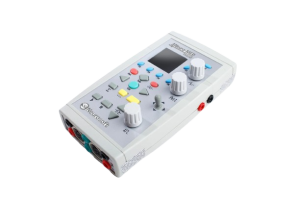 | 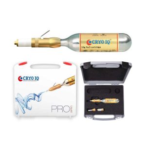 | 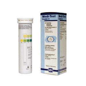 | 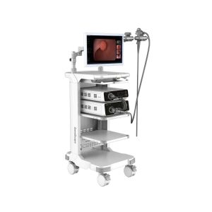 |  |  | |||||||||||||||||||||||||||||||||||||||||||||||||||||||||||||||||||||||||||||||||||||||||||||||||||||||
| SKU | SF1033560130187-16 | SF1033560096-6 | SF1033560084-231 | SF1033560012-24 | SF1033560012-20 | SF1033560084-222 | |||||||||||||||||||||||||||||||||||||||||||||||||||||||||||||||||||||||||||||||||||||||||||||||||||||||
| Rating | |||||||||||||||||||||||||||||||||||||||||||||||||||||||||||||||||||||||||||||||||||||||||||||||||||||||||||||
| Price |
|
|
|
| $4,620.00 | $11.00 | |||||||||||||||||||||||||||||||||||||||||||||||||||||||||||||||||||||||||||||||||||||||||||||||||||||||
| Stock | |||||||||||||||||||||||||||||||||||||||||||||||||||||||||||||||||||||||||||||||||||||||||||||||||||||||||||||
| Availability | |||||||||||||||||||||||||||||||||||||||||||||||||||||||||||||||||||||||||||||||||||||||||||||||||||||||||||||
| Add to cart | |||||||||||||||||||||||||||||||||||||||||||||||||||||||||||||||||||||||||||||||||||||||||||||||||||||||||||||
| Description | Shipped From Abroad
In coloproctology centers, EMG of external sphincter and pelvic floor muscles is often required. Portable EMG and NCS system Neuro-MEP-Micro packed with the necessary techniques is the right solution for these needs.
Delivery & Availability:
Typically 10-21 working days – excluding furniture and heavy/bulky equipment. Please contact us for further information.
| Shipped from Abroad
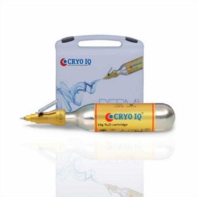
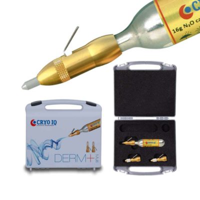
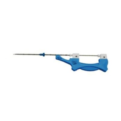
CryoIQ® PRO Contact is based on our contact freezing system (closed gold plated metal contact tips with diameters of 1, 3, 5, or 7 mm). It provides precise treatment of the affected skin areas only in the size of the contact applicator without damage to the surrounding healthy tissue.
| In stock
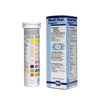
Medi-Test Combi 2 urine test strips from Macherey-Nagel are meant for use in urinalysis to quickly and easily detect the presence of glucose and protein in urine. These strips are very flexible, so the test can be carried out even with small samples. Thanks to the high resistance to interferences caused by ascorbic acid, there is usually no need to perform the test again.
| Shipped from Abroad Delivery & Availability: Typically 21 working days – excluding furniture and heavy/bulky equipment. Please contact us for further information. | Shipped from Abroad SonoScape has developed a new probe and function for the E1 Exp. With these additions the E1 Exp will bring users a more efficient examination experience with satisfying image quality and a smooth workflow. Delivery & Availability: Typically 5-7 working days – excluding furniture and heavy/bulky equipment. Please contact us for further information. | In stock
| |||||||||||||||||||||||||||||||||||||||||||||||||||||||||||||||||||||||||||||||||||||||||||||||||||||||
| Content | FeaturesSpecific techniques for neurophysiological assessment of external sphincter and other pelvic floor neuromuscular structures The system is used to perform various neurophysiological examinations of pelvic floor for diagnostic and scientific purposes, i.e.:
| CryoIQ® PRO Contact is based on our contact freezing system (closed gold plated metal contact tips with diameters of 1, 3, 5, or 7 mm). It provides precise treatment of the affected skin areas only in the size of the contact applicator without damage to the surrounding healthy tissue. The most versatile model for contact freezing treatment of warts and common skin lesions. Its unique construction with a built-in non-return valve prevents gas leakage, thus giving maximum use of the gas content. With the new PRO model, you now have the possibility to change the tip for different areas of application, such as dermatology, dentistry, podiatry, gynecology/urology, and veterinary.
Features:
| Medi-Test Combi 2 urine test strips from Macherey-Nagel are meant for use in urinalysis to quickly and easily detect the presence of glucose and protein in urine. These strips are very flexible, so the test can be carried out even with small samples. Thanks to the high resistance to interferences caused by ascorbic acid, there is usually no need to perform the test again.
Product Details for Medi-Test Combi 2
| HD-500 VIDEO PROCESSOR
Click Here To Download Catalogue | DETAILS
Efficient Diagnosis
μ-Scan, Speckle Reduction & Edge Enhancement
Spatial Compound Imaging
PIH - Pure Inversion Harmonic
Wide Scan - Enlarged Image Area
Tissue-Specific Imaging
SR Flow
Ergonomic Designs
Up to 2 Transducer Ports
Light Weight and Compact
15.6 inch Anti-flickering HD LED Screen
Tilting Monitor Angle Adjustment
Backlit Keyboard and Intelligent Panel
Long-lasting Battery for 90 mins
Ease of Use
Quick Boot Up
Auto-Brightness Adjustment
Auto Image Optimization
Auto IMT
Auto Trace
Equipped Accessories
Wi-Fi and Bluetooth Available
DICOM
500GB Hard Disk
Height Adjustable Trolley
Durable, Carry-on Site Suitcase
Click Here To Download Catalogue |
| |||||||||||||||||||||||||||||||||||||||||||||||||||||||||||||||||||||||||||||||||||||||||||||||||||||||
| Weight | N/A | N/A | N/A | N/A | N/A | N/A | |||||||||||||||||||||||||||||||||||||||||||||||||||||||||||||||||||||||||||||||||||||||||||||||||||||||
| Dimensions | N/A | N/A | N/A | N/A | N/A | N/A | |||||||||||||||||||||||||||||||||||||||||||||||||||||||||||||||||||||||||||||||||||||||||||||||||||||||
| Additional information | |||||||||||||||||||||||||||||||||||||||||||||||||||||||||||||||||||||||||||||||||||||||||||||||||||||||||||||


Reviews
There are no reviews yet.