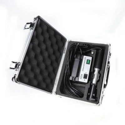
NEVRO MICROSCOPE
$0.00
Shipped From Abroad
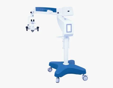
NEVRO Microscope, a line of equipment specially designed in detail, to meet the most demanding standards of surgical procedures,
for the areas of Neurosurgery and ENT, head and neck.
All functions can be activated via Gand Grip, with total comfort and lightness. This ensures less effort and ease during the procedures.
It has electromagnetic brakes, which control any and all positioning accurately, ensuring comfort, ease and time optimization.
Through the LCD touch screen, the same controls made via Hand Grip can also be performed with a simple touch.
Microsurgeries currently require cutting-edge technology with the ability to perform extremely complex procedures in an agile and simple way.
For optimizing surgical processes, the NEVRO microscope is the most efficient and advanced.
Delivery & Availability:
Typically 10-21 working days – excluding furniture and heavy/bulky equipment. Please contact us for further information.
Typically 10-21 working days – excluding furniture and heavy/bulky equipment. Please contact us for further information.
Description
Description
Control via Hand Grip
All microscope functions can be activated via Hand Grip, with total accuracy, comfort and lightness. This ensures less effort and ease during all surgical procedures.
Electromagnetic Brakes
Through the electromagnetic brakes, any and all positioning of the microscope can be done with total precision and comfort. Guaranteeing less effort, ease and time optimization.
LCD screen
The touch screen LCD display is used to view and activate all equipment settings. With its touch screen, the same controls made via Hand Grip can also be performed with a simple touch.
Quick Comparison
| NEVRO MICROSCOPE remove | Pantoscopic Ophthalmoscope remove | Tonometer remove | Auto Lensmeter TL-6500B remove | Auto Refractometer remove | Slit Lamp remove | |||||||||||||||||||||||||||||||||||
|---|---|---|---|---|---|---|---|---|---|---|---|---|---|---|---|---|---|---|---|---|---|---|---|---|---|---|---|---|---|---|---|---|---|---|---|---|---|---|---|---|
| Name | NEVRO MICROSCOPE remove | Pantoscopic Ophthalmoscope remove | Tonometer remove | Auto Lensmeter TL-6500B remove | Auto Refractometer remove | Slit Lamp remove | ||||||||||||||||||||||||||||||||||
| Image |  | 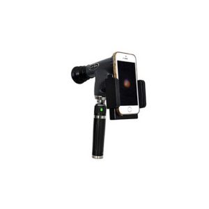 | 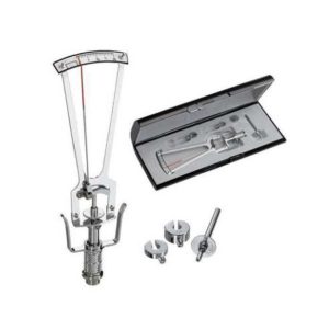 |  | 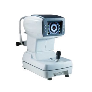 | 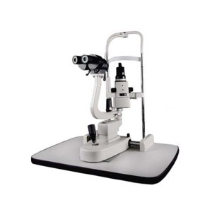 | ||||||||||||||||||||||||||||||||||
| SKU | SF103356013091-12 | SF1033560107-3 | SF1033560107-9 | SF1033560107-20 | SF1033560107-14 | SF1033560107-15 | ||||||||||||||||||||||||||||||||||
| Rating | ||||||||||||||||||||||||||||||||||||||||
| Price |
|
| $220.00 | $770.00 | $2,035.00 | $1,375.00 | ||||||||||||||||||||||||||||||||||
| Stock | ||||||||||||||||||||||||||||||||||||||||
| Availability | ||||||||||||||||||||||||||||||||||||||||
| Add to cart | ||||||||||||||||||||||||||||||||||||||||
| Description | Shipped From Abroad
NEVRO Microscope, a line of equipment specially designed in detail, to meet the most demanding standards of surgical procedures,
for the areas of Neurosurgery and ENT, head and neck.
All functions can be activated via Gand Grip, with total comfort and lightness. This ensures less effort and ease during the procedures.
It has electromagnetic brakes, which control any and all positioning accurately, ensuring comfort, ease and time optimization.
Through the LCD touch screen, the same controls made via Hand Grip can also be performed with a simple touch.
Microsurgeries currently require cutting-edge technology with the ability to perform extremely complex procedures in an agile and simple way.
For optimizing surgical processes, the NEVRO microscope is the most efficient and advanced.
Delivery & Availability:
Typically 10-21 working days – excluding furniture and heavy/bulky equipment. Please contact us for further information.
| Shipped from abroad
The brand-new Pantoscopic Ophthalmoscope is a portable digital imaging device which makes it possible to view and take pictures of the eyes.
| Shipped from abroad
This product is used to measure the intraocular pressure (IOP) by measuring the depth produced on the surface of the cornea by a load of a known weight. Each division on the scale corresponds to 1/20mm corneal depth.
| Shipped from abroad
| Shipped from abroad
| Shipped from abroad
| ||||||||||||||||||||||||||||||||||
| Content | DescriptionControl via Hand Grip All microscope functions can be activated via Hand Grip, with total accuracy, comfort and lightness. This ensures less effort and ease during all surgical procedures. Electromagnetic Brakes Through the electromagnetic brakes, any and all positioning of the microscope can be done with total precision and comfort. Guaranteeing less effort, ease and time optimization. LCD screen The touch screen LCD display is used to view and activate all equipment settings. With its touch screen, the same controls made via Hand Grip can also be performed with a simple touch. | The brand-new Pantoscopic Ophthalmoscope is a portable digital imaging device which makes it possible to view and take pictures of the eyes. The optical access of the Pantoscopic Ophthalmoscope is aligned to the visual axis of the smartphone camera by the adaptor which allows to you take pictures of the fundus and retinal nerve in high resolution. You could save pictures for each patient or email and print as needed. The Pantoscopic Ophthalmoscope provides a 5X larger view of the fundus compared with the standard ophthalmoscope. It has a wider view field of 230. Without dilating the pupil, the fundus imagines could be captured at any time and places.
Features:
| This product is used to measure the intraocular pressure (IOP) by measuring the depth produced on the surface of the cornea by a load of a known weight. Each division on the scale corresponds to 1/20mm corneal depth.
Features:
| Features:
| Features:
| Slit Lamp Features:
| ||||||||||||||||||||||||||||||||||
| Weight | N/A | N/A | N/A | N/A | N/A | N/A | ||||||||||||||||||||||||||||||||||
| Dimensions | N/A | N/A | N/A | N/A | N/A | N/A | ||||||||||||||||||||||||||||||||||
| Additional information |


Reviews
There are no reviews yet.