Nio Color 3MP (MDNC‑3521)
$0.00
Shipped From Abroad
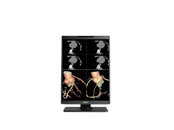
The Barco Nio Color 3MP (MDNC-3521) is a high-fidelity diagnostic display offering 3-megapixel resolution, IPS-SFT color panel, uniform luminance, and advanced quality-control features—ideal for general radiology and multi-modality image review.
Typically 10-21 working days – excluding furniture and heavy/bulky equipment. Please contact us for further information.
Description
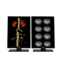
The Barco Nio Color 3MP MDNC-3521 is engineered for diagnostic imaging with a 21.3″ IPS-SFT color LCD and a native resolution of 2048 × 1536 (3MP). Its 4:3 aspect ratio and color/grayscale support make it suitable for PACS, CT, MRI, and general radiology workflows. The display includes uniformity correction, front-sensor quality assurance (I-Guard), high luminance, and 701 JND (Just Noticeable Difference) precision for subtle detail discrimination. It balances compact form factor with medical-grade performance and integrated diagnostic capabilities.
Our Nio Color 3MP display is part of a range of modern radiology monitors that give you what you need. No fluff, no overload of functionalities. But a sleek thin bezel design and just those tools and technologies that help you process cases effortlessly and efficiently.
A radiology monitor that fits your daily reading like a glove
Nio 3MP offers you exactly those tools that will make a difference. It boasts no less than 701 JNDs, so you can read detailed images with confidence. Its high luminance, I-Guard, and Uniform Luminance technologies offer you a bright and stable screen quality. And the display also contains our SteadyColor and SteadyGray technologies for stable colors and grays. In any imaging modality.
Clean desk, clear head
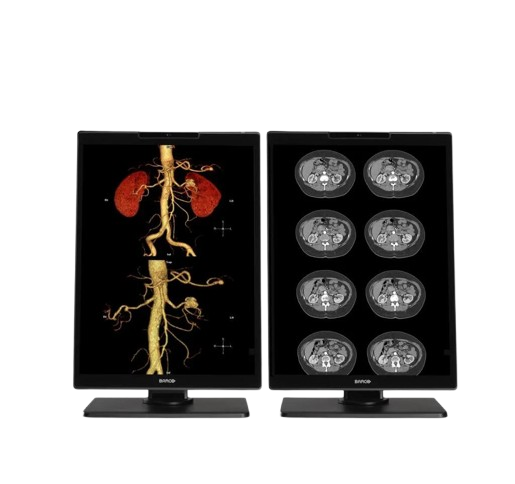
All of the above is built into an elegant design with thin bezels that fit both your hospital and your home office desks. Moreover, you can rotate your Nio 3MP monitor between portrait and landscape formats. Combine it in multi-head arrangements, two or three or four heads, neatly arranged in an arc embracing you.
A bright future
With Nio 3MP, you get a stable performance and a complete, industry-leading 5-year warranty that guarantees 20,000 backlight hours. Furthermore, our unique set of Intuitive Workflow Tools offers you a series of productivity hacks for more focus, flexibility, and comfort. For example, did you know that SpotView has been proven to decrease reading time by no less than 15.5%? In short, you can rest easy with this young companion. For many years to come.
Always a clear view with QAWeb Enterprise
QAWeb Enterprise helps you manage quality and assure compliance of your expanding healthcare enterprise with less effort, lower cost, and complete confidence. This fully automated and secure system supports consistent image quality, stable performance, and uptime for all PACS display systems across your enterprise. You can install QAWeb Enterprise for free on all our diagnostic and clinical review displays.
Ensuring diagnostic confidence with MDR Class IIa
Our radiology displays are MDR-certified as Class IIa. Their product information has been reviewed and cleared by independent medical and technical experts, and is audited yearly. In other words, we ensure diagnostic confidence and peace of mind for our users.
Key Features
-
IPS-SFT color LCD panel technology
-
3MP resolution: 2048 × 1536 pixels
-
Aspect ratio 4:3, matching many medical imaging formats
-
Uniform luminance correction to ensure brightness consistency
-
I-Guard front sensor for calibration / QA
-
701 JND contrast precision
-
Designed for use in radiology, PACS, and multi-modality image display
-
Color and grayscale imaging support
-
Compact 21.3″ diagonal active area
Specifications
| Category | Specification |
|---|---|
| Screen Technology | IPS-SFT Color LCD |
| Active Screen Size (Diagonal) | 541 mm (21.3″) |
| Active Screen Size (H × V) | 433 × 325 mm (17.1″ × 12.8″) |
| Aspect Ratio | 4:3 |
| Resolution | 3MP (2048 × 1536 pixels) |
| Pixel Pitch | 0.2115 mm |
| Color Imaging / Gray Imaging | Yes / Yes |
| Bit Depth | 30-bit |
| Viewing Angle (H/V) | 178° |
| Uniformity Correction | ULT |
| SteadyGray / SteadyColor | Yes (with Barco Display Controller and QAWeb Enterprise) |
| RapidFrame | Yes |
| Ambient Light Presets / Sensor | Yes / Yes |
| Front Sensor / Presence Sensor | Yes (I-Guard) / Yes |
| Maximum Luminance (Typical) | 1050 cd/m² |
| DICOM Calibrated Luminance | 600 cd/m² |
| Contrast Ratio | 2000:1 |
| Response Time | 12 ms (gray-to-gray average) |
| Housing Color | Black (RAL 9004) / White (RAL 9003) |
| Video Input Signals | 2 × DisplayPort 1.4 |
| Video Output Signals | N/A |
| USB Ports | 2 × USB-B 2.0 upstream; 5 × USB-A 2.0 downstream (1 charge port) |
| KVM Switch | Yes |
| Power Rating | 24 VDC, 4 A |
| Power Requirements | Adapter Technology Co., Ltd., type ATM160T-P240 <br> Input: 100–240 VAC, 1.8–0.9 A, 50–60 Hz <br> Output: 24 VDC, 6.6 A |
| Power Consumption | 45 W (nominal); <0.35 W (hibernate); <0.30 W (off) |
| Dimensions with Stand (W × H × D) | Portrait: 351 × 531~631 × 225 mm <br> Landscape: 491 × 462~562 × 225 mm |
| Dimensions without Stand (W × H × D) | Portrait: 351 × 491 × 64 mm <br> Landscape: 491 × 351 × 64 mm |
| Dimensions Packaged (W × H × D) | 455 × 210 × 770 mm |
| Net Weight with Stand | xPxx: 8.8 kg; xNxx: 7.7 kg |
| Net Weight without Stand | xPxx: 5.8 kg; xNxx: 4.7 kg |
| Net Weight Packaged | xPxx: 12.2 kg; xNxx: 11.2 kg |
| Tilt / Swivel / Pivot | -10° to +30° / ±30° / 90° |
| Height Adjustment Range | 100 mm |
| Mounting Standard | VESA (100 mm) |
| Screen Protection | SPES/SPES DE: Protective, anti-reflective glass; SNES: N/A |
| Recommended Modalities | All digital images except digital mammography |
| Certifications | CE0123, FDA 510(k) K230520, CCC, KC, BSMI, INMETRO, BIS, EAC |
| Safety Standards | IEC/EN/UL/CSA 60950-1, 62368-1, 60601-1, AAMI ES 60601-1 |
| EMI Standards | IEC/EN 60601-1-2, FCC Part 15 Class B, ICES-001 Level B, VCCI |
| Environmental Compliance | EU RoHS, China RoHS, REACH, Canada Health, WEEE, Packaging Directive |
| Supplied Accessories | User Guide, Documentation disc, System sheet, Video cables, Mains cable(s), USB cable, External power supply |
| Optional Accessories | Display controller, QA software (QAWeb Enterprise) |
| Warranty | 5 years (includes 20,000 hrs backlight warranty) |
| Operating Temperature | 0–35°C (specs: 20–30°C) |
| Storage Temperature | -20–60°C |
| Operating Humidity | 8–80% RH (non-condensing) |
| Storage Humidity | 5–85% RH (non-condensing) |
| Operating Pressure | 70 kPa |
| Storage Pressure | 50–106 kPa |
Quick Comparison
| Nio Color 3MP (MDNC‑3521) remove | Sonoscape P10 Ultrasound Machine remove | DrGem Ceiling Mounted Digital X-ray remove | Sonoscape P15 Ultrasound Machine With Four Probes remove | LED Double X-ray Viewing Box remove | Sonoscape P20 Ultrasound Machine remove | |||||||||||||||||||||||||||||||||||||||||||||||||||||||||||||||||||||||||||||||||||||||||||||||||||||||
|---|---|---|---|---|---|---|---|---|---|---|---|---|---|---|---|---|---|---|---|---|---|---|---|---|---|---|---|---|---|---|---|---|---|---|---|---|---|---|---|---|---|---|---|---|---|---|---|---|---|---|---|---|---|---|---|---|---|---|---|---|---|---|---|---|---|---|---|---|---|---|---|---|---|---|---|---|---|---|---|---|---|---|---|---|---|---|---|---|---|---|---|---|---|---|---|---|---|---|---|---|---|---|---|---|---|---|---|---|
| Name | Nio Color 3MP (MDNC‑3521) remove | Sonoscape P10 Ultrasound Machine remove | DrGem Ceiling Mounted Digital X-ray remove | Sonoscape P15 Ultrasound Machine With Four Probes remove | LED Double X-ray Viewing Box remove | Sonoscape P20 Ultrasound Machine remove | ||||||||||||||||||||||||||||||||||||||||||||||||||||||||||||||||||||||||||||||||||||||||||||||||||||||
| Image |  |  |  |  |  |  | ||||||||||||||||||||||||||||||||||||||||||||||||||||||||||||||||||||||||||||||||||||||||||||||||||||||
| SKU | SF1033560012-7 | SF1033560074-4 | SF1033560012-8 | SF1033560084-193 | SF1033560012-9 | |||||||||||||||||||||||||||||||||||||||||||||||||||||||||||||||||||||||||||||||||||||||||||||||||||||||
| Rating | ||||||||||||||||||||||||||||||||||||||||||||||||||||||||||||||||||||||||||||||||||||||||||||||||||||||||||||
| Price |
| $9,350.00 |
| $13,900.00 | $151.00 |
| ||||||||||||||||||||||||||||||||||||||||||||||||||||||||||||||||||||||||||||||||||||||||||||||||||||||
| Stock | ||||||||||||||||||||||||||||||||||||||||||||||||||||||||||||||||||||||||||||||||||||||||||||||||||||||||||||
| Availability | ||||||||||||||||||||||||||||||||||||||||||||||||||||||||||||||||||||||||||||||||||||||||||||||||||||||||||||
| Add to cart | ||||||||||||||||||||||||||||||||||||||||||||||||||||||||||||||||||||||||||||||||||||||||||||||||||||||||||||
| Description | Shipped From Abroad
The Barco Nio Color 3MP (MDNC-3521) is a high-fidelity diagnostic display offering 3-megapixel resolution, IPS-SFT color panel, uniform luminance, and advanced quality-control features—ideal for general radiology and multi-modality image review.
Delivery & Availability:
Typically 10-21 working days – excluding furniture and heavy/bulky equipment. Please contact us for further information.
| Shipped from Abroad The P10 color Doppler ultrasound system is a new generation product from SonoScape. It is designed to give high quality images, rich probe configurations, various clinical tools and automatic analysis software to provide you with comprehensive solutions for your growing demand for clinical applications. Delivery & Availability: Typically 5-7 working days – excluding furniture and heavy/bulky equipment. Please contact us for further information. | In Stock The GXR-SD is a diagnostic digital radiography system that provides reliable high quality digital radiographic images with a reduced dose. The GXR-SD DR systems offer comprehensive digital solutions to all radiography needs, featuring ACQUIDR digital imaging system with stationary or portable digital flat-panel detectors as well as reliable high-frequency x-ray generators that are known worldwide for their excellent performance, lifetime and stability. Patient tables and wall stands are also offered. Delivery & Availability: Typically 21 working days – excluding furniture and heavy/bulky equipment. Please contact us for further information. | In Stock A feature-rich system inheriting the Wi-Sono high-end platform, the P15 uses an array of advanced tools to help enhance the image quality. It's a cost-effective, simplified console with an intuitive user interface and multiple intelligent functions. Delivery & Availability: Typically 2 working days – excluding furniture and heavy/bulky equipment. Please contact us for further information. | In stock
Double x-ray film viewer, Compact, Solid with Backlight of LED’s based panel, Long Life Approximate LED’s life 50, 000 Hrs., Uniform Light at the total surface area, No Heat Emission, Wall Mounted, Can be used for tracing on X-Ray, Auto-sensor, Screen. Size: 430 mm x 710 mm.
| Shipped from Abroad Incorporating innovative technologies, P20’s user-friendly design with a simple operation panel, intuitive user interface and a variety of intelligent auxiliary scanning tools, will significantly improve your daily examination experience. Besides general imaging applications, P20 has entitled with diagnostic 4D technology which has an extraordinary performance in obstetrics and gynecology applications. Delivery & Availability: Typically 5-7 working days – excluding furniture and heavy/bulky equipment. Please contact us for further information. | ||||||||||||||||||||||||||||||||||||||||||||||||||||||||||||||||||||||||||||||||||||||||||||||||||||||
| Content | The Barco Nio Color 3MP MDNC-3521 is engineered for diagnostic imaging with a 21.3″ IPS-SFT color LCD and a native resolution of 2048 × 1536 (3MP). Its 4:3 aspect ratio and color/grayscale support make it suitable for PACS, CT, MRI, and general radiology workflows. The display includes uniformity correction, front-sensor quality assurance (I-Guard), high luminance, and 701 JND (Just Noticeable Difference) precision for subtle detail discrimination. It balances compact form factor with medical-grade performance and integrated diagnostic capabilities.
Our Nio Color 3MP display is part of a range of modern radiology monitors that give you what you need. No fluff, no overload of functionalities. But a sleek thin bezel design and just those tools and technologies that help you process cases effortlessly and efficiently. A radiology monitor that fits your daily reading like a glove Nio 3MP offers you exactly those tools that will make a difference. It boasts no less than 701 JNDs, so you can read detailed images with confidence. Its high luminance, I-Guard, and Uniform Luminance technologies offer you a bright and stable screen quality. And the display also contains our SteadyColor and SteadyGray technologies for stable colors and grays. In any imaging modality. Clean desk, clear head All of the above is built into an elegant design with thin bezels that fit both your hospital and your home office desks. Moreover, you can rotate your Nio 3MP monitor between portrait and landscape formats. Combine it in multi-head arrangements, two or three or four heads, neatly arranged in an arc embracing you. A bright future With Nio 3MP, you get a stable performance and a complete, industry-leading 5-year warranty that guarantees 20,000 backlight hours. Furthermore, our unique set of Intuitive Workflow Tools offers you a series of productivity hacks for more focus, flexibility, and comfort. For example, did you know that SpotView has been proven to decrease reading time by no less than 15.5%? In short, you can rest easy with this young companion. For many years to come. Always a clear view with QAWeb Enterprise QAWeb Enterprise helps you manage quality and assure compliance of your expanding healthcare enterprise with less effort, lower cost, and complete confidence. This fully automated and secure system supports consistent image quality, stable performance, and uptime for all PACS display systems across your enterprise. You can install QAWeb Enterprise for free on all our diagnostic and clinical review displays. Ensuring diagnostic confidence with MDR Class IIa Our radiology displays are MDR-certified as Class IIa. Their product information has been reviewed and cleared by independent medical and technical experts, and is audited yearly. In other words, we ensure diagnostic confidence and peace of mind for our users. Key Features
Specifications
| DETAILS
B + Compound
B + Compound utilizes several lines of sight for optimal contrast resolution, speckle reduction and border detection, with which P10 is ideal for superficial and abdominal imaging with better clarity and improved continuity of structures.
μ-Scan
The new generation μ-Scan imaging technology gives you better image quality by reducing noise, improving signal strength and improving visualization.
P10 offers a comprehensive selection of electronic probes to maximize its capabilities to meet a wide range of applications including abdomen, pediatric, OB/GYN, cardiovascular, musculoskeletal, etc. The advanced probe technologies also effectively enhance the image quality and confidence in reaching clinical diagnoses, even in difficult patients.
Convex Probe 3C-A
Ideal for an abundant of application such as abdomen, gynecology, obstetrics, urology and even abdomen biopsy.
Linear Probe L741
This linear probe is designed to satisfy vascular, breast, thyroid, and other small parts diagnosis, and its adjustable parameters could also present users a clear view of MSK and deep vessels.
Phase Array Probe 3P-A
For the purpose of adult and pediatric cardiology and emergency, the phase array probe provides elaborate presets for different exam modes, even for difficult patients.
Intracavitary Probe 6V1
Intracavitary probe could face application of gynecology, urology, prostate, and its temperature detection technology not only protects the patient but also extends the service life.
Click Here To Download Catalogue | DrGem Ceiling Mounted Digital X-ray is a diagnostic digital radiography system that provides reliable high quality digital radiographic images with a reduced dose. The GXR-SD DR systems offer comprehensive digital solutions to all radiography needs, featuring ACQUIDR digital imaging system with stationary or portable digital flat-panel detectors as well as reliable high-frequency x-ray generators that are known worldwide for their excellent performance, lifetime and stability. Patient tables and wall stands are also offered.
Features:
Click Here To Download Catalogue | DETAILS
Super Wide-bandwidth Platform
Inheriting Wi-sono's ultra-wide system platform and with the advanced probe technology, high-resolution and deep penetration images are provided for precision medicine.
Spatial Compound Imaging
Spatial Compound Imaging utilizes several lines of sight for optimal contrast resolution, speckle reduction and border detection, with which P15 is ideal for superficial and abdominal imaging with better clarity and improved continuity of structures.
μ-Scan+
The new generation μ-Scan imaging technology gives you better image quality by reducing noise, improving signal strength and improving visualization.
Dynamic Color
Dynamic color improves upon already existing color Doppler technologies for a clearer capture of color flow and detailed visualization of even tiny veins with lower velocities.
Real-time Panoramic
With real-time panoramic, you can acquire an extended field of view for large organs or long vessels for easy measurement and diagnostic efficiency. Accomplished in real-time for the convenience of the sonographers, any mistake can also be easily back tracked and corrected without interrupting the scan.
3D/4D
Outstanding volume performance with speed and convenience makes P15 outshine others on volume imaging.
Tissue Doppler Imaging
Tissue Doppler Imaging allows clinical doctors to quantitatively evaluate local myocardial movements and functions, facilitating them with the ability to analyze and compare the motions of the different parts of the patient's heart.
Auto IMT
Quick measurement of intra-media vessel thickness ensures good reproducibility and high diagnostic efficiency.
Click Here To Download Catalogue | Double x-ray film viewer, Compact, Solid with Backlight of LED’s based panel, Long Life Approximate LED’s life 50, 000 Hrs., Uniform Light at the total surface area, No Heat Emission, Wall Mounted, Can be used for tracing on X-Ray, Auto-sensor, Screen. Size: 430 mm x 710 mm. | DETAILS
Upgraded Images with More Clarity
SonoScape never stops making progress in improving the image quality of its ultrasound products to enhance the confidence of diagnosis for doctors. With extraordinary images provided by P20, the anatomy structures are clearer than ever.
C-Xlasto Imaging
With C-xlasto Imaging, P20 enables comprehensive quantitative elastic analysis. Meanwhile, C-xlasto on P20 is supported by linear, convex and transvaginal probes, to ensure good reproducibility and highly consistent quantitative elastic results.
S-Live
S-Live allows for detailed visualization of subtle anatomical features, thereby enabling intuitive diagnosis with real-time 3D images and enriching patient communication.
Pelvic Floor 4D
Transperineal 4D pelvic floor ultrasound can provide useful clinical values in assessing the vaginal delivery impact on the female anterior compartment, judging whether the pelvic organs are prolapsed or not and the extent, determining if the pelvic muscles were torn accurately.
Anatomic M Mode
Anatomic M Mode helps you observe the myocardial motion at different phases by freely placing sample lines. It accurately measures the myocardial thickness and the heart size of even difficult patients and supports the myocardial function and LV wall-motion assessment.
Tissue Doppler Imaging
P20 is endowed with Tissue Doppler Imaging which provides velocities and other clinical information on myocardial functions, facilitating clinical doctors with the ability to analyze and compare the motions of different parts of the patient's heart.
Click Here To Download Catalogue | ||||||||||||||||||||||||||||||||||||||||||||||||||||||||||||||||||||||||||||||||||||||||||||||||||||||
| Weight | N/A | N/A | N/A | N/A | N/A | N/A | ||||||||||||||||||||||||||||||||||||||||||||||||||||||||||||||||||||||||||||||||||||||||||||||||||||||
| Dimensions | N/A | N/A | N/A | N/A | N/A | N/A | ||||||||||||||||||||||||||||||||||||||||||||||||||||||||||||||||||||||||||||||||||||||||||||||||||||||
| Additional information |


Reviews
There are no reviews yet.