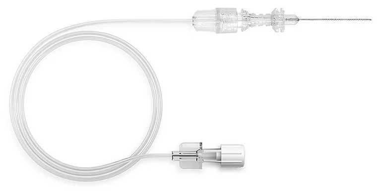
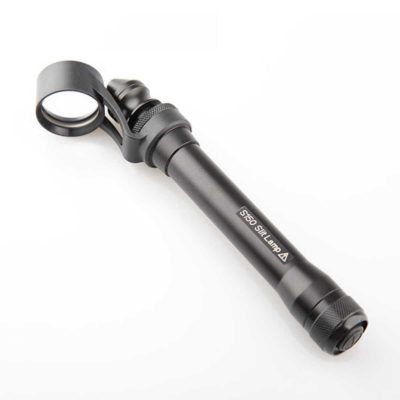
SonoEye-Echogenic Needle for Ultrasound-Guided Retrobulbar Blocks
$0.00
Shipped From Abroad
Ultrasound-guided ophthalmic blocks ensure excellent anesthesia with high success rates for eye surgeries. PAJUNK’s SonoEye needle combines the proven Atkinson tip with the innovative Cornerstone Reflectors for optimum needle visibility under ultrasound.
Delivery & Availability:
Typically 10-21 working days – excluding furniture and heavy/bulky equipment. Please contact us for further information.
Typically 10-21 working days – excluding furniture and heavy/bulky equipment. Please contact us for further information.
Description
SonoEye Features & Advantages
Cornerstone Reflectors
360° graduations on the first 20 mm of the needle in two sections:
- Optimized ultrasound visibility of the needle shaft4
- Reliable and optimized needle echogenicity at higher insertion angles.
Echogenic Needle Tip
- Atkinson tip
- Improved needle tip visibility under ultrasound
Quick Comparison
| SonoEye-Echogenic Needle for Ultrasound-Guided Retrobulbar Blocks remove | Handheld Digital Auto-refractometer remove | Portable Rebound Tonometer remove | Tonometer remove | ENT/Neurosurgery Operating Microscope remove | Manual lensmeter remove | ||||||||||||||||||||||||||||||||||||||||||
|---|---|---|---|---|---|---|---|---|---|---|---|---|---|---|---|---|---|---|---|---|---|---|---|---|---|---|---|---|---|---|---|---|---|---|---|---|---|---|---|---|---|---|---|---|---|---|---|
| Name | SonoEye-Echogenic Needle for Ultrasound-Guided Retrobulbar Blocks remove | Handheld Digital Auto-refractometer remove | Portable Rebound Tonometer remove | Tonometer remove | ENT/Neurosurgery Operating Microscope remove | Manual lensmeter remove | |||||||||||||||||||||||||||||||||||||||||
| Image |  | 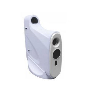 | 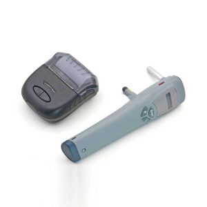 | 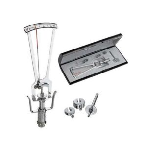 |  | 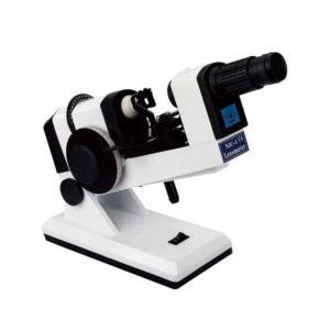 | |||||||||||||||||||||||||||||||||||||||||
| SKU | SF1033560130169-9 | SF1033560107-2 | SF1033560107-17 | SF1033560107-9 | SF1033560109-1 | SF1033560107-22 | |||||||||||||||||||||||||||||||||||||||||
| Rating | |||||||||||||||||||||||||||||||||||||||||||||||
| Price |
|
| $1,815.00 | $220.00 |
|
| |||||||||||||||||||||||||||||||||||||||||
| Stock | |||||||||||||||||||||||||||||||||||||||||||||||
| Availability | |||||||||||||||||||||||||||||||||||||||||||||||
| Add to cart | |||||||||||||||||||||||||||||||||||||||||||||||
| Description | Shipped From Abroad
Ultrasound-guided ophthalmic blocks ensure excellent anesthesia with high success rates for eye surgeries. PAJUNK’s SonoEye needle combines the proven Atkinson tip with the innovative Cornerstone Reflectors for optimum needle visibility under ultrasound.
Delivery & Availability:
Typically 10-21 working days – excluding furniture and heavy/bulky equipment. Please contact us for further information.
| Shipped from abroad
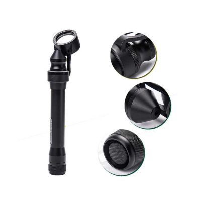
AutoSight 900 is a portable vision screener for patients at any age. Its working principle is the refraction of light.
| Shipped from abroad
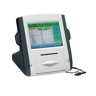
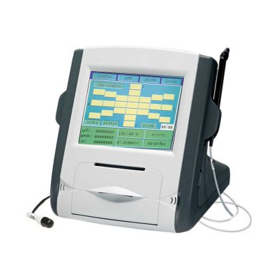
Tonometer SW-500 with vertical and horizontal two working modes, wireless output print data.
| Shipped from abroad
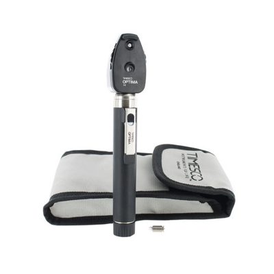
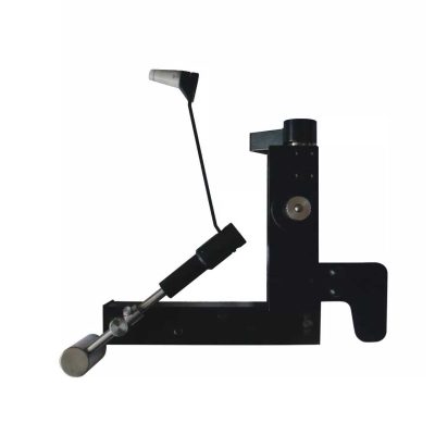
This product is used to measure the intraocular pressure (IOP) by measuring the depth produced on the surface of the cornea by a load of a known weight. Each division on the scale corresponds to 1/20mm corneal depth.
| Shipped from abroad
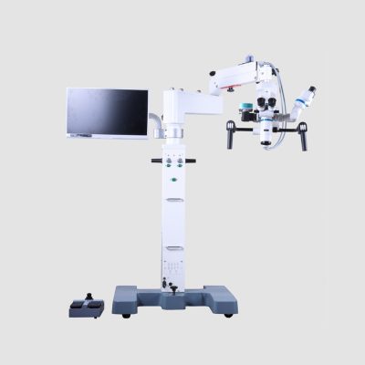
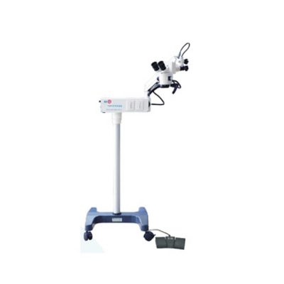
Corder Microscope has Fluid, Responsive and Accurate.Fluid. Responsive. Accurate. These were a few of the principles guiding every phase in the design of the Corder Microscope. With the choicest mechanical machined components, the Corder Microscope has the grace and agility to adjust to every desired position on command. Well designed Apochromatic optics treated with Corder's Mcoatings produce true-to life sharp images with high depth, definition and contrast. | Shipped from abroad
The NJC-4 Lensmeter is used to measure the diopters of spherical lens and cylinder lens, the axis of cylinder lens, the strength and the baseline direction of shuttle lens, it can also stamp the optical center of lens, the axis of cylinder len and the base direction of shuttle lens.
| |||||||||||||||||||||||||||||||||||||||||
| Content | SonoEye Features & AdvantagesCornerstone Reflectors
360° graduations on the first 20 mm of the needle in two sections:
Echogenic Needle Tip
| Handheld Digital Auto-refractometer(AutoSight 900) is a portable vision screener for patients at any age. Its working principle is the refraction of light. Optical rays are focused on a sensor after passing through the eye's refractive system. The spherical power, cylindrical power, and axis of both eyes can be obtained by digital signal processing.
Features of Handheld Digital Auto-refractometer:
| Tonometer SW-500 with vertical and horizontal two working modes, wireless output print data. The equipment is used to measure intraocular pressure, using the principle of: the probe hits the surfaces of different hardness at a certain speed, has a different reaction when the probe rebounds. Be of advantages of high accuracy, portable, without anesthesia, without the cross-infection, etc.
Features:
| This product is used to measure the intraocular pressure (IOP) by measuring the depth produced on the surface of the cornea by a load of a known weight. Each division on the scale corresponds to 1/20mm corneal depth.
Features:
| Features:Corder Microscope has Fluid, Responsive and Accurate.Fluid. Responsive. Accurate. These were a few of the principles guiding every phase in the design of the Corder Microscope. With the choicest mechanical machined components, the Corder Microscope has the grace and agility to adjust to every desired position on command. Well designed Apochromatic optics treated with Corder's Mcoatings produce true-to life sharp images with high depth, definition and contrast. More comfortable operation Tiltable binocular tubes available, which can incline more than 60° depending on the posture and physique of the operating surgeon. Movable range: 30° (straight) to 90° (inclined) Corder microscope configured with XYZ motorized movement operated through a comfortable foot /Handle control, a veryeffective co-axial illumnation and 50W halogen light source makes it ideal for Neuro surgeries.Doctor-patient communication is easierTo address digital documentation needs, a host of digital SLR, video camera, and CCD adapters are made available with the ProLine in addition to Corder's proprietary iVu multi-functional imaging solution. 1080P full hd image quality, efficient image management during the operation. Integrate your digital workflow to facilitate case management and facilitate more intuitive patient communication. Technical Permeants: Magnification: motorized zoom system, 1:6 zoom ratio, magnification 3x~16x Focusing range: 50mm Binocular tube: 30°~90° tiltable tube ,(0° ~200° optional) Eyepiece: 12.5x / 10x Objective lens: F 300mm(175mm, 250mm, 350mm optional) pupil distance: 55mm~75mm diopter adjustment: +6D ~ -6D Field of view: Φ74~Φ12mm X-Y translator: Motorized by foot switch or handle controller, ±30mm Assistant tube: 360° Rotating assistant tube Reset functions: YES Illumination System: Coaxial illumination Light source: Halogen lamp Light intensity adjustment: Continuous brightness adjustment 0-100000lux Fiber optic illumination: Dual fiber Field of illumination: Φ50mm Filter: Red free filter, small spot Accessories CCD Camera system: Beam splitter, CCD adapter, CCD, Display XENON LAMP: 150000lux Integrated Video Adapter: SONY / CANON CameraClick Here To Download Catalogue | The NJC-4 Lensmeter is used to measure the diopters of the spherical lens and cylinder lens, the axis of cylinder lens, the strength and the baseline direction of shuttle lens, it can also stamp the optical center of the lens, the axis of cylinder lens, and the base direction of shuttle lens. This instrument (Both Ac and Dc are permitted Two cells When Dc) has clear readings and graduations as well as high objective precision and reliabilities except that it can be operated easily and conveniently, AU the lenses and the made glasses can be measured by it, therefore, it is a required and ideal measuring instrument for glasses manufactures, glasses stores and ophthalmological hospitals.
Technical Specifications:
| |||||||||||||||||||||||||||||||||||||||||
| Weight | N/A | N/A | N/A | N/A | N/A | N/A | |||||||||||||||||||||||||||||||||||||||||
| Dimensions | N/A | N/A | N/A | N/A | N/A | N/A | |||||||||||||||||||||||||||||||||||||||||
| Additional information | |||||||||||||||||||||||||||||||||||||||||||||||


Reviews
There are no reviews yet.