SonoEye-Echogenic Needle for Ultrasound-Guided Retrobulbar Blocks
$0.00
Shipped From Abroad
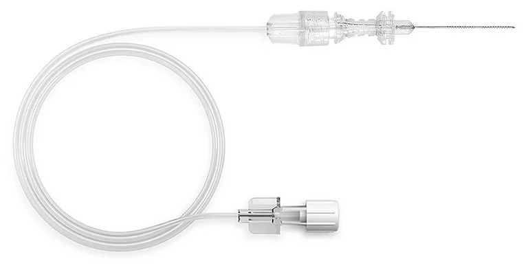
Ultrasound-guided ophthalmic blocks ensure excellent anesthesia with high success rates for eye surgeries. PAJUNK’s SonoEye needle combines the proven Atkinson tip with the innovative Cornerstone Reflectors for optimum needle visibility under ultrasound.
Delivery & Availability:
Typically 10-21 working days – excluding furniture and heavy/bulky equipment. Please contact us for further information.
Typically 10-21 working days – excluding furniture and heavy/bulky equipment. Please contact us for further information.
Description
SonoEye Features & Advantages
Cornerstone Reflectors
360° graduations on the first 20 mm of the needle in two sections:
- Optimized ultrasound visibility of the needle shaft4
- Reliable and optimized needle echogenicity at higher insertion angles.
Echogenic Needle Tip
- Atkinson tip
- Improved needle tip visibility under ultrasound
Quick Comparison
| SonoEye-Echogenic Needle for Ultrasound-Guided Retrobulbar Blocks remove | Ophthalmic AB Scan Machine remove | Pantoscopic Ophthalmoscope remove | Retinoscope remove | Handheld Digital Auto-refractometer remove | Applanation Tonometer remove | |||||||||||||||||||||||||||||||||||||||||
|---|---|---|---|---|---|---|---|---|---|---|---|---|---|---|---|---|---|---|---|---|---|---|---|---|---|---|---|---|---|---|---|---|---|---|---|---|---|---|---|---|---|---|---|---|---|---|
| Name | SonoEye-Echogenic Needle for Ultrasound-Guided Retrobulbar Blocks remove | Ophthalmic AB Scan Machine remove | Pantoscopic Ophthalmoscope remove | Retinoscope remove | Handheld Digital Auto-refractometer remove | Applanation Tonometer remove | ||||||||||||||||||||||||||||||||||||||||
| Image |  | 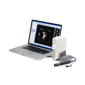 | 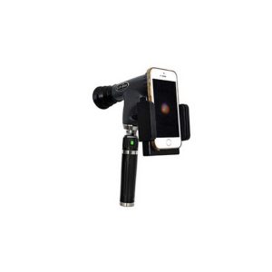 | 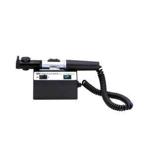 | 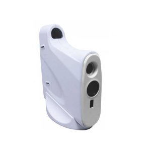 | 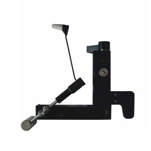 | ||||||||||||||||||||||||||||||||||||||||
| SKU | SF1033560130169-9 | SF1033560107-8 | SF1033560107-3 | SF1033560107-12 | SF1033560107-2 | SF1033560107-1 | ||||||||||||||||||||||||||||||||||||||||
| Rating | ||||||||||||||||||||||||||||||||||||||||||||||
| Price |
| $4,895.00 |
| $165.00 |
|
| ||||||||||||||||||||||||||||||||||||||||
| Stock | ||||||||||||||||||||||||||||||||||||||||||||||
| Availability | ||||||||||||||||||||||||||||||||||||||||||||||
| Add to cart | ||||||||||||||||||||||||||||||||||||||||||||||
| Description | Shipped From Abroad
Ultrasound-guided ophthalmic blocks ensure excellent anesthesia with high success rates for eye surgeries. PAJUNK’s SonoEye needle combines the proven Atkinson tip with the innovative Cornerstone Reflectors for optimum needle visibility under ultrasound.
Delivery & Availability:
Typically 10-21 working days – excluding furniture and heavy/bulky equipment. Please contact us for further information.
| Shipped from abroad
| Shipped from abroad
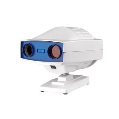
The brand-new Pantoscopic Ophthalmoscope is a portable digital imaging device which makes it possible to view and take pictures of the eyes.
| Shipped from abroad
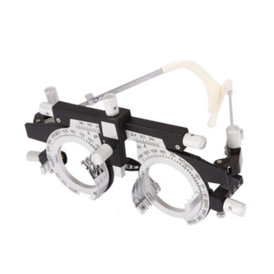
The product can quickly and precisely measure the astigmatism axis and is one of the necessary instruments in optometry inspection.
| Shipped from abroad
AutoSight 900 is a portable vision screener for patients at any age. Its working principle is the refraction of light.
| Shipped from abroad
The product is designed on the principle basis of Goldman tonometer. It can be connected with slit lamp(Carl Zeiss type).
| ||||||||||||||||||||||||||||||||||||||||
| Content | SonoEye Features & AdvantagesCornerstone Reflectors
360° graduations on the first 20 mm of the needle in two sections:
Echogenic Needle Tip
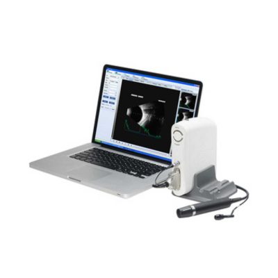
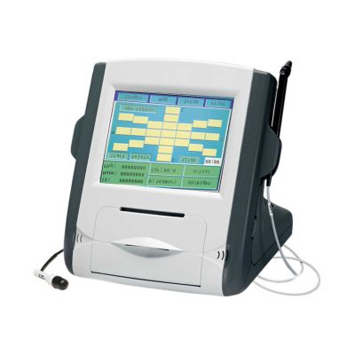
| Functions of Ophthalmic AB Scan Machine:
| The brand-new Pantoscopic Ophthalmoscope is a portable digital imaging device which makes it possible to view and take pictures of the eyes. The optical access of the Pantoscopic Ophthalmoscope is aligned to the visual axis of the smartphone camera by the adaptor which allows to you take pictures of the fundus and retinal nerve in high resolution. You could save pictures for each patient or email and print as needed. The Pantoscopic Ophthalmoscope provides a 5X larger view of the fundus compared with the standard ophthalmoscope. It has a wider view field of 230. Without dilating the pupil, the fundus imagines could be captured at any time and places.
Features:
| The product can quickly and precisely measure the astigmatism axis and is one of the necessary instruments in optometry inspection.
Features:
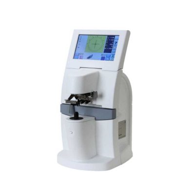
| Handheld Digital Auto-refractometer(AutoSight 900) is a portable vision screener for patients at any age. Its working principle is the refraction of light. Optical rays are focused on a sensor after passing through the eye's refractive system. The spherical power, cylindrical power, and axis of both eyes can be obtained by digital signal processing.
Features of Handheld Digital Auto-refractometer:
| Applanation Tonometer is designed on the principle basis of Goldman tonometer. It can be connected with slit lamp(Carl Zeiss type).
Features of Applanation Tonometer:
| ||||||||||||||||||||||||||||||||||||||||
| Weight | N/A | N/A | N/A | N/A | N/A | N/A | ||||||||||||||||||||||||||||||||||||||||
| Dimensions | N/A | N/A | N/A | N/A | N/A | N/A | ||||||||||||||||||||||||||||||||||||||||
| Additional information | ||||||||||||||||||||||||||||||||||||||||||||||


Reviews
There are no reviews yet.