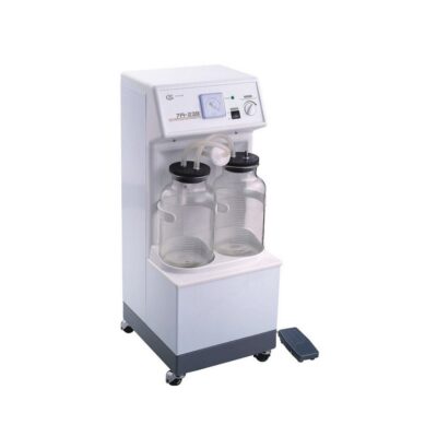
System for measuring hourly diuresis MISURO
$0.00
Shipped From Abroad
Typically 10-21 working days – excluding furniture and heavy/bulky equipment. Please contact us for further information.
Description
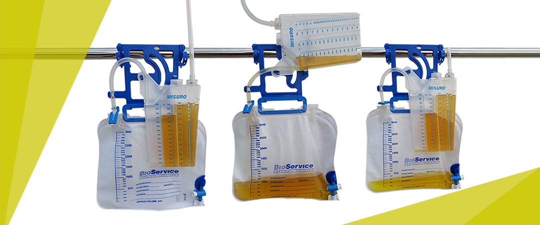
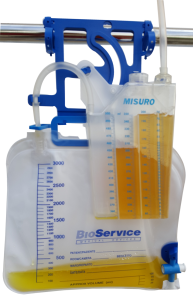
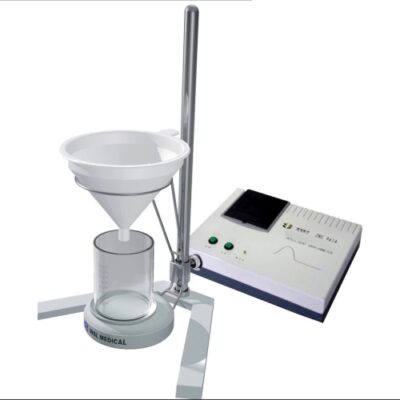
The device is made up of :
- a graduated rigid plastic container ( 500 ml capacity ) for measuring waste fluid , and is equipped with a pre- drip chamber antibacterial filter and a non-return valve.
- drainage tube with wide lumen and smooth internal surface to guarantee high flow.
- graded catheter cone with universal connector with drawing point and protective cap.
- Tube-clamp plate for vesical maneuvers and sheet clip for proper positioning of the tube.
- Collection bag in various capacities with non-return valve , antibacterial filter and drainage tap with rigid storage support.
- System for attachement to bed with two clamps ( optionally the device can be supplied with a hook ) .
| MISURO | |||
|---|---|---|---|
| MODEL | BAG | CODE | CODE (with hook) |
| MISURO 15 | 1500 ml (without drainage tap) | H90033E | H90033E-G |
| MISURO 20 | 2000 ml (with drainage tap) | H90034E | H90034E-G |
| MISURO 30 | 3000 ml (with drainage tap) | H90035E | H90035E-G |
| MISURO 35 | 3500 ml (without drainage tap) | H90037E | H90037E-G |
| MISURO 50 | 5000 ml (without drainage tap) | H90036E | H90036E-G |
All the diveces are available in multi-packs of 10 pieces
MISURO ACCESSORIES
The bag needs to be replaced without having to disconnect the urinometer from the patient , BioService offers a series of spare bags in various capacities.
Collection bag replacement is very practical and fast.
The device is supplied in individual packaging sterilized by EtO
| SPARE BAG | |||
|---|---|---|---|
| CODE | CAPACITY | PACKAGING | |
| H90111E | 1500 ml | 100 pcs | |
Click here to download Catalogue
Quick Comparison
| System for measuring hourly diuresis MISURO remove | Vigeo VTek Reusable Biopsy Device remove | Sonoscape P10 Ultrasound Machine remove | Ecleris C100-FID Binocular Colposcope remove | Sonoscape P50 Ultrasound Machine remove | Sonoscape S22 Ultrasound Machine remove | |||||||||||||||||||||||||||||||||||||||
|---|---|---|---|---|---|---|---|---|---|---|---|---|---|---|---|---|---|---|---|---|---|---|---|---|---|---|---|---|---|---|---|---|---|---|---|---|---|---|---|---|---|---|---|---|
| Name | System for measuring hourly diuresis MISURO remove | Vigeo VTek Reusable Biopsy Device remove | Sonoscape P10 Ultrasound Machine remove | Ecleris C100-FID Binocular Colposcope remove | Sonoscape P50 Ultrasound Machine remove | Sonoscape S22 Ultrasound Machine remove | ||||||||||||||||||||||||||||||||||||||
| Image |  | 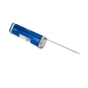 |  | 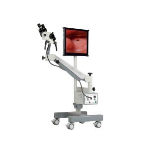 |  |  | ||||||||||||||||||||||||||||||||||||||
| SKU | SF1033560130101-4 | SF10335601282 | SF1033560012-7 | SF1033560087-1 | SF1033560012-11 | SF1033560012-3 | ||||||||||||||||||||||||||||||||||||||
| Rating | ||||||||||||||||||||||||||||||||||||||||||||
| Price |
|
| $9,350.00 | $5,042.00 |
| $9,350.00 | ||||||||||||||||||||||||||||||||||||||
| Stock | ||||||||||||||||||||||||||||||||||||||||||||
| Availability | ||||||||||||||||||||||||||||||||||||||||||||
| Add to cart | ||||||||||||||||||||||||||||||||||||||||||||
| Description | Shipped From Abroad
Delivery & Availability:
Typically 10-21 working days – excluding furniture and heavy/bulky equipment. Please contact us for further information.
| Shipped From Abroad
Features:
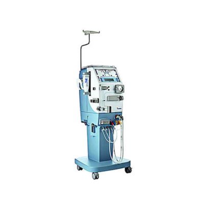
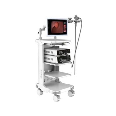
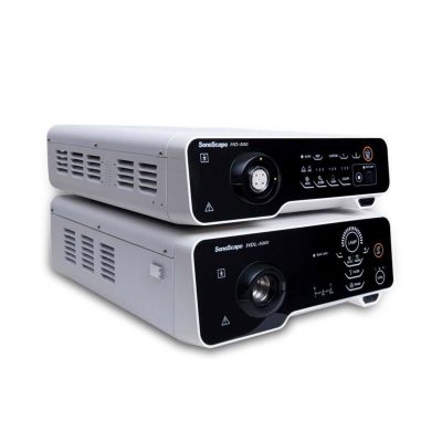
| Shipped from Abroad The P10 color Doppler ultrasound system is a new generation product from SonoScape. It is designed to give high quality images, rich probe configurations, various clinical tools and automatic analysis software to provide you with comprehensive solutions for your growing demand for clinical applications. Delivery & Availability: Typically 5-7 working days – excluding furniture and heavy/bulky equipment. Please contact us for further information. | Shipped from Abroad Content Includes: Forearm, Pantographic Arm, Floor Stand (H Shaped Base and Column), 5 Magnifications Head, Green Filter and Inclined Binocular, LED Light Source, Fiber Optic Cable, 110/220V Power Cable and User`s Guide, Stand for LCD Monitor and Printer, Digital Capturing System for Images, Videos and Sounds, USB 2.0. Includes Main Unit Processor with three Camera Inputs, USB Cable, Software, Hands Free Microphone and Footswitch. Delivery & Availability: Typically 10 working days – excluding furniture and heavy/bulky equipment. Please contact us for further information. | Shipped from Abroad Easily accomplish more with SonoScape’s new P50 ultrasound system. Incorporating single crystal clarity, automatic corrections and calculation, and user defined flexibility promises a confident diagnostic experience as well as opening new doors of opportunity for ultrasound use. Delivery & Availability: Typically 7-14 working days – excluding furniture and heavy/bulky equipment. Please contact us for further information. | Shipped from Abroad As SonoScape steps forward to add value and efficiency to ultrasound, the latest S22 was designed in a user-friendly platform to address current and future demanding needs. It represents an excellent mix in performance and price. Delivery & Availability: Typically 5-7 working days – excluding furniture and heavy/bulky equipment. Please contact us for further information. | ||||||||||||||||||||||||||||||||||||||
| Content | The device is made up of :
Click here to download Catalogue | Vigeo VTek Reusable Biopsy Device Features:
Click Here To Download Catalogue | DETAILS
B + Compound
B + Compound utilizes several lines of sight for optimal contrast resolution, speckle reduction and border detection, with which P10 is ideal for superficial and abdominal imaging with better clarity and improved continuity of structures.
μ-Scan
The new generation μ-Scan imaging technology gives you better image quality by reducing noise, improving signal strength and improving visualization.
P10 offers a comprehensive selection of electronic probes to maximize its capabilities to meet a wide range of applications including abdomen, pediatric, OB/GYN, cardiovascular, musculoskeletal, etc. The advanced probe technologies also effectively enhance the image quality and confidence in reaching clinical diagnoses, even in difficult patients.
Convex Probe 3C-A
Ideal for an abundant of application such as abdomen, gynecology, obstetrics, urology and even abdomen biopsy.
Linear Probe L741
This linear probe is designed to satisfy vascular, breast, thyroid, and other small parts diagnosis, and its adjustable parameters could also present users a clear view of MSK and deep vessels.
Phase Array Probe 3P-A
For the purpose of adult and pediatric cardiology and emergency, the phase array probe provides elaborate presets for different exam modes, even for difficult patients.
Intracavitary Probe 6V1
Intracavitary probe could face application of gynecology, urology, prostate, and its temperature detection technology not only protects the patient but also extends the service life.
Click Here To Download Catalogue | Ecleris Colposcope Series C-100 was designed to cover all the diagnosis and therapeutic needs of modern gynecology. All models can be transformed into video colposcopes with our high resolution video camera. Digital handling of patients and image filing is achieved through the endoDIGI software which is easily adaptable to a portable PC or desktop computer. Great and accurate quality images, improved clearness, resolution and focal range can be obtained through our new C-100 Colposcope optic system. Floor and wall-mounted models include 5 magnifications (4, 6, 10, 16 and 25x).
The C-100 model, with its pantographic arm has enhanced maneuverability, as the arms are mounted on bearings and guarantee smooth movements and greater stability (WBS, weight balance system). Its state-of-the-art designed base allows for easy transport. We provide accessories that allow using the microscope light source and video camera also to carry out endoscopic studies, without need to have two light sources and two video cameras at the doctor’s office.
A new dimension in microsurgery
Ecleris 3D Splitter
Click Here To Download Catalogue | DETAILS
Powerful Compact Precision
Taking into consideration the evolving expectations and needs for ultrasound, the P50 is a slim and unobtrusive trolley system that is comfortable in tight, congested spaces with little room to work in. Providing everything you need for a comfortable examination in a small space for both you and your patient.
Single Crystal Transducer
Wideband single crystal probes greatly improve the signal ratio, acquire stunning images and provide superior sensitivity and resolution for both the near and far-fields.
μ-Scan+
The new generation μ-Scan imaging technologies give you better image quality by reducing noise, improving signal strength and improving visualization.
Dynamic Color
Dynamic colour improves upon already existing colour Doppler technologies for clear capture of colour flow and detail visualization of even tiny veins with lower velocities.
Solution for Radiology
P50, is a leading-edge ultrasound system that can meet the demands of any clinical setting. You can experience a superior performance in multi-dimensional imaging for a full range of clinical applications – abdominal, breast and cardiovascular.
C-xlasto Imaging
By understanding that tissue stiffness varies depending on the type of tissue, we can use C-xlasto Imaging to easily find abnormalities and tumours within soft tissue. The differences in tissue responses are detected and visualized in real-time by the elastography algorithms through different representations, which can be particularly helpful in analyzing breast, thyroid and musculoskeletal structures. Predominately used only in linear probes, SonoScape’s new transvaginal and bi-plane probe for gynaecology and urology are breaking the mould and expanding elastography applications.
Real-time Color Panoramic
With the combination of colour flow and real-time panoramic, visualizing the blood flow of an entire vein or artery is now an easy task. Accomplished in real-time for the convenience of the sonographers, any mistakes can also be easily backtracked and corrected without interrupting the scan.
Contrast Imaging
Contrast Imaging on P50 makes full use of the infra harmonic signal and second harmonic signal to improve the image resolution and deep penetration. What’s more, the Dynamic Acoustic Control technology effectively controls the acoustic pressure for the contrast agent, decreasing the required agent dose and assures uniform image quality, guaranteeing longer contrast agent duration and better lesion perfusion of delayed phase observation.
Solution for OB/GYN
P50 has superior image quality, automated measurement tools, and a variety of volume technologies to provide ideal solutions for clinical examinations such as pregnancy examinations, and gynecologic disease diagnosis. With a new 4D transvaginal probe, P50 helps you to see and detect fetal abnormalities and significantly improves your diagnostic confidence during your examinations.
S-Live Silhouette
A unique transparent 3D anatomical image of the fetus for improved initial anatomical review. By using this new application, the system can create completely different fetal images from conventional ultrasound images, which can depict the fetal's intracorporeal anatomical structure.
Pelvic Floor 4D
Working in conjunction with SonoScape’s latest transvaginal probes, trans-perineal 4D pelvic floor ultrasound provides a useful clinical assessment of the impact of vaginal delivery on the female anterior compartment. Allowing doctors to judge whether the pelvic organs prolapsed or not, the extent of prolapse, and determining whether the pelvic muscles tore correctly.
S-Guide
S-Guide gives the user an extensive list of example obstetric ultrasound images as reference guides and a convenient checklist system to keep track of their progress during their obstetrics examination.
Auto Face
Automatically removes masking layers in front of the fetus’s face for a clearer vision of the fetus’s face.
AVC Follicle
AVC Follicle automatically identifies how many follicles are present and calculates their individual volumes.
Solution for Cardiology
P50 provides clear 2D clinical images and Doppler sensitivity to assess critical cardiac performance. Compatible with SonoScape’s single crystal probes, the P50 can provide images with better resolution and penetration in Cardiac diagnosis.
Tissue Doppler Imaging
Tissue Doppler Imaging allows clinical doctors to quantitatively evaluate local myocardial movements and functions, facilitating them with the ability to analyze and compare the motions of the different parts of the patient’s heart.
Stress Echo
Stress echocardiography is the combination of 2D echocardiography with physical, pharmacological or electrical stress of the patient. It also then provides users with report management tools such as configurable template editor, multiple loops to select one for storage, wall motion scoring, stress echo report, etc
Auto IMT
Auto IMT is used when determining the level of vascular sclerosis present in the patient by automatically tracing and calculating the thickness of the carotid vessels. What distinguishes the P50 is that it provides an instant and accurate Mean and Max index at the touch of a single button.
Auto EF
Automated 2D Cardiac Quantification is a fully intelligent trace function for endocardium with 19 easily-adjustable points providing rapid access to proven 2D EF and volumes.
Click Here To Download Catalogue | DETAILS
As SonoScape steps forward to add value and efficiency to ultrasound, the latest S22 was designed in a user-friendly platform to address current and future demanding needs. It represents an excellent mix in performance and price.
S22, is a shared service ultrasound system with a slim and elegant package that has combined mobility with utility to fit in specific clinical situations including emergency department, ICU, operating room and so on. Furthermore, its ergonomic design, easy operating and flexible data management will give you a memorable experience.
SPECIFICATION
• Large high-resolution widescreen LED
• Sensitive touch screen
• Four transducer sockets plus one socket for pencil probe
• A comprehensive selection of probes: linear, Convex, Micro-convex, Volumetric, Endocavity, Bi-plane, Phased Array, TEE, Intraoperative, Pencil
• Premium application technology: 4D, μ-scan speckle reduction, compound imaging, Pulse Inversion Harmonic Imaging, Color M-Mode, Steer M-Mode, PDI, TDI, Real-time Panoramic Imaging, Trapezoid Imaging, Auto-IMT…
• Full patient database and image management solutions: DICOM 3.0, AVI/JPG, USB 2.0, HDD, DVD, PDF report
• Multi-Language Input Keyboard
• Built-in battery
Click Here To Download Catalogue | ||||||||||||||||||||||||||||||||||||||
| Weight | N/A | N/A | N/A | N/A | N/A | N/A | ||||||||||||||||||||||||||||||||||||||
| Dimensions | N/A | N/A | N/A | N/A | N/A | N/A | ||||||||||||||||||||||||||||||||||||||
| Additional information | ||||||||||||||||||||||||||||||||||||||||||||


Reviews
There are no reviews yet.