
Wireless Endoscopy Camera CW-2000
$0.00
Shipped From Abroad
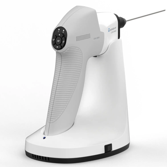
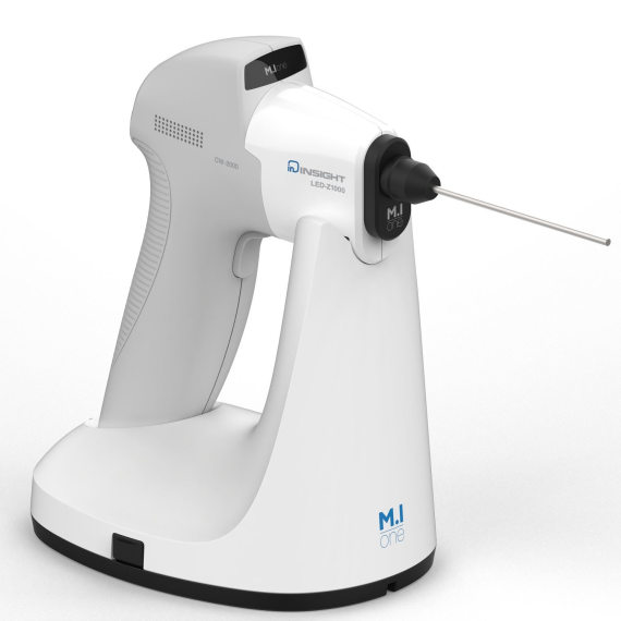
The CW-2000 is a wireless endoscopic camera system developed by M.I. One Co., Ltd., is designed to enhance efficiency and convenience in various endoscopic procedures. Its ergonomic design ensures a comfortable grip, eliminating the hassle of wires, and it offers Full HD image transmission at 1920 x 1080 resolution with a frame rate of 60p. The system features auto white balance, auto-focusing, and a zoom function ranging from 1.0 to 2.5 times magnification. An integrated LED light source provides illumination with a luminance of 220,000 lux. The wireless receiver maintains a connection range exceeding 3 meters, improving the work efficiency of medical staff. Additionally, the system includes an auto-rechargeable charging cradle for convenience.
Typically 10-21 working days – excluding furniture and heavy/bulky equipment. Please contact us for further information.
Description
Features
- Ergonomic design for comfortable grip, No hassle of wire
- Full HD Images (1920×1080 60p) transmission
- Auto White Balance
- Auto Focusing
- Zoom In /Out (1.0 ~ x 2.5 image )to enhance operating efficiency
- Integrated LED Light Source
- Changeable endoscope for each suitable diagnosis
- Smart On/Off System
- Auto rechargeable Charging cradle system
Technical Details
- Camera Size Resolution:
- Full-HD(1920×1080 60p)
- Image sensor: 1/2.8” CMOS
- White balance: AWB, preset manual
- Digital Zoom : x1.0~x2.5(0.1 step)
- Freeze : 1(Full) / 2 split view
- Wireless frequency: 60GHz
- Wireless Range : 3m
- Receiver video out: HDMI
- Light source color temperature: 5700K
- Light source luminance: 220,000 lux
- Battery Operation Time: approx. 2 Hrs
- Full recharging time: approx. 4 Hrs
- Camera Weight: approx. 220 g (without Endoscope)
- Cradle Weight: approx. 400 g
- Adaptor Input: AC 100-240V 50/60Hz
- Adaptor output: : DC 12V, 2A
- Power Consumption:6W
Quick Comparison
| Wireless Endoscopy Camera CW-2000 remove | Durable Dental Chair Unit remove | Hearing Aid remove | Rigid scopes remove | M.I One ENT Treatment Unit ( IU-3000) remove | MIR Minispir New Spirometer remove | |||||||||||||||||||||||||
|---|---|---|---|---|---|---|---|---|---|---|---|---|---|---|---|---|---|---|---|---|---|---|---|---|---|---|---|---|---|---|
| Name | Wireless Endoscopy Camera CW-2000 remove | Durable Dental Chair Unit remove | Hearing Aid remove | Rigid scopes remove | M.I One ENT Treatment Unit ( IU-3000) remove | MIR Minispir New Spirometer remove | ||||||||||||||||||||||||
| Image | 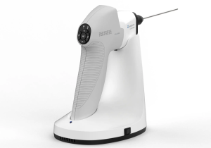 | 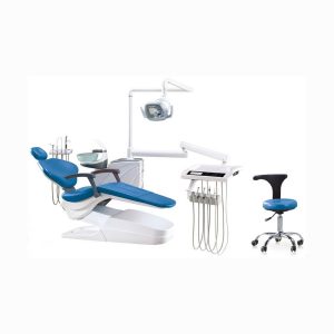 |  | 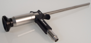 | 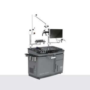 | 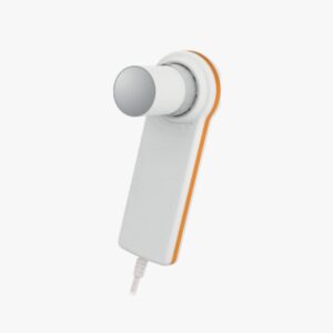 | ||||||||||||||||||||||||
| SKU | SF10335601264 | SF1033560067 | SF1033560084-119 | SF103356013089-3 | SF10335601261 | SF10335601295 | ||||||||||||||||||||||||
| Rating | ||||||||||||||||||||||||||||||
| Price |
| $1,089.00 | $16.90 |
|
|
| ||||||||||||||||||||||||
| Stock | ||||||||||||||||||||||||||||||
| Availability | ||||||||||||||||||||||||||||||
| Add to cart | ||||||||||||||||||||||||||||||
| Description | Shipped From Abroad
The CW-2000 is a wireless endoscopic camera system developed by M.I. One Co., Ltd., is designed to enhance efficiency and convenience in various endoscopic procedures. Its ergonomic design ensures a comfortable grip, eliminating the hassle of wires, and it offers Full HD image transmission at 1920 x 1080 resolution with a frame rate of 60p. The system features auto white balance, auto-focusing, and a zoom function ranging from 1.0 to 2.5 times magnification. An integrated LED light source provides illumination with a luminance of 220,000 lux. The wireless receiver maintains a connection range exceeding 3 meters, improving the work efficiency of medical staff. Additionally, the system includes an auto-rechargeable charging cradle for convenience.
Delivery & Availability:
Typically 10-21 working days – excluding furniture and heavy/bulky equipment. Please contact us for further information.
| Shipped From Abroad
Feature:
1. All controlled by the electric valve
2. DC Motor.(2000N)
3. Automatic thermostatic water supply system
4. Handpiece tubing with standard fittings (3sets),
5.Three way syringe (one for hot, one for cold)(2sets),
6.Water suction and saliva ejector (with switch)(1set),
7. Powerful suction apparatus (1set each)
8. Easy cleaning Integral and turn able toughened glass spittoon
Delivery & Availability:
Typically 14 working days – excluding furniture and heavy/bulky equipment. Please contact us for further information.
| In stock
| Shipped From Abroad
Delivery & Availability:
Typically 10-21 working days – excluding furniture and heavy/bulky equipment. Please contact us for further information.
| Shipped From Abroad
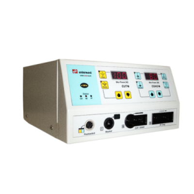
The IU-3000 ENT treatment unit is ergonomically designed to simplify ENT procedures, vastly improve efficiency and reduce user fatigue in the ENT consulting / treatment room.
Its additional storage space, articulated arm for Microscopy, CCU Camera, and various integrated functions makes it suitable for ENT consulting rooms
Delivery & Availability:
Typically 14-21 Working Days – excluding furniture and heavy/bulky equipment. Please contact us for further information.
| In Stock
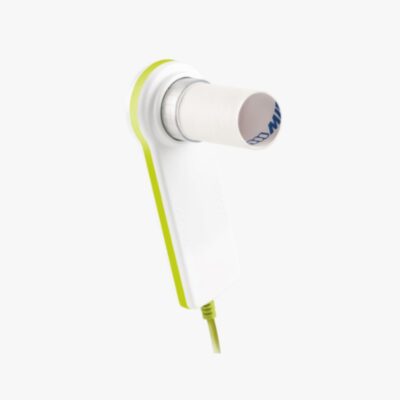
The MiniSpir New is a PC-based spirometer for complete airway analysis with extended spirometry test interpretation. The PC-Spirometer is delivered as standard with the WinspiroPRO software,
Delivery & Availability:
Typically 7-14 working days – excluding furniture and heavy/bulky equipment. Please contact us for further information.
| ||||||||||||||||||||||||
| Content |
https://youtu.be/CwuPlUQKWTg
Features
Technical Details
| Feature:
1. All controlled by the electric valve
2. DC Motor.(2000N)
3. Automatic thermostatic water supply system
4. Handpiece tubing with standard fittings (3sets),
5.Three way syringe (one for hot, one for cold)(2sets),
6.Water suction and saliva ejector (with switch)(1set),
7. Powerful suction apparatus (1set each)
8. Easy cleaning Integral and turn able toughened glass spittoon
9. Strong and weak dual-purpose operation light (1set),
10. X-film viewer (1 set)
11. Spirit lock tight Equilibrium device
12. Suitable for adult and children headrest.
13. Doctor stool (1set),
14. Foot pedal (1set)
Specifications:
Click To Download Catalogue |
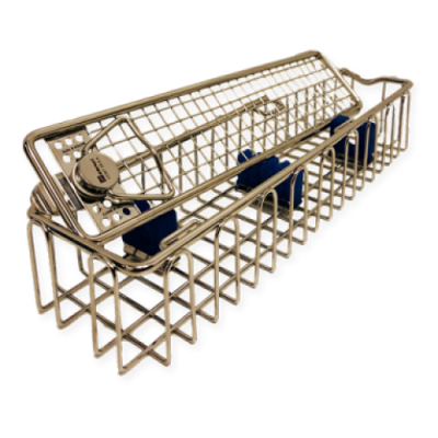
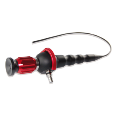
| They are available in a large range of external diameters, from 1 to 12 mm. Commonly, rigid endoscopes have a series of high-resolution optical glass rod lenses.Features
| The IU-3000 ENT treatment unit is ergonomically designed to simplify ENT procedures, vastly improve efficiency and reduce user fatigue in the ENT consulting / treatment room.
Its additional storage space, articulated arm for Microscopy, CCU Camera, and various integrated functions makes it suitable for ENT consulting rooms
Feature:
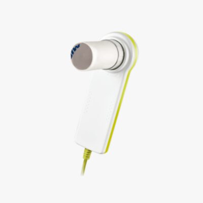
| The MiniSpir New is a PC-based spirometer for complete airway analysis with extended spirometry test interpretation. The PC-Spirometer is delivered as standard with the WinspiroPRO software, which is characterized by its high user-friendliness. Thanks to various, child-friendly animations, children can be motivated to play with their children. The MiniSpir New enables you to use cost-effective COPD and asthma screening and can be operated absolutely intuitively.
Features:
MIR Minispir Technical details
Click Here To Download Catalogue | ||||||||||||||||||||||||
| Weight | N/A | N/A | N/A | N/A | N/A | N/A | ||||||||||||||||||||||||
| Dimensions | N/A | N/A | N/A | N/A | N/A | N/A | ||||||||||||||||||||||||
| Additional information |


Reviews
There are no reviews yet.