

Anke Anatom 32 Fit Multi-Slice Spiral CT Scan
$0.00
Shipped from Abroad
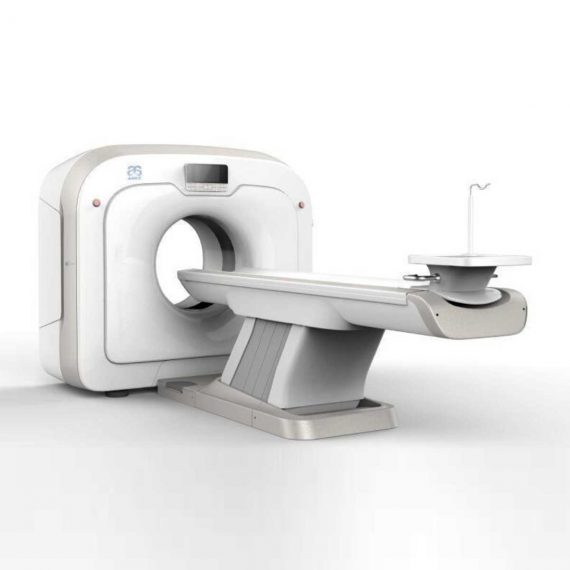
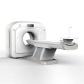
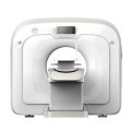
This Machine gives a possibility to perform computed tomography without any problems and on high quality level. This device is used to conduct exams of internal organs and their functioning. With its help, a physician has a possibility to assess the condition of the human body as a whole.
Delivery & Availability:
Typically 90 working days – excluding furniture and heavy/bulky equipment. Please contact us for further information.
Description
This Machine gives a possibility to perform computed tomography without any problems and on high quality level. This device is used to conduct exams of internal organs and their functioning. With its help, a physician has a possibility to assess the condition of the human body as a whole.
Features:
- It is easy to use;
- Convenience;
- Multi functionality;
- Obtained images are of high definition;
- High-definition 3D images of the area under study;
- The procedure is pain-free;
- The data is processed fast;
- The image can be stored in the computer memory;
- The diagnostics does not take a lot of time;
- Acceptable radiation dose.
Technical Specifications:
| No. | Technical Features | Descriptions |
| 1 | Gantry | |
| 1.01 | Gantry type | Low voltage slip-ring |
| 1.02 | Gantry driven type | Strap-driven |
| 1.03 | Patient opening | 70cm |
| 1.04 | Gantry tilt mode | Digital gantry tilt |
| 1.05 | Digital tilt capability | ±50° |
| 1.06 | Detector type | OptiWave rare-earth ceramic detector |
| 1.07 | Numbers of detector rows | 16 |
| 1.08 | Width of Z-axle detector | 20mm |
| 1.09 | Detector columns of channels per row | 848 |
| 1.10 | Numbers of detector columns | 13568 |
| 1.11 | Data-transfer type | RF, optical fiber communication |
| 1.12 | Distance of focus-ISO-center | 53cm |
| 1.13 | Distance of focus-detector | 94cm |
| 1.14 | 3D laser orientation | Provided |
| 1.15 | 13″ integrated display panel | Provided |
| 1.16 | Adose automatic exposure control (mA
Modulation) |
Provided |
| 1.17 | Auto-voice manager | Breath Graphical Display
Hold Message (Record/Playback) Breath Message (Record/Playback) |
| 1.18 | AccuSaving energy conservation management | Provided |
| 2 | HVPS and X-ray tube | |
| 2.01 | Maximum continuous output of HVgenerator | 42kW |
| 2.02 | Tube kV selections | 70kV, 80kV, 100 kV, 120 kV, 140 kV |
| 2.03 | Tube mA range | 10~350mA |
| 2.04 | Tube anode heat capacity | 3.5MHU |
| 2.05 | Max. anode cooling rate | 735kHU/min |
| 2.06 | Type of cooling | Oil cooling + Air cooling |
| 2.07 | Tube focus | Large: 1.2mm×1.4mm
Small: 0.7mm×0.8mm |
| 2.08 | Collimator width selection | 4-level election |
| 2.09 | Focus spot tracking technology | Provided |
| 3 | Patient table | |
| 3.01 | Maximum horizontal-movable range | 1850mm |
| 3.02 | Table horizontal-scannablerange | 1800mm |
| 3.03 | Table horizontal-position repeatability | ±0.25mm |
| 3.04 | Minimum height above floor | 430mm |
| 3.05 | Maximum vertical-movable range | 500mm |
| 3.06 | Maximum speed of vertical movement | 35mm |
| 3.07 | Maximum speed of horizontal movement | 150mm/s |
| 3.08 | Maximum patient weight | 205kg |
| 3.09 | Foot pedal of patient table control | Provided |
| 4 | Computer | |
| 4.01 | CPU | 3.5GHz |
| 4.02 | Memory | 32GB |
| 4.03 | Storage of hard-disk | 1TB×2 |
| 4.04 | Monitor | 24’’ LCD Monitor |
| 4.05 | Resolution of monitor | 1920×1200 |
| 4.06 | Image-data external storage type | CD/DVD/USB |
| 4.07 | Time of image reconstruction (512×512) | 33.3ms/image |
| 4.08 | Speed of image reconstruction (512×12) | 30fps |
| 4.09 | DICOM 3.0 interface | Provided |
| 4.10 | Printer DICOM 3.0 interface | Provided |
| 4.11 | Auto filming | Provided |
| 4.12 | Worklist function | Provided |
| 5 | Scan parameters | |
| 5.01 | Shortest 360 degree rotation time | 0.75s |
| 5.02 | Allowed rotation times | 0.75s, 1.0s, 1.5s, 2.0s, 3.0s, 4.0s |
| 5.03 | Maximum slice numbers per rotation | 32 |
| 5.04 | Minimum slice thickness of scan | 1.25mm |
| 5.05 | Minimum slice thickness of reconstruction | 0.625mm |
| 5.06 | Maximum slice thickness of scan | 20mm |
| 5.07 | Nominal reconstruction slice thickness | 0.625mm, 1.25mm, 2.5mm, 5.0mm, 7.5mm,
10mm, 20mm |
| 5.08 | Speed of image reconstruction (512×512) | 30 frames/s |
| 5.09 | Scan FOV | 50cm |
| 5.10 | Image reconstruction matrix | 512×512, 1024×1024 (Optional) |
| 5.11 | Image reconstruction matrix | 512×512, 1024×1024 (Optional) |
| 5.12 | Image display matrix | 512×512, 1024×1024 (Optional) |
| 5.13 | Maximum continuous scan duration | 120s |
| 5.14 | Maximum continuous scan length | 180cm |
| 5.15 | Direction of TOPO | Front-back, Left-right |
| 5.16 | Max. length of TOPO | 180cm |
| 5.17 | Range of pitch | 0.5~1.5 |
| 5.18 | Scan mode | Scout scan
Axial scan Helical scan Cine scan |
| 6 | Image Quality | |
| 6.01 | High contrast resolution | 21lp/cm@0%MTF |
| 6.02 | Low contrast resolution | 2.0mm@0.30% |
| 6.03 | Isotropic imaging resolution | 0.24mm |
| 6.04 | Range of CT numbers | -32767~32768 |
| 6.05 | Image noise | ≤0.29@28mGy |
| 7 | Advanced application | |
| 7.01 | Multi-Planar Reconstruction (MPR) | Provided |
| 7.02 | Curve Multi-Planar Reconstruction (CPR) | Provided |
| 7.03 | Surface Shaded Display (SSD) | Provided |
| 7.04 | Volume Rendering (VR) | Provided |
| 7.05 | Maximum Intensity Projection (MIP) | Provided |
| 7.06 | Minimum Intensity Projection (MinIP) | Provided |
| 7.07 | Virtual Endoscopy (VE) | Provided |
| 7.08 | CT angiography (CTA) | Provided |
| 7.09 | Tissue segmentation | Provided |
| 7.10 | One click bone remove | Provided |
| 7.11 | One click patient table remove | Provided |
| 7.12 | Bolus-tracking Technology | Provided |
| 7.13 | Spiral auto start | Provided |
| 7.14 | Cine display | Provided |
| 7.15 | AbastTM bone artifact suppression technology | Provided |
| 7.16 | AmastTM metal artifact suppression technology | Provided |
| 7.17 | Admir3D all-domain iterative reconstruction | Provided |
| 7.18 | Low-dose pediatric scan technology | Provided |
| 7.19 | Low-dose lung scan technology | Provided |
| 7.20 | AccuHead grey-white matter enhanced
technology |
Provided |
| 7.21 | AccuOrgan lung high resolution scan technology | Provided |
| 7.22 | AccuOrgan inner-ear high resolution scan
technology |
Provided |
| 7.23 | AccuOrgan body high resolution scan technology | Provided |
| 7.24 | AccuOrgan bone high resolution scan technology | Provided |
| 7.25 | AccuMatter dual-energy with Admir3D for new
application |
Provided |
Click Here To Download Catalogue
Additional information
| Model | Advanced, Advanced Plus, Basic, Smart |
|---|
Quick Comparison
| Anke Anatom 32 Fit Multi-Slice Spiral CT Scan remove | Jade Mobile X-ray machine (Analogue) remove | ASPEL AsCARD Coral PC Based ECG Machine remove | Sonoscape P10 Ultrasound Machine remove | Sonoscape E2 Ultrasound Machine remove | DrGem Diamond All-In-One Digital X-ray Machine remove | |||||||||||||||||||||||||||||||||||||||||||||||||||||||||||||||||||||||||||||||||||||||||||||||||||||||||||||||||||||||||||||||||||||||||||||||||||||||||||||||||||||||||||||||||||||||||||||||||||||||||||||||||||||||||||||||||||||||||||||||||||||||||||||||||||||||||||||||||||||||||||||||||||||||||||||||||||||||||
|---|---|---|---|---|---|---|---|---|---|---|---|---|---|---|---|---|---|---|---|---|---|---|---|---|---|---|---|---|---|---|---|---|---|---|---|---|---|---|---|---|---|---|---|---|---|---|---|---|---|---|---|---|---|---|---|---|---|---|---|---|---|---|---|---|---|---|---|---|---|---|---|---|---|---|---|---|---|---|---|---|---|---|---|---|---|---|---|---|---|---|---|---|---|---|---|---|---|---|---|---|---|---|---|---|---|---|---|---|---|---|---|---|---|---|---|---|---|---|---|---|---|---|---|---|---|---|---|---|---|---|---|---|---|---|---|---|---|---|---|---|---|---|---|---|---|---|---|---|---|---|---|---|---|---|---|---|---|---|---|---|---|---|---|---|---|---|---|---|---|---|---|---|---|---|---|---|---|---|---|---|---|---|---|---|---|---|---|---|---|---|---|---|---|---|---|---|---|---|---|---|---|---|---|---|---|---|---|---|---|---|---|---|---|---|---|---|---|---|---|---|---|---|---|---|---|---|---|---|---|---|---|---|---|---|---|---|---|---|---|---|---|---|---|---|---|---|---|---|---|---|---|---|---|---|---|---|---|---|---|---|---|---|---|---|---|---|---|---|---|---|---|---|---|---|---|---|---|---|---|---|---|---|---|---|---|---|---|---|---|---|---|---|---|---|---|---|---|---|---|---|---|---|---|---|---|---|---|---|---|---|---|---|---|---|---|---|---|---|
| Name | Anke Anatom 32 Fit Multi-Slice Spiral CT Scan remove | Jade Mobile X-ray machine (Analogue) remove | ASPEL AsCARD Coral PC Based ECG Machine remove | Sonoscape P10 Ultrasound Machine remove | Sonoscape E2 Ultrasound Machine remove | DrGem Diamond All-In-One Digital X-ray Machine remove | ||||||||||||||||||||||||||||||||||||||||||||||||||||||||||||||||||||||||||||||||||||||||||||||||||||||||||||||||||||||||||||||||||||||||||||||||||||||||||||||||||||||||||||||||||||||||||||||||||||||||||||||||||||||||||||||||||||||||||||||||||||||||||||||||||||||||||||||||||||||||||||||||||||||||||||||||||||||||
| Image | 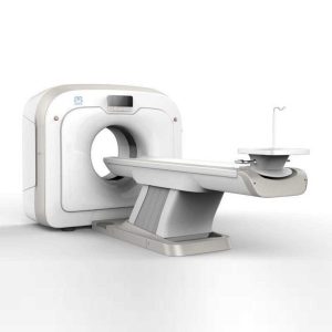 | 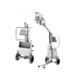 |  | 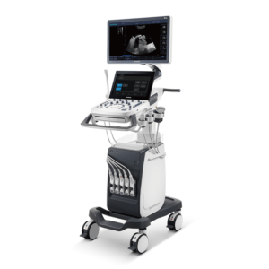 | 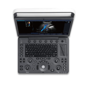 | 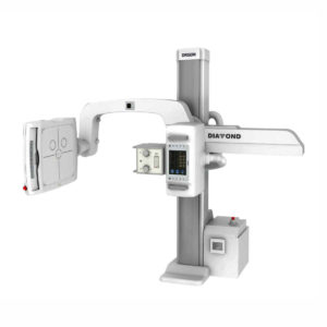 | ||||||||||||||||||||||||||||||||||||||||||||||||||||||||||||||||||||||||||||||||||||||||||||||||||||||||||||||||||||||||||||||||||||||||||||||||||||||||||||||||||||||||||||||||||||||||||||||||||||||||||||||||||||||||||||||||||||||||||||||||||||||||||||||||||||||||||||||||||||||||||||||||||||||||||||||||||||||||
| SKU | SF1033560092-1 | SF1033560074-2 | SF1033560075-11 | SF1033560012-7 | SF1033560012-17 | SF1033560074-3 | ||||||||||||||||||||||||||||||||||||||||||||||||||||||||||||||||||||||||||||||||||||||||||||||||||||||||||||||||||||||||||||||||||||||||||||||||||||||||||||||||||||||||||||||||||||||||||||||||||||||||||||||||||||||||||||||||||||||||||||||||||||||||||||||||||||||||||||||||||||||||||||||||||||||||||||||||||||||||
| Rating | ||||||||||||||||||||||||||||||||||||||||||||||||||||||||||||||||||||||||||||||||||||||||||||||||||||||||||||||||||||||||||||||||||||||||||||||||||||||||||||||||||||||||||||||||||||||||||||||||||||||||||||||||||||||||||||||||||||||||||||||||||||||||||||||||||||||||||||||||||||||||||||||||||||||||||||||||||||||||||||||
| Price |
|
|
|
|
|
| ||||||||||||||||||||||||||||||||||||||||||||||||||||||||||||||||||||||||||||||||||||||||||||||||||||||||||||||||||||||||||||||||||||||||||||||||||||||||||||||||||||||||||||||||||||||||||||||||||||||||||||||||||||||||||||||||||||||||||||||||||||||||||||||||||||||||||||||||||||||||||||||||||||||||||||||||||||||||
| Stock | ||||||||||||||||||||||||||||||||||||||||||||||||||||||||||||||||||||||||||||||||||||||||||||||||||||||||||||||||||||||||||||||||||||||||||||||||||||||||||||||||||||||||||||||||||||||||||||||||||||||||||||||||||||||||||||||||||||||||||||||||||||||||||||||||||||||||||||||||||||||||||||||||||||||||||||||||||||||||||||||
| Availability | ||||||||||||||||||||||||||||||||||||||||||||||||||||||||||||||||||||||||||||||||||||||||||||||||||||||||||||||||||||||||||||||||||||||||||||||||||||||||||||||||||||||||||||||||||||||||||||||||||||||||||||||||||||||||||||||||||||||||||||||||||||||||||||||||||||||||||||||||||||||||||||||||||||||||||||||||||||||||||||||
| Add to cart | ||||||||||||||||||||||||||||||||||||||||||||||||||||||||||||||||||||||||||||||||||||||||||||||||||||||||||||||||||||||||||||||||||||||||||||||||||||||||||||||||||||||||||||||||||||||||||||||||||||||||||||||||||||||||||||||||||||||||||||||||||||||||||||||||||||||||||||||||||||||||||||||||||||||||||||||||||||||||||||||
| Description | Shipped from Abroad
This Machine gives a possibility to perform computed tomography without any problems and on high quality level. This device is used to conduct exams of internal organs and their functioning. With its help, a physician has a possibility to assess the condition of the human body as a whole.
Delivery & Availability: Typically 90 working days – excluding furniture and heavy/bulky equipment. Please contact us for further information. | In Stock JADE is one of the lightest portable X-ray systems on the market, allowing it to be used in any imaginable way including bedside, operating rooms, intensive care units and in veterinary fields. With a simple, easy-to-use operator console, three-way control, two-step foldable stand and auto lock system, JADE is a user-friendly portable X-ray system. Delivery & Availability: Typically 21 working days – excluding furniture and heavy/bulky equipment. Please contact us for further information. | Shipped from Abroad AsCARD Coral electrocardiograph is a 3-, 6-, 12-channel ECG equipped with CardioTEKA software allows transmission of full 12 ECG leads to the user PC through USB interface. It is intended for carrying out ECG examinations in adults and pediatric patients in all types of health care centres. ECG procedures can be performed by qualified personnel only. AsCARD Coral can cooperate also with CardioTEST system as 12-channel ECG device allows transmission of full 12 ECG leads to the user PC through USB interface. Delivery & Availability: Typically 10 working days – excluding furniture and heavy/bulky equipment. Please contact us for further information. | Shipped from Abroad The P10 color Doppler ultrasound system is a new generation product from SonoScape. It is designed to give high quality images, rich probe configurations, various clinical tools and automatic analysis software to provide you with comprehensive solutions for your growing demand for clinical applications. Delivery & Availability: Typically 5-7 working days – excluding furniture and heavy/bulky equipment. Please contact us for further information. | Shipped from Abroad Sonoscape E2 portable ultrasound machine is a color Doppler ultrasound system that reaches beyond your expectations due to its compact and fashionable appearance. It fulfills GI, OB/GYN, Cardiac and POC applications to fit your routine scanning needs while its color mode will help you for more accurate and efficient diagnosis of lesions. E2 provides a wide range of applications to assist users with routine scanning. E2 provides automatic calculations to enhance your diagnostic confidence and save you time for patient communication. Delivery & Availability: Typically 14 working days – excluding furniture and heavy/bulky equipment. Please contact us for further information. | Shipped from Abroad DrGem Diamond All-In-One Digital X-ray Machine is a fully automatic digital radiography system providing state-of-the-art image quality, image processing and user interface. With a wide selection of anatomical studies on the imaging software, DIAMOND automatically sets up the x-ray generator’s preprogrammed exposure technique settings, motorized radiographic stand positioning, x-ray collimation and post-image processing for the selected study. Specifically designed to increase workflow, this fully digital system offers convenient auto-positioning and advanced image processing to achieve big performance with little effort. Delivery & Availability: Typically 21 working days – excluding furniture and heavy/bulky equipment. Please contact us for further information. | ||||||||||||||||||||||||||||||||||||||||||||||||||||||||||||||||||||||||||||||||||||||||||||||||||||||||||||||||||||||||||||||||||||||||||||||||||||||||||||||||||||||||||||||||||||||||||||||||||||||||||||||||||||||||||||||||||||||||||||||||||||||||||||||||||||||||||||||||||||||||||||||||||||||||||||||||||||||||
| Content | This Machine gives a possibility to perform computed tomography without any problems and on high quality level. This device is used to conduct exams of internal organs and their functioning. With its help, a physician has a possibility to assess the condition of the human body as a whole.
Features:
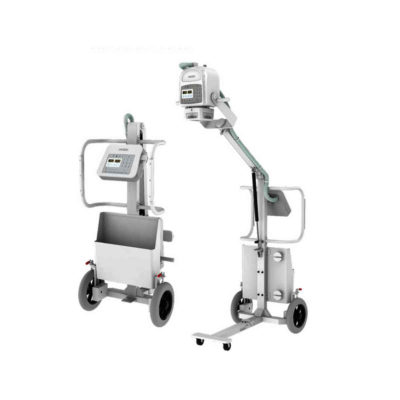
Click Here To Download Catalogue | JADE Mobile X-ray machine is one of the lightest portable X-ray systems on the market, allowing it to be used in any imaginable way including bedside, operating rooms, intensive care units and veterinary fields. With a simple, easy-to-use operator console, three-way control, two-step foldable stand and auto-lock system, the JADE Mobile X-ray machine is a user-friendly portable X-ray system.
Convenient & Intuitive Operation:
JADE is one of the lightest portable X-ray systems on the market, allowing it to be used in any imaginable way including bedside, operating rooms, intensive care units and in veterinary fields. With a simple, easy-to-use operator console, three-way control, two-step foldable stand and auto-lock system, JADE is a user-friendly portable X-ray system.
Compact & Powerful Design:
JADE Mobile X-ray machine is an innovative, highly versatile portable X-ray system suitable for a variety of clinical uses. Utilizing the unique technology used in DRGEM’s universally recognized X-ray generators, JADE is a compact but powerful unit with a 4kW output and thoughtfully designed components to increase efficiency and maximize workflow. The core part of X-ray source adopts high-quality tube assembly, X-ray collimator and high frequency X-ray generator with excellent performance, lifetime and stability.
Features:
Click Here To Download Catalogue |
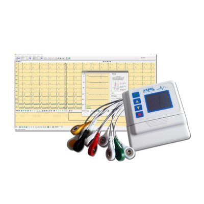
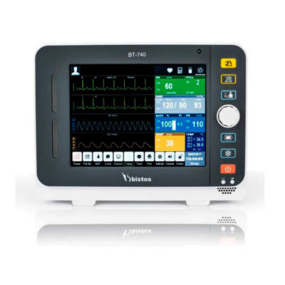
AsCARD Coral electrocardiograph is a 3-, 6-, 12-channel ECG equipped with CardioTEKA software allows transmission of full 12 ECG leads to the user PC through USB interface. It is intended for carrying out ECG examinations in adults and pediatric patients in all types of health care centres. ECG procedures can be performed by qualified personnel only. AsCARD Coral can cooperate also with CardioTEST system as 12-channel ECG device allows transmission of full 12 ECG leads to the user PC through USB interface.
Technical Specification:
Click Here To Download Catalogue | DETAILS
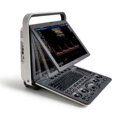
B + Compound
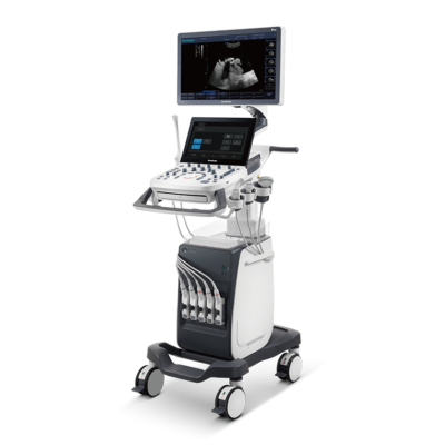
B + Compound utilizes several lines of sight for optimal contrast resolution, speckle reduction and border detection, with which P10 is ideal for superficial and abdominal imaging with better clarity and improved continuity of structures.
μ-Scan
The new generation μ-Scan imaging technology gives you better image quality by reducing noise, improving signal strength and improving visualization.
P10 offers a comprehensive selection of electronic probes to maximize its capabilities to meet a wide range of applications including abdomen, pediatric, OB/GYN, cardiovascular, musculoskeletal, etc. The advanced probe technologies also effectively enhance the image quality and confidence in reaching clinical diagnoses, even in difficult patients.
Convex Probe 3C-A
Ideal for an abundant of application such as abdomen, gynecology, obstetrics, urology and even abdomen biopsy.
Linear Probe L741
This linear probe is designed to satisfy vascular, breast, thyroid, and other small parts diagnosis, and its adjustable parameters could also present users a clear view of MSK and deep vessels.
Phase Array Probe 3P-A
For the purpose of adult and pediatric cardiology and emergency, the phase array probe provides elaborate presets for different exam modes, even for difficult patients.
Intracavitary Probe 6V1
Intracavitary probe could face application of gynecology, urology, prostate, and its temperature detection technology not only protects the patient but also extends the service life.
Click Here To Download Catalogue | SONOSCAPE E2 DETAILS
Auto Image Optimization
A portable ultrasound machine with the press of a button, the image is automatically adjusted and optimized, saving you time with parameter adjustments. Additionally, with Auto Focus on, the focus area follows the depth of the ROI box as it is moved in the scanning field, providing users with excellent image quality in the desired area of interest.
Automated Calculation
Auto IMT is used when determining the level of vascular sclerosis present in the patient by automatically tracing the thickness of the carotid vessels.
Auto trace provides users sensitive and accurate wave tracing, avoiding the error of manual trace and giving out calculation result in no time
In-Build Battery pack
This portable ultrasound machine was equipped with an in-build battery pack which enable the user to perform image scanning when AC power is not available.
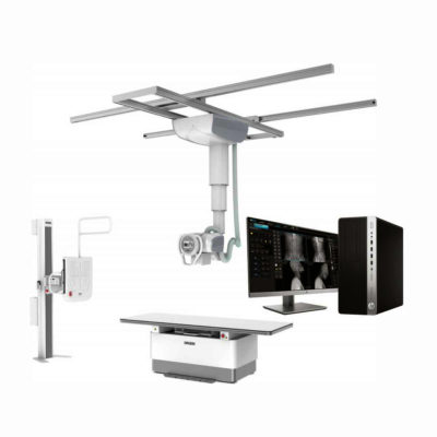
Click Here To Download Catalogue | DrGem Diamond All-In-One Digital X-ray Machine is a fully automatic digital radiography system providing state-of-the-art image quality, image processing and user interface. With a wide selection of anatomical studies on the imaging software, DIAMOND automatically sets up the x-ray generator’s pre-programmed exposure technique settings, motorized radiographic stand positioning, x-ray collimation and post-image processing for the selected study. Specifically designed to increase workflow, this fully digital system offers convenient auto-positioning and advanced image processing to achieve big performance with little effort.
Features of DrGem Diamond All-In-One Digital X-ray Machine:
Outstanding Image Quality -
Digital radiography via at panel detector improves your workflow, exam speed and comfort with efficiency. Digital at panel detector with Csl screen provides excellent spatial resolution, MTF, DQE and stability based on ne pixel pitch. A 3-field ion-chamber is provided for AEC function.
Automatic Collimation –
Automatic x-ray eld size control of the motorized collimator corresponds to dierent SIDs. Includes user adjustable lamp timer with on/oswitch.
Automatic Positioning –
Click Here To Download Catalogue | ||||||||||||||||||||||||||||||||||||||||||||||||||||||||||||||||||||||||||||||||||||||||||||||||||||||||||||||||||||||||||||||||||||||||||||||||||||||||||||||||||||||||||||||||||||||||||||||||||||||||||||||||||||||||||||||||||||||||||||||||||||||||||||||||||||||||||||||||||||||||||||||||||||||||||||||||||||||||
| Weight | N/A | N/A | N/A | N/A | N/A | N/A | ||||||||||||||||||||||||||||||||||||||||||||||||||||||||||||||||||||||||||||||||||||||||||||||||||||||||||||||||||||||||||||||||||||||||||||||||||||||||||||||||||||||||||||||||||||||||||||||||||||||||||||||||||||||||||||||||||||||||||||||||||||||||||||||||||||||||||||||||||||||||||||||||||||||||||||||||||||||||
| Dimensions | N/A | N/A | N/A | N/A | N/A | N/A | ||||||||||||||||||||||||||||||||||||||||||||||||||||||||||||||||||||||||||||||||||||||||||||||||||||||||||||||||||||||||||||||||||||||||||||||||||||||||||||||||||||||||||||||||||||||||||||||||||||||||||||||||||||||||||||||||||||||||||||||||||||||||||||||||||||||||||||||||||||||||||||||||||||||||||||||||||||||||
| Additional information |
|

Reviews
There are no reviews yet.