
Anke MRI Openmark 5000 Permanent System
Ask for Price$0.00
Shipped from Abroad
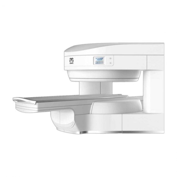
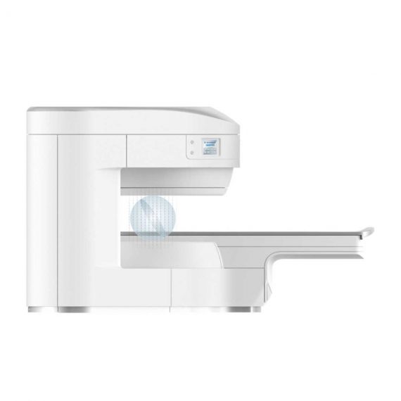
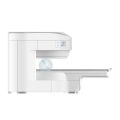
OPENMARK 5000 is 0.51T MRI. It’s approved by FDA and has CE mark. It adopts two-pillar magnet design with 280 degree openness and equipped with powerful
RF and gradient system, together with advanced imaging technology, making it as a high-end system which is comparable to high-field MRI.
Delivery & Availability:
Typically 90 working days – excluding furniture and heavy/bulky equipment. Please contact us for further information.
Description
OPENMARK 5000 is 0.51T MRI. It’s approved by FDA and has CE mark. It adopts two-pillar magnet design with 280 degree openness and equipped with powerful
RF and gradient system, together with advanced imaging technology, making it as a high-end system which is comparable to high-field MRI.
Features:
- With the highest system stability and the highest homogeneity of the
magnet field in permanent MRI - Screens on both sides facilitate positioning; 280 degree two-pillar magnet
design ensures stable magnet structure and facilitates interventional
treatment. - Active and passive shimming calibrate technology ensures the magnetic
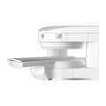
field uniformity - Motor-driven patient couch makes it easier for patients to access and for
positioning - Powerful hardware and software platforms ensure the scan speed, image
quality and make it possible for advanced imaging functions - Fast scan speed eliminates motion artifact
- Rich scan sequences, advanced imaging technology and powerful postprocessing
technology ensure image quality, extend more applications,
which can fully satisfy the clinical needs - Intelligent user-friendly operating system ensures you easy operation
Technical Specifications:
| No. | Technique Description | Parameter |
| 1 | Magnet System | |
| 1.1 | Magnet Type | Permanent Magnet
Automatic constant temperature system |
| 1.2 | Field Strength | 0.51T |
| 1.3 | Magnet Shape | Dual-pillar shape |
| 1.4 | Homogeneity(40cm,DSV,VRMS) | ≤1.6ppm |
| 1.5 | Shim Method | Active/Passive |
| 1.6 | Magnet Vertical Gap (Cover) | 40cm |
| 1.7 | Magnetic Pole Dia. (Exclude Cover) | 145cm |
| 1.8 | Accessibility(Horizontal Opening Angle, | 280° |
| 1.9 | 5 Gauss fringe field | X-axis:horizontal ≤2.5m
Y-axis:Vertical ≤2.5m Z-axis:horizontal ≤2.5m |
| 2 | Patient Couch and Communication | |
| 2.1 | Patient Couch Driven mode | Motor-driven |
| 2.2 | Max. Patient Weight | ≥200kg(440lbs) |
| 2.3 | Patient Positioning Tools | Laser Light Localizer for positioning of center slice Motor-driven transfer to center of imaging volume |
| 2.4 | Position accuracy | ±1mm |
| 2.5 | Emergency Call Key | Yes |
| 2.6 | Intercom System | Yes |
| 3 | Gradient System | |
| 3.1 | Gradient Field Strength(Single Axis) | ≥30mT/m |
| 3.2 | Gradient Slew Rate (Single Axis) | ≥100mT/m/ms |
| 3.3 | Rise Time | ≤0.3ms |
| 3.4 | Gradient Cooling System ( Gradient coils
and Power electronics) |
Air Cooling |
| 4 | RF System | |
| 4.1 | RF System Type | Digital Transmit and
Receive signal |
| 4.2 | Number of RF Channels | 4 |
| 4.3 | Transmitter Amplifier Peak Power | 6kW |
| 4.4 | RF Bandwidth of Receiver | ≥1.25MHz |
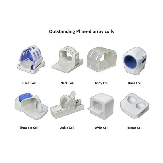
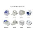
| 4.5 | Head Coil | Yes |
| 4.6 | Neck Coil | Yes |
| 4.7 | Body/Spine Coil (17 inch) | Yes |
| 4.8 | Body/Spine Coil (21 inch) | Yes |
| 4.9 | Knee Coil | Yes |
| 4.10 | Shoulder Coil | Yes |
| 4.11 | Flexible Coil | Optional |
| 4.12 | Breast Coil | Optional |
| 5 | Computer System | |
| 5.1 | Host Computer | DELL Computer (for MR) |
| 5.2 | System Software | Windows XP |
| 5.3 | Operation Software | APEX |
| 5.4 | CPU Clock rate | 3.0GHz |
| 5.5 | Main Memory | 4GB |
| 5.6 | Color LCD Monitor | 19” |
| 5.7 | Keyboard and Mouse | Standard |
| 5.8 | Image Reconstruction Speed(256 x 256
Matrix) |
200 frame/Sec. |
| 5.9 | Hard Disk | 500GB |
| 5.10 | Image Storage Capacity(256 x 256
Matrix) |
500,000 |
| 5.11 | Media Driver | DVD RW |
| 5.12 | DICOM 3.0 | Yes |
| 5.13 | Ethernet | Yes |
| 5.14 | Operation Console | Yes |
| 5.15 | Operation Chair | Yes |
| 6 | Scanning Parameter | |
| 6.1 | Max. FOV | 410mm |
| 6.2 | Min. FOV | 5mm |
| 6.3 | Min. TE(SE) | 5ms |
| 6.4 | Min. TR(SE) | 11ms |
| 6.5 | Min. TE(GR) | 1ms |
| 6.6 | Min. TR(GR) | 3ms |
| 6.7 | Min. 2D Thickness | 1.0mm |
| 6.8 | Min. 3D Thickness | 0.1mm |
| 6.9 | Max. Image Matrix | 512×512 |
| 7 | Scanning Sequence & Imaging Technique | |
| 7.1 | Spin Echo 2D/3D (SE 2D/3D) | Yes |
| 7.2 | DE/QE | Yes |
| 7.3 | Fast Spin Echo 2D/3D(FSE 2D/3D) | Yes |
| 7.4 | Single Shot FSE 2D/3D | Yes |
| 7.5 | Inversion Recovery(IR) | Yes |
| 7.6 | Fast Inversion Recovery(FIR) | Yes |
| 7.7 | Gradient Echo 2D/3D(GR 2D/3D) | Yes |
| 7.8 | Fast GR 2D/3D | Yes |
| 7.9 | SPGR | Yes |
| 7.10 | FLAIR | Yes |
| 7.11 | Fat Imaging | Yes |
| 7.12 | Fat Suppression imaging | Yes |
| 7.13 | Water-Fat Separation imaging | Yes |
| 7.14 | TOF MRA(2D/3D) | Yes |
| 7.15 | MRCP(2D/3D) | Yes |
| 7.16 | MRU (2D/3D) | Yes |
| 7.17 | MRM | Yes |
| 7.18 | Fast Hydrograph Imaging | Yes |
| 7.19 | Diffusion Weighted Imaging(DWI) | Yes |
| 7.20 | Max. b Value | 1000s/mm2 |
| 7.21 | Breath Hold Technology | Yes |
| 7.22 | Magnetization Transfer Contrast(MTC) | Yes |
| 7.23 | Multi-slice and Angle-free Presaturation | Yes |
| 7.24 | Saturation Tracking | Yes |
| 7.25 | Maximum Intensity Projection(MIP) | Yes |
| 7.26 | Multi-Angle Projection(MAP) | Yes |
| 7.27 | 3D Reconstruction | Yes |
| 7.28 | Multi-planar Reconstruction(MPR) | Yes |
| 7.29 | Multi-Artifacts Eliminating technology | Yes |
| 7.30 | Checking with Part Metal Implant | Yes |
| 7.31 | Online Image Filtration | Yes |
| 7.32 | Online Post Procession | Yes |
| 7.33 | 3D Scout | Yes |
| 7.34 | Scanning Protocol Preset | Yes |
| 7.35 | Scanning Protocol Queue Waiting | Yes |
| 7.36 | Advanced Image Post Processing | Yes |
| 7.37 | Image Fusion Technology of Vascular | Yes |
| 7.38 | Image Fusion Technology of Spine | Yes |
Click Here To Download Catalogue
Additional information
| Model | Advanced, Advanced Plus, Basic, Smart |
|---|
Quick Comparison
| Settings | Anke MRI Openmark 5000 Permanent System remove | Sonoscape S11 Ultrasound Machine remove | Bistos BT-770-12.1" Touchscreen Patient Monitor remove | Sonoscape P50 Ultrasound Machine remove | ASPEL AsPEKT 712 Holter Monitor and Software remove | Topaz Digital X-ray Machine remove | |||||||||||||||||||||||||||||||||||||||||||||||||||||||||||||||||||||||||||||||||||||||||||||||||||||||||||||||||||||||||||||||||||||||||||||||||||||||||||||||||||||||||||||||||||||||||||||||||||||||||||||||||||||||||||||||||||||||||||||||||||||||||||||||||||||||||||||||||||||||||||||||||||||||||||||||
|---|---|---|---|---|---|---|---|---|---|---|---|---|---|---|---|---|---|---|---|---|---|---|---|---|---|---|---|---|---|---|---|---|---|---|---|---|---|---|---|---|---|---|---|---|---|---|---|---|---|---|---|---|---|---|---|---|---|---|---|---|---|---|---|---|---|---|---|---|---|---|---|---|---|---|---|---|---|---|---|---|---|---|---|---|---|---|---|---|---|---|---|---|---|---|---|---|---|---|---|---|---|---|---|---|---|---|---|---|---|---|---|---|---|---|---|---|---|---|---|---|---|---|---|---|---|---|---|---|---|---|---|---|---|---|---|---|---|---|---|---|---|---|---|---|---|---|---|---|---|---|---|---|---|---|---|---|---|---|---|---|---|---|---|---|---|---|---|---|---|---|---|---|---|---|---|---|---|---|---|---|---|---|---|---|---|---|---|---|---|---|---|---|---|---|---|---|---|---|---|---|---|---|---|---|---|---|---|---|---|---|---|---|---|---|---|---|---|---|---|---|---|---|---|---|---|---|---|---|---|---|---|---|---|---|---|---|---|---|---|---|---|---|---|---|---|---|---|---|---|---|---|---|---|---|---|---|---|---|---|---|---|---|---|---|---|---|---|---|---|---|---|---|---|---|---|---|---|---|---|---|---|---|---|---|---|---|---|---|---|---|---|---|---|---|---|---|---|---|---|---|---|---|---|---|---|---|---|---|---|
| Name | Anke MRI Openmark 5000 Permanent System remove | Sonoscape S11 Ultrasound Machine remove | Bistos BT-770-12.1" Touchscreen Patient Monitor remove | Sonoscape P50 Ultrasound Machine remove | ASPEL AsPEKT 712 Holter Monitor and Software remove | Topaz Digital X-ray Machine remove | |||||||||||||||||||||||||||||||||||||||||||||||||||||||||||||||||||||||||||||||||||||||||||||||||||||||||||||||||||||||||||||||||||||||||||||||||||||||||||||||||||||||||||||||||||||||||||||||||||||||||||||||||||||||||||||||||||||||||||||||||||||||||||||||||||||||||||||||||||||||||||||||||||||||||||||||
| Image |  |  | 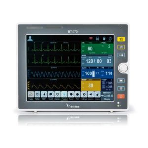 |  |  |  | |||||||||||||||||||||||||||||||||||||||||||||||||||||||||||||||||||||||||||||||||||||||||||||||||||||||||||||||||||||||||||||||||||||||||||||||||||||||||||||||||||||||||||||||||||||||||||||||||||||||||||||||||||||||||||||||||||||||||||||||||||||||||||||||||||||||||||||||||||||||||||||||||||||||||||||||
| SKU | SF1033560092-3 | SF1033560012-1 | SF1033560059-1 | SF1033560012-11 | SF1033560075-4 | SF1033560074-1 | |||||||||||||||||||||||||||||||||||||||||||||||||||||||||||||||||||||||||||||||||||||||||||||||||||||||||||||||||||||||||||||||||||||||||||||||||||||||||||||||||||||||||||||||||||||||||||||||||||||||||||||||||||||||||||||||||||||||||||||||||||||||||||||||||||||||||||||||||||||||||||||||||||||||||||||||
| Rating | |||||||||||||||||||||||||||||||||||||||||||||||||||||||||||||||||||||||||||||||||||||||||||||||||||||||||||||||||||||||||||||||||||||||||||||||||||||||||||||||||||||||||||||||||||||||||||||||||||||||||||||||||||||||||||||||||||||||||||||||||||||||||||||||||||||||||||||||||||||||||||||||||||||||||||||||||||||
| Price | Ask for Price | $6,950.00 | $758.00 | Ask for Price | Ask for Price | $52,000.00 | |||||||||||||||||||||||||||||||||||||||||||||||||||||||||||||||||||||||||||||||||||||||||||||||||||||||||||||||||||||||||||||||||||||||||||||||||||||||||||||||||||||||||||||||||||||||||||||||||||||||||||||||||||||||||||||||||||||||||||||||||||||||||||||||||||||||||||||||||||||||||||||||||||||||||||||||
| Stock | |||||||||||||||||||||||||||||||||||||||||||||||||||||||||||||||||||||||||||||||||||||||||||||||||||||||||||||||||||||||||||||||||||||||||||||||||||||||||||||||||||||||||||||||||||||||||||||||||||||||||||||||||||||||||||||||||||||||||||||||||||||||||||||||||||||||||||||||||||||||||||||||||||||||||||||||||||||
| Availability | |||||||||||||||||||||||||||||||||||||||||||||||||||||||||||||||||||||||||||||||||||||||||||||||||||||||||||||||||||||||||||||||||||||||||||||||||||||||||||||||||||||||||||||||||||||||||||||||||||||||||||||||||||||||||||||||||||||||||||||||||||||||||||||||||||||||||||||||||||||||||||||||||||||||||||||||||||||
| Add to cart | |||||||||||||||||||||||||||||||||||||||||||||||||||||||||||||||||||||||||||||||||||||||||||||||||||||||||||||||||||||||||||||||||||||||||||||||||||||||||||||||||||||||||||||||||||||||||||||||||||||||||||||||||||||||||||||||||||||||||||||||||||||||||||||||||||||||||||||||||||||||||||||||||||||||||||||||||||||
| Description | Shipped from Abroad
OPENMARK 5000 is 0.51T MRI. It's approved by FDA and has CE mark. It adopts two-pillar magnet design with 280 degree openness and equipped with powerful
RF and gradient system, together with advanced imaging technology, making it as a high-end system which is comparable to high-field MRI.
Delivery & Availability: Typically 90 working days – excluding furniture and heavy/bulky equipment. Please contact us for further information. | In Stock A Value Choice beyond Your Expectation. SonoScape’s trolley color Doppler system S11 redefines price and performance with practical design. The S11 will go beyond your expectations but not your budget. Delivery & Availability: Typically 2 working days – excluding furniture and heavy/bulky equipment. Please contact us for further information. | Shipped from Abroad The Bistos BT-770 patient monitor is equipped with a 12.1" touchscreen display, which allows for an easy operation and readability with a powerful rechargeable battery guaranteeing a continuous operation of 5 hours to monitor ECG, SpO2, NIBP, temperature and respiration Delivery & Availability: Typically 14 working days – excluding furniture and heavy/bulky equipment. Please contact us for further information. | Shipped from Abroad Easily accomplish more with SonoScape’s new P50 ultrasound system. Incorporating single crystal clarity, automatic corrections and calculation, and user defined flexibility promises a confident diagnostic experience as well as opening new doors of opportunity for ultrasound use. Delivery & Availability: Typically 7-14 working days – excluding furniture and heavy/bulky equipment. Please contact us for further information. | Shipped from Abroad The Holta Monitor allows quick analysis of ECG examination and detection, reviewing and editing capability in the qualitative assessment of VE, VT, Single SVE, PSVT, Pauses, Irregular Rhythm, VT, IVR, Brady - and Tachycardia, Couplets, ST-segment elevation and depression, Maximum, Minimum and averaged Heart Rates, artifacts Delivery & Availability: Typically 10 working days – excluding furniture and heavy/bulky equipment. Please contact us for further information. | In Stock DRGEM’s TOPAZ X-ray machine is a state-of-the-art mobile digital radiography system, designed with maximum comfort for patients and users in mind. From its user-friendly software to smooth movements, TOPAZ is made to improve your workflow and provide you with high-quality images. Delivery & Availability: Typically 21 working days – excluding furniture and heavy/bulky equipment. Please contact us for further information. | |||||||||||||||||||||||||||||||||||||||||||||||||||||||||||||||||||||||||||||||||||||||||||||||||||||||||||||||||||||||||||||||||||||||||||||||||||||||||||||||||||||||||||||||||||||||||||||||||||||||||||||||||||||||||||||||||||||||||||||||||||||||||||||||||||||||||||||||||||||||||||||||||||||||||||||||
| Content | OPENMARK 5000 is 0.51T MRI. It's approved by FDA and has CE mark. It adopts two-pillar magnet design with 280 degree openness and equipped with powerful
RF and gradient system, together with advanced imaging technology, making it as a high-end system which is comparable to high-field MRI.
Features:
Click Here To Download Catalogue | DETAILS
SonoScape’s trolley colour Doppler system S11 redefines price and performance with practical design. The S11 will go beyond your expectations but not your budget. As an easy-to-use ultrasound system, the S11 is integrated with a new software platform, especially optimized for a smooth workflow and convenient operation. The system speeds up the exam process and makes file management easier.
SPECIFICATION
- 15-inch high definition LCD monitor with articulating arm
- Compact and agile trolley design
- 3 active transducer sockets available for a wide range of applications
- Duplex, Color Doppler, DPI, PW Doppler, tissue harmonic imaging, μ-scan speckle reduction imaging, compound imaging, trapezoidal imaging
- Customized settings based on your own working style
- Full patient database and image management solutions
Click Here To Download Catalogue |
Bistos BT-770 is a 12.1" touchscreen patient monitor designed for easy operations.
SPECIFICATIONS
Click Here To Download Catalogue | DETAILS
Powerful Compact Precision
Taking into consideration the evolving expectations and needs for ultrasound, the P50 is a slim and unobtrusive trolley system that is comfortable in tight, congested spaces with little room to work in. Providing everything you need for a comfortable examination in a small space for both you and your patient.
Single Crystal Transducer
Wideband single crystal probes greatly improve the signal ratio, acquire stunning images and provide superior sensitivity and resolution for both the near and far-fields.
μ-Scan+
The new generation μ-Scan imaging technologies give you better image quality by reducing noise, improving signal strength and improving visualization.
Dynamic Color
Dynamic colour improves upon already existing colour Doppler technologies for clear capture of colour flow and detail visualization of even tiny veins with lower velocities.
Solution for Radiology
P50, is a leading-edge ultrasound system that can meet the demands of any clinical setting. You can experience a superior performance in multi-dimensional imaging for a full range of clinical applications – abdominal, breast and cardiovascular.
C-xlasto Imaging
By understanding that tissue stiffness varies depending on the type of tissue, we can use C-xlasto Imaging to easily find abnormalities and tumours within soft tissue. The differences in tissue responses are detected and visualized in real-time by the elastography algorithms through different representations, which can be particularly helpful in analyzing breast, thyroid and musculoskeletal structures. Predominately used only in linear probes, SonoScape’s new transvaginal and bi-plane probe for gynaecology and urology are breaking the mould and expanding elastography applications.
Real-time Color Panoramic
With the combination of colour flow and real-time panoramic, visualizing the blood flow of an entire vein or artery is now an easy task. Accomplished in real-time for the convenience of the sonographers, any mistakes can also be easily backtracked and corrected without interrupting the scan.
Contrast Imaging
Contrast Imaging on P50 makes full use of the infra harmonic signal and second harmonic signal to improve the image resolution and deep penetration. What’s more, the Dynamic Acoustic Control technology effectively controls the acoustic pressure for the contrast agent, decreasing the required agent dose and assures uniform image quality, guaranteeing longer contrast agent duration and better lesion perfusion of delayed phase observation.
Solution for OB/GYN
P50 has superior image quality, automated measurement tools, and a variety of volume technologies to provide ideal solutions for clinical examinations such as pregnancy examinations, and gynecologic disease diagnosis. With a new 4D transvaginal probe, P50 helps you to see and detect fetal abnormalities and significantly improves your diagnostic confidence during your examinations.
S-Live Silhouette
A unique transparent 3D anatomical image of the fetus for improved initial anatomical review. By using this new application, the system can create completely different fetal images from conventional ultrasound images, which can depict the fetal's intracorporeal anatomical structure.
Pelvic Floor 4D
Working in conjunction with SonoScape’s latest transvaginal probes, trans-perineal 4D pelvic floor ultrasound provides a useful clinical assessment of the impact of vaginal delivery on the female anterior compartment. Allowing doctors to judge whether the pelvic organs prolapsed or not, the extent of prolapse, and determining whether the pelvic muscles tore correctly.
S-Guide
S-Guide gives the user an extensive list of example obstetric ultrasound images as reference guides and a convenient checklist system to keep track of their progress during their obstetrics examination.
Auto Face
Automatically removes masking layers in front of the fetus’s face for a clearer vision of the fetus’s face.
AVC Follicle
AVC Follicle automatically identifies how many follicles are present and calculates their individual volumes.
Solution for Cardiology
P50 provides clear 2D clinical images and Doppler sensitivity to assess critical cardiac performance. Compatible with SonoScape’s single crystal probes, the P50 can provide images with better resolution and penetration in Cardiac diagnosis.
Tissue Doppler Imaging
Tissue Doppler Imaging allows clinical doctors to quantitatively evaluate local myocardial movements and functions, facilitating them with the ability to analyze and compare the motions of the different parts of the patient’s heart.
Stress Echo
Stress echocardiography is the combination of 2D echocardiography with physical, pharmacological or electrical stress of the patient. It also then provides users with report management tools such as configurable template editor, multiple loops to select one for storage, wall motion scoring, stress echo report, etc
Auto IMT
Auto IMT is used when determining the level of vascular sclerosis present in the patient by automatically tracing and calculating the thickness of the carotid vessels. What distinguishes the P50 is that it provides an instant and accurate Mean and Max index at the touch of a single button.
Auto EF
Automated 2D Cardiac Quantification is a fully intelligent trace function for endocardium with 19 easily-adjustable points providing rapid access to proven 2D EF and volumes.
Click Here To Download Catalogue | The Holter Monitor allows quick analysis of ECG examination (arrhythmias and ST segment).
Technical specifications:
HolCARD 24W Software:
Click Here To Download Catalogue | TOPAZ X-ray machine is among the high end X-ray machine manufactured by DRGEM, a digital X-ray system that provides quality images with little or no effort.
It begins with Advanced Technology
Integrating high technology and over a decade of experience in conventional and digital radiography systems, DRGEM’s TOPAZ X-ray machine is a state-of-the-art mobile digital radiography system, designed with maximum comfort for patients and users. From its user-friendly software to smooth movements, TOPAZ X-ray machine is made to improve your workflow and provide you with high-quality images.
Full Featured Imaging Software & Excellent Digital Image Processing
With a high-performance, built-in touchscreen, TOPAZ X-ray machine offers a user-friendly interface and powerful software for easy operation and increased workflow. The anatomical view-based digital image processing, automatically optimizes and enhances the quality of the image. it also comes with automatic image storage and print with DICOM 3.0 networking capability. additionally, the system offers increasing exam throughput while decreasing examination time.
Click Here To Download Catalogue | |||||||||||||||||||||||||||||||||||||||||||||||||||||||||||||||||||||||||||||||||||||||||||||||||||||||||||||||||||||||||||||||||||||||||||||||||||||||||||||||||||||||||||||||||||||||||||||||||||||||||||||||||||||||||||||||||||||||||||||||||||||||||||||||||||||||||||||||||||||||||||||||||||||||||||||||
| Weight | N/A | N/A | N/A | N/A | N/A | N/A | |||||||||||||||||||||||||||||||||||||||||||||||||||||||||||||||||||||||||||||||||||||||||||||||||||||||||||||||||||||||||||||||||||||||||||||||||||||||||||||||||||||||||||||||||||||||||||||||||||||||||||||||||||||||||||||||||||||||||||||||||||||||||||||||||||||||||||||||||||||||||||||||||||||||||||||||
| Dimensions | N/A | N/A | N/A | N/A | N/A | N/A | |||||||||||||||||||||||||||||||||||||||||||||||||||||||||||||||||||||||||||||||||||||||||||||||||||||||||||||||||||||||||||||||||||||||||||||||||||||||||||||||||||||||||||||||||||||||||||||||||||||||||||||||||||||||||||||||||||||||||||||||||||||||||||||||||||||||||||||||||||||||||||||||||||||||||||||||
| Additional information |
|


Reviews
There are no reviews yet.