
ASPEL AsCARD Green ECG Machine
$0.00
Shipped from Abroad
AsCARD Green v.06.101 is a 1-, 3-, 6- and 12-channel ECG unit which enables to make electrocardiogram in full 12 leads. Intended for ECG examinations of adult and paediatric patients aimed at identification of cardiological abnormalities, myocardial ischaemia or infarction. The device is intended for use in healthcare facilities by duly trained personnel. ECG examination may be recorded in manual or automatic mode with the ability to perform the analysis and interpretation.
Delivery & Availability:
Typically 10 working days – excluding furniture and heavy/bulky equipment. Please contact us for further information.

Description
AsCARD Green v.06.101 is a 1-, 3-, 6- and 12-channel ECG unit which enables to make electrocardiogram in full 12 leads. Intended for ECG examinations of adult and paediatric patients aimed at identification of cardiological abnormalities, myocardial ischaemia or infarction. The device is intended for use in healthcare facilities by duly trained personnel. ECG examination may be recorded in manual or automatic mode with the ability to perform the analysis and interpretation.
Electrocardiograph is based on advanced microprocessor technology .It is equipped with a thermal printer with high-resolution head and 4,3″ LCD display. A touch panel and high-tech membrane keyboard makes this device intuitive in usage and its menu navigation exceptionally easy. This light-weight, small-footprint and battery powered cause that device can be easily transported to any location. With plastic casing and foil covered keyboard, the device is neat and easy to clean.
Technical Specifications:
- dimensions: 220x153x55 mm (LxWxH)
- weight: < 0,6 kg
- recording of standard 12 ECG leads
- saving the ECG signal simultaneously from all the 12 leads during the automatic recording, together with the date and time of the examination, filter settings, examination recording time and, optionally, patient’s data, in the internal memory
- printout of the automatic ECG recording from internal memory, in groups of three leads
- printout of the analysis, interpretation of automatic ECG examination
- display of 1, 3, 6 or 12 lead ECG recordings
- ECG printout in 1-, 3-, 6- or 12-channel modes
- attaching the name of the patient to the ECG printout
- LCD TFT (4,3″, 480×282) touch screen
- membrane alphanumeric keyboard with functional keys
- graphical menu displayed on the screen for easy operation using keyboard
- automatic analysis and interpretation in compliance with EN 60601-2-25 (CSE database) – interpretation dependable on age & sex of a patient
- memory of the last automatic examinations, with a limit from 5 to 1000
- up to 130 automatic examinations in battery mode
- continuous heart rate (HR) measurement and display
- automatic detection of QRS complex
- adapted to direct operation on an open heart
- power line interference filter: 50 Hz, 60 Hz
- muscle interference filter; filters available: 25 Hz, 35 Hz, 45 Hz
- contour line filters; filters available: 0.15 Hz, 0.45 Hz, 0.75 Hz, 1.5 Hz
- detection of electrode detachment, independent for each channel
- selectable channel to detect heart rate
- thickness of the printing ECG waveforms line to choose from: normal or bold
- presentation of the curves in the standard layout or Cabrera layout
- multilingual menu
- external USB communication port to connect a PC and the CardioTEKA software, for real-time transmission of ECG signal
- acoustic signalling of detected beats
- protection against defibrillation pulse
- detection and presentation of stimulating pulses on printout
- automatic examination with the printout of patient data and clinic data
- battery power saving functions
Standard Equipment:
- clamp electrodes (set of 4)
- chest electrodes (set of 6)
- KEKG-30R ECG cable
- medical power connector M12-15
- ECG paper RB1 58 mm wide (1 roll)
- ECG gel
- operation manual
Click Here To Catalogue Download
Review(1)
Quick Comparison
| ASPEL AsCARD Green ECG Machine remove | DrGem Ceiling Mounted Digital X-ray remove | Bistos BT-770-12.1" Touchscreen Patient Monitor remove | ASPEL Ambulatory BP Machine remove | ASPEL AsPEKT 712 Holter Monitor and Software remove | Sonoscape E1 Ultrasound Machine With Two Probes remove | |
|---|---|---|---|---|---|---|
| Name | ASPEL AsCARD Green ECG Machine remove | DrGem Ceiling Mounted Digital X-ray remove | Bistos BT-770-12.1" Touchscreen Patient Monitor remove | ASPEL Ambulatory BP Machine remove | ASPEL AsPEKT 712 Holter Monitor and Software remove | Sonoscape E1 Ultrasound Machine With Two Probes remove |
| Image | 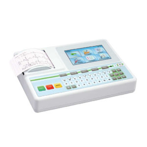 |  | 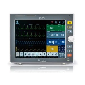 | 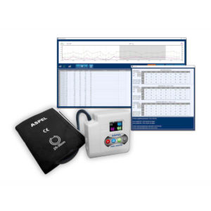 |  |  |
| SKU | SF1033560075-9 | SF1033560074-4 | SF1033560059-1 | SF1033560075-13 | SF1033560075-4 | SF1033560012-20 |
| Rating | ||||||
| Price |
|
| $902.00 | $920.00 | $1,991.00 | $4,620.00 |
| Stock | ||||||
| Availability | ||||||
| Add to cart | ||||||
| Description | Shipped from Abroad AsCARD Green v.06.101 is a 1-, 3-, 6- and 12-channel ECG unit which enables to make electrocardiogram in full 12 leads. Intended for ECG examinations of adult and paediatric patients aimed at identification of cardiological abnormalities, myocardial ischaemia or infarction. The device is intended for use in healthcare facilities by duly trained personnel. ECG examination may be recorded in manual or automatic mode with the ability to perform the analysis and interpretation. Delivery & Availability: Typically 10 working days – excluding furniture and heavy/bulky equipment. Please contact us for further information. | In Stock The GXR-SD is a diagnostic digital radiography system that provides reliable high quality digital radiographic images with a reduced dose. The GXR-SD DR systems offer comprehensive digital solutions to all radiography needs, featuring ACQUIDR digital imaging system with stationary or portable digital flat-panel detectors as well as reliable high-frequency x-ray generators that are known worldwide for their excellent performance, lifetime and stability. Patient tables and wall stands are also offered. Delivery & Availability: Typically 21 working days – excluding furniture and heavy/bulky equipment. Please contact us for further information. | Shipped from Abroad The Bistos BT-770 patient monitor is equipped with a 12.1" touchscreen display, which allows for an easy operation and readability with a powerful rechargeable battery guaranteeing a continuous operation of 5 hours to monitor ECG, SpO2, NIBP, temperature and respiration Delivery & Availability: Typically 14 working days – excluding furniture and heavy/bulky equipment. Please contact us for further information. | Shipped from Abroad ASPEL Ambulatory BP Machine - is a recorder of long-term records of non-invasive measurement of blood pressure intended for use in clinics, hospitals, outpatient centers and specialist surgeries. The recorder enables the assessment of blood pressure by the oscillometric method in adult patients, pregnant women, including preeclampsia and pediatric patients (from 3 years of age). Blood pressure is assessed by using an inflatable cuff, an accurate pressure transducer, and a deflation valve. Delivery & Availability: Typically 10 working days – excluding furniture and heavy/bulky equipment. Please contact us for further information. | Shipped from Abroad The Holta Monitor allows quick analysis of ECG examination and detection, reviewing and editing capability in the qualitative assessment of VE, VT, Single SVE, PSVT, Pauses, Irregular Rhythm, VT, IVR, Brady - and Tachycardia, Couplets, ST-segment elevation and depression, Maximum, Minimum and averaged Heart Rates, artifacts Delivery & Availability: Typically 10 working days – excluding furniture and heavy/bulky equipment. Please contact us for further information. | Shipped from Abroad SonoScape has developed a new probe and function for the E1 Exp. With these additions the E1 Exp will bring users a more efficient examination experience with satisfying image quality and a smooth workflow. Delivery & Availability: Typically 5-7 working days – excluding furniture and heavy/bulky equipment. Please contact us for further information. |
| Content | AsCARD Green v.06.101 is a 1-, 3-, 6- and 12-channel ECG unit which enables to make electrocardiogram in full 12 leads. Intended for ECG examinations of adult and paediatric patients aimed at identification of cardiological abnormalities, myocardial ischaemia or infarction. The device is intended for use in healthcare facilities by duly trained personnel. ECG examination may be recorded in manual or automatic mode with the ability to perform the analysis and interpretation.
Electrocardiograph is based on advanced microprocessor technology .It is equipped with a thermal printer with high-resolution head and 4,3" LCD display. A touch panel and high-tech membrane keyboard makes this device intuitive in usage and its menu navigation exceptionally easy. This light-weight, small-footprint and battery powered cause that device can be easily transported to any location. With plastic casing and foil covered keyboard, the device is neat and easy to clean.
Technical Specifications:
Click Here To Catalogue Download | DrGem Ceiling Mounted Digital X-ray is a diagnostic digital radiography system that provides reliable high quality digital radiographic images with a reduced dose. The GXR-SD DR systems offer comprehensive digital solutions to all radiography needs, featuring ACQUIDR digital imaging system with stationary or portable digital flat-panel detectors as well as reliable high-frequency x-ray generators that are known worldwide for their excellent performance, lifetime and stability. Patient tables and wall stands are also offered.
Features:
Click Here To Download Catalogue |
Bistos BT-770 is a 12.1" touchscreen patient monitor designed for easy operations.
SPECIFICATIONS
Click Here To Download Catalogue | ASPEL Ambulatory BP Machine - is a recorder of long-term records of non-invasive measurement of blood pressure intended for use in clinics, hospitals, outpatient centers and specialist surgeries. The recorder enables the assessment of blood pressure by the oscillometric method in adult patients, pregnant women, including preeclampsia and pediatric patients (from 3 years of age). Blood pressure is assessed by using an inflatable cuff, an accurate pressure transducer, and a deflation valve.
Features:
Save-2-Safe: Double security system
Thanks to the use of two independent measuring systems with an additional valve, it meets the highest standards and takes care of patient safety even better.
Start-Easy: Quick start in two moves
The quick launch function allows you to use the device instantly, easily allows you to start recording in holter mode.
Memo-Care: Cuff pressure memory
Recorder remembers the pressure in the cuff. Thanks to the use of Intelligent Solutions, it adapts individually to the patient.
Power-Usb: USB connection
The device can work without batteries: by connecting to a computer via a USB cable.
Technical Specification:
Click Here To Download Catalogue | The Holter Monitor allows quick analysis of ECG examination (arrhythmias and ST segment).
Technical specifications:
HolCARD 24W Software:
Click Here To Download Catalogue | DETAILS
Efficient Diagnosis
μ-Scan, Speckle Reduction & Edge Enhancement
Spatial Compound Imaging
PIH - Pure Inversion Harmonic
Wide Scan - Enlarged Image Area
Tissue-Specific Imaging
SR Flow
Ergonomic Designs
Up to 2 Transducer Ports
Light Weight and Compact
15.6 inch Anti-flickering HD LED Screen
Tilting Monitor Angle Adjustment
Backlit Keyboard and Intelligent Panel
Long-lasting Battery for 90 mins
Ease of Use
Quick Boot Up
Auto-Brightness Adjustment
Auto Image Optimization
Auto IMT
Auto Trace
Equipped Accessories
Wi-Fi and Bluetooth Available
DICOM
500GB Hard Disk
Height Adjustable Trolley
Durable, Carry-on Site Suitcase
Click Here To Download Catalogue |
| Weight | N/A | N/A | N/A | N/A | N/A | N/A |
| Dimensions | N/A | N/A | N/A | N/A | N/A | N/A |
| Additional information |


WAHID
Dear
I have ASPEL AsCARD Green ECG Machine v. 04.202
I need the manual because I have some difficulties
thanks