


Ceitek Digital Otoscope HD Resolution DO382
$0.00
Shipped From Abroad
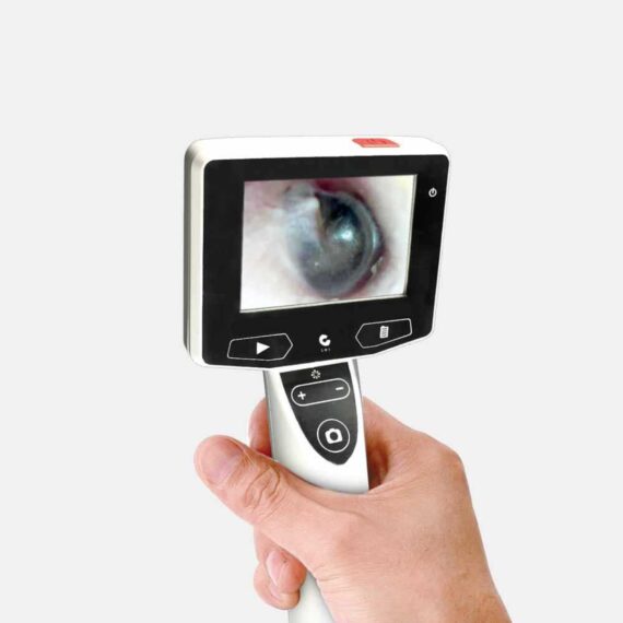
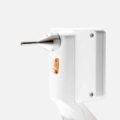
The Digital otoscope has higher magnification and extensive field of view. It’s easily to capture ear canal and tympanic membrane. Better visualisation can enhance learning, decision-making and patient outcomes.
Delivery & Availability:
Typically 14-21 Working Days – excluding furniture and heavy/bulky equipment. Please contact us for further information.
Description
The Digital otoscope has higher magnification and extensive field of view. It’s easy to capture ear canal and tympanic membrane. Better visualisation can enhance learning, decision-making and patient outcomes.
Features:
- 3” TFT LCD screen
- Camera resolution: HD (960 x 720 pixels)
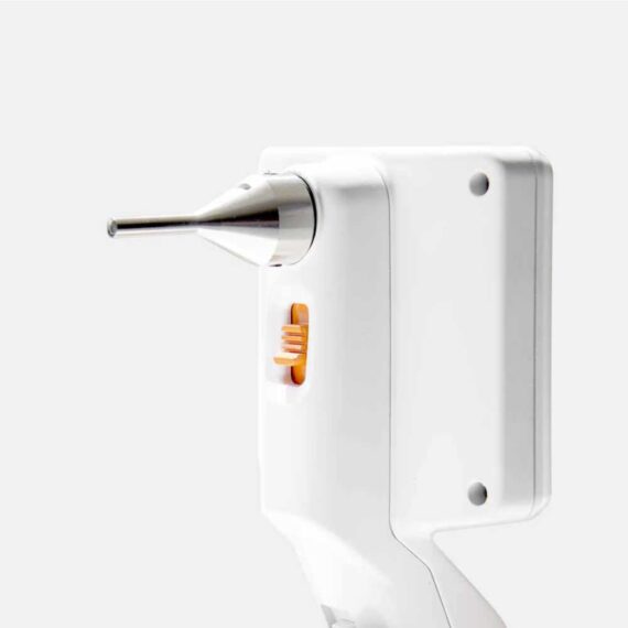
- Easily to capture ear canal and tympanic membrane
- Higher magnification and resolution
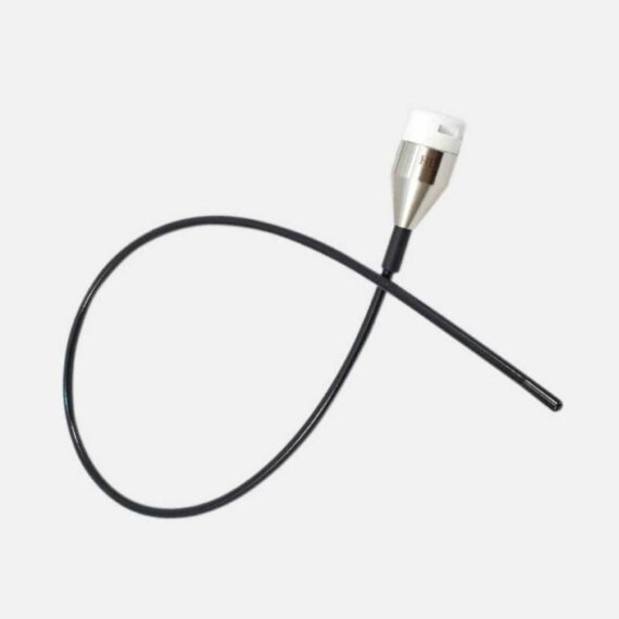
- Data transferring with micro USB connection
- 10 languages available per user’s preference
- Brightness control
- Camera preview, snapshots, and video recording
- rechargeable and replaceable Li-Ion battery
Technical Specification:
Display: 3″ TFT LCD
Display Material: Tempered Glass
Interface: Micro USB 2.0 / AV out (2.5mm)
Battery: Replaceable & Rechargeable Lithium Battery (3.78V)
Power adapter: 100-240V AC in, 50/60Hz 0.3-0.12A 5.0V DC out, 2A
File storage: 32GB Memory
Storage Capacity: Up to 60,000 pieces pictures or video recording 300 minutes(approx.)
Photo Storage Format: JPEG (960 x 720)
Resolution: HD (960 x 720 Pixels)
Depth of Field (DOF): 10 – 30 mm
Video Recording Format: MOV (960 x 720)
Video Out Format: NTSC & PAL
Frame rate: 30 fps
Compression Format: MPEG4
Video size: 10 min. (150 MB)
Functions: Snapshot, Video recording, Image (Photo & video)
review on LCD screen, TV Out, Photo & video transfer
from flash memory to PC (or Mac)
Operating time: 8 hours (average)
Charging time: 4 hours (average)
Working & Storage Temperature: -10°C ~ 65 °C (14°F ~ 149°F)
Quick Comparison
| Ceitek Digital Otoscope HD Resolution DO382 remove | DrGem Ceiling Analogue X-ray Machine remove | Topaz Digital X-ray Machine remove | DrGem Floor Mounted Analogue X-ray remove | DrGem GXR-SD 400mA Floor Mounted Digital X-ray remove | ASPEL AsPEKT 712 Holter Monitor and Software remove | |
|---|---|---|---|---|---|---|
| Name | Ceitek Digital Otoscope HD Resolution DO382 remove | DrGem Ceiling Analogue X-ray Machine remove | Topaz Digital X-ray Machine remove | DrGem Floor Mounted Analogue X-ray remove | DrGem GXR-SD 400mA Floor Mounted Digital X-ray remove | ASPEL AsPEKT 712 Holter Monitor and Software remove |
| Image | 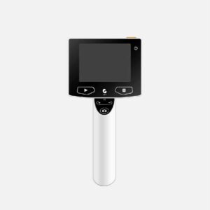 |  |  |  |  |  |
| SKU | SF10335601274 | SF1033560074-7 | SF1033560074-1 | SF1033560074-6 | SF1033560074-5 | SF1033560075-4 |
| Rating | ||||||
| Price |
|
|
|
|
| $1,991.00 |
| Stock | ||||||
| Availability | ||||||
| Add to cart | ||||||
| Description | Shipped From Abroad
The Digital otoscope has higher magnification and extensive field of view. It's easily to capture ear canal and tympanic membrane. Better visualisation can enhance learning, decision-making and patient outcomes.
Delivery & Availability:
Typically 14-21 Working Days – excluding furniture and heavy/bulky equipment. Please contact us for further information.
| Shipped from abroad The DrGem Ceiling Analogue X-ray Machine is a diagnostic radiography system that provides reliable high quality radiographic images with a reduced dose. The reliable high-frequency x-ray generators that are known worldwide for their excellent performance, lifetime and stability. Patient tables and wall stands are also offered. Delivery & Availability: Typically 21 working days – excluding furniture and heavy/bulky equipment. Please contact us for further information. | In Stock DRGEM’s TOPAZ X-ray machine is a state-of-the-art mobile digital radiography system, designed with maximum comfort for patients and users in mind. From its user-friendly software to smooth movements, TOPAZ is made to improve your workflow and provide you with high-quality images. Delivery & Availability: Typically 21 working days – excluding furniture and heavy/bulky equipment. Please contact us for further information. | In Stock GXR Analogue X-ray system matches with a radiographic room which perfectly fits your workow and can be easily upgraded to DR system with the help of DR interface and PC interface in GXR generator as well as Bucky suitable to Flat Panel Detector. GXR X-ray system is equipped with a high frequency X-ray generator which consistently produces high quality radiograph in favor of high quality X-ray output with a very small kV ripple and accurate mA and mAs. GXR X-ray system is designed to provide convenience to operator and comfort to patient. Delivery & Availability: Typically 21 working days – excluding furniture and heavy/bulky equipment. Please contact us for further information. | In Stock The GXR-SD Digital X-ray is a diagnostic digital radiography system that provides reliable high quality digital radiographic images with a reduced dose. The GXR-SD DR systems offer comprehensive digital solutions to all radiography needs, featuring ACQUIDR digital imaging system with stationary or portable digital flat-panel detectors as well as reliable high-frequency x-ray generators that are known worldwide for their excellent performance, lifetime and stability. Patient tables and wall stands are also offered. Delivery & Availability: Typically 21 working days – excluding furniture and heavy/bulky equipment. Please contact us for further information. | Shipped from Abroad The Holta Monitor allows quick analysis of ECG examination and detection, reviewing and editing capability in the qualitative assessment of VE, VT, Single SVE, PSVT, Pauses, Irregular Rhythm, VT, IVR, Brady - and Tachycardia, Couplets, ST-segment elevation and depression, Maximum, Minimum and averaged Heart Rates, artifacts Delivery & Availability: Typically 10 working days – excluding furniture and heavy/bulky equipment. Please contact us for further information. |
| Content | The Digital otoscope has higher magnification and extensive field of view. It's easy to capture ear canal and tympanic membrane. Better visualisation can enhance learning, decision-making and patient outcomes.
Features:
| DrGem Ceiling Analogue X-ray Machine is a diagnostic radiography system X-ray Machine that provides reliable high quality radiographic images with a reduced dose. The reliable high-frequency x-ray generators that are known worldwide for their excellent performance, lifetime and stability. Patient tables and wall stands are also offered.
Features of DrGem Ceiling Analogue X-ray Machine
Click Here To Download Catalogue | TOPAZ X-ray machine is among the high end X-ray machine manufactured by DRGEM, a digital X-ray system that provides quality images with little or no effort.
It begins with Advanced Technology
Integrating high technology and over a decade of experience in conventional and digital radiography systems, DRGEM’s TOPAZ X-ray machine is a state-of-the-art mobile digital radiography system, designed with maximum comfort for patients and users. From its user-friendly software to smooth movements, TOPAZ X-ray machine is made to improve your workflow and provide you with high-quality images.
Full Featured Imaging Software & Excellent Digital Image Processing
With a high-performance, built-in touchscreen, TOPAZ X-ray machine offers a user-friendly interface and powerful software for easy operation and increased workflow. The anatomical view-based digital image processing, automatically optimizes and enhances the quality of the image. it also comes with automatic image storage and print with DICOM 3.0 networking capability. additionally, the system offers increasing exam throughput while decreasing examination time.
Click Here To Download Catalogue | DrGem GXR Floor Mounted Analogue X-ray system matches with a radiographic room which perfectly fits your workflow and can be easily upgraded to DR system with the help of DR interface and PC interface in GXR generator as well as Bucky suitable to Flat Panel Detector. GXR (Analogue X-ray)system is equipped with a high frequency X-ray generator which consistently produces high quality radiograph in favor of high quality X-ray output with a very small kV ripple and accurate mA and mAs. GXR (Analogue X-ray) system is designed to provide convenience to operator and comfort to patient.
Features of DrGem GXR Floor Mounted Analogue X-ray:
Click Here To Download Catalogue | DrGem GXR-SD 400mA Floor Mounted Digital X-ray system matches with a radiographic room which perfectly fits your workow and can be easily upgraded to DR system with the help of DR interface and PC interface in GXR generator as well as Bucky suitable to Flat Panel Detector. GXR X-ray system is equipped with a high frequency X-ray generator which consistently produces high quality radiograph in favor of high quality X-ray output with a very small kV ripple and accurate mA and mAs. GXR X-ray system is designed to provide convenience to operator and comfort to patient
Features of DrGem GXR-SD 400mA Floor Mounted Digital X-ray:
Click Here To Download Catalogue | The Holter Monitor allows quick analysis of ECG examination (arrhythmias and ST segment).
Technical specifications:
HolCARD 24W Software:
Click Here To Download Catalogue |
| Weight | N/A | N/A | N/A | N/A | N/A | N/A |
| Dimensions | N/A | N/A | N/A | N/A | N/A | N/A |
| Additional information |


Reviews
There are no reviews yet.