| Description | Shipped From Abroad
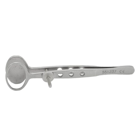
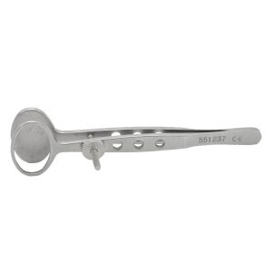
Oval Plate, 23×16mm
Delivery & Availability:
Typically 10-21 working days – excluding furniture and heavy/bulky equipment. Please contact us for further information.
| Ship from abroad
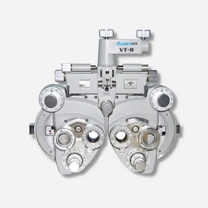
- Equipped with comprehensive measuring functions, it provides SPH, CYL, AXIS and pupil distance optometry
- Durable and easy to operate
- Easily and intuitively read the sphere focal scale value
- High eco-friendly materials
- Design fitting the face curve and no stimulation
Delivery & Availability:
Typically 14 working days – excluding furniture and heavy/bulky equipment. Please contact us for further information.
| In Stock
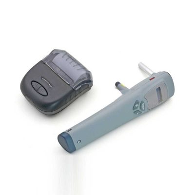
Features:
●Professional
Same ear wax removal tool as those used by doctors, you can easily eliminate ear wax buildup at home, really save your money and time on medical visiting. Safe and Environmentally Friendly.
●Quick & Easy
This ear wax removal kit is a quick, effective treatment for excess ear wax buildup. Fill the bottle with solution, Twist on the disposable tip, Use the trigger handle to spray solution into the ear canal. So Easy.
Delivery & Availability:
Typically 7-14 working days – excluding furniture and heavy/bulky equipment. Please contact us for further information.
| Shipped from abroad
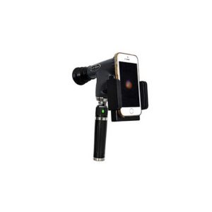
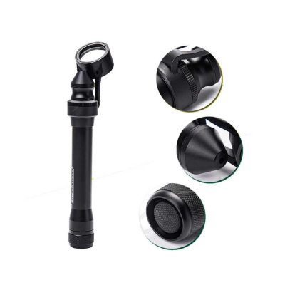
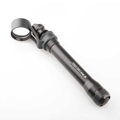
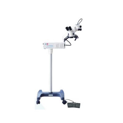
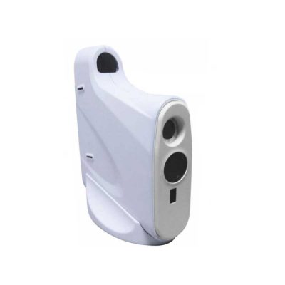
The brand-new Pantoscopic Ophthalmoscope is a portable digital imaging device which makes it possible to view and take pictures of the eyes.
Delivery & Availability:
Typically 14 working days – excluding furniture and heavy/bulky equipment. Please contact us for further information.
| Shipped from abroad
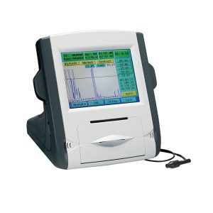
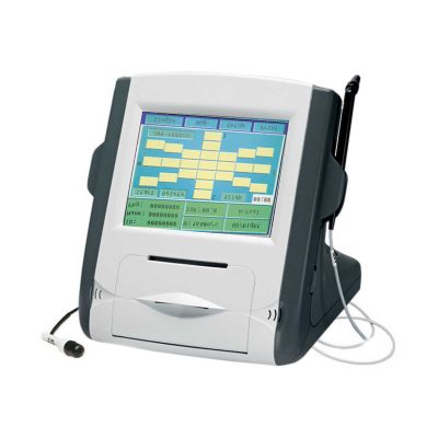
- Large color liquid-crystal screen.
- Touch screen input, easy operation.
- Curve freezing: Manual/Auto mode, controlled by pedal.
- Built-in speed thermal printer.
- Can display ultrasound waveform when measuring.
Delivery & Availability:
Typically 14 working days – excluding furniture and heavy/bulky equipment. Please contact us for further information.
| Shipped from abroad
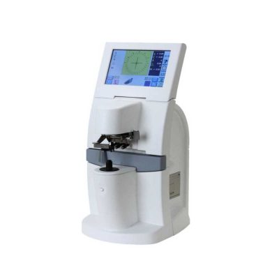
- Large color liquid-crystal screen
- Touch screen input, easy operation
- Curve freezing: Manual/Auto mode, controlled by pedal
- Built-in speed thermal printer
- Enter the name & ID; easy to check archive
Delivery & Availability:
Typically 14 working days – excluding furniture and heavy/bulky equipment. Please contact us for further information.
|
| Content | | Features:
- Equipped with comprehensive measuring functions, it provides SPH, CYL, AXIS and pupil distance optometry
- Durable and easy to operate
- Easily and intuitively read the sphere focal scale value
- High eco-friendly materials
- Design fitting the face curve and no stimulation
- Easy to take and clean
- Free switch between the cross-cylindrical lens and the rotary prism
- When the rotating risk is turning by the sphere, it can make sphere power adjust 3.00D for big scope.
- It is designed expediently and smartly for a particular cross cylinder. Supporting supplementary lens could increase scope of measurement.
Technical Specifications:
|
Sphere |
Range:-19.00~+16.75m-1 Step: 0.25m-1, 3.00m-1 |
|
Cylinder |
Range: 0.00~-6.00m-1(Measuring Range With Accessories0.00~-8.00m-1) Step: 0.25m-1 |
|
Cylinder Axis |
Range: 0~180°, Step:5° |
|
Distance of Optical center (also known as Pupil) |
Range: 50~75mmStep: 1mm |
|
Sight Switch |
Range:∞~380mm (distance of Optical center is64mm) |
|
Front Chin Test |
Range: 0~16mm |
|
Distance (from cornea vertex to the lens surface) |
16mm |
|
Standard Accessories Lens |
two pieces of Auxiliary Cylinder -2.00m-1 and -0.12m-1 respectively |
|
Standard Accessories |
one piece of M2 Hexagon wrench , one piece of a Myopia Standard Card, two piece of Myopia Standard Card , one piece of standard card holder , a dust cover |
|
Auxiliary Lens |
“O”:Open aperture
“R”:Retinoscope lens
“R”:Retinoscope lens
“R”:Retinoscope lens
*Lens of +1.50m-1 ,It is suit for the distance of 67 centimeters
“P”:Polaroid
* it is used for examining the dioptric balance of eyes , Implicit strabismus and stereo vision
“RMV”:Red Vertical maddox
*Be used to examine Implicit strabismus
“RMH”:Red horizontal maddox
*Be used to examine Implicit strabismus
“WMV”:Plane Vertical Maddox
*Be used to examine Implicit strabismus
“WMH”:Plane horizontal maddox
*Be used to examine Implicit strabismus
“RL”:Red lens
*Be used to examine eye function, Blending function and Implicit strabismus
“GL”:Green lens
*Be used to examine eye function, Blending function and Implicit strabismus
“+”:Test mark of optical center adjustment
“+.12”:Dioptric of the Spherical Lens is +0.12m-1
*Be used for the semi-adjustment of sphere lens, 0.25m-1
“PH”:1mmPinhole lens
*Be used to exclude visual non-refractive errors of the tested eye
“6ΔU”:6ΔBottom-up prism
*Be used to examine the rotating prism with the detection of nearly horizontal squint
“10ΔI”:10ΔBottom-up prism
*Be used to examine the rotating prism with the detection of nearly horizontal squint
“±0.50”:Cross-cylindrical lens
*Be used to examine the corrected dioptric of the Presbyopia and spherical lens
“OC”:Black lens
|
|
size |
338(L)×99(W)×292(H)mm |
|
NW |
about 5kg |
| Features:
●Professional
Same ear wax removal tool as those used by doctors, you can easily eliminate ear wax buildup at home, really save your money and time on medical visiting. Safe and Environmentally Friendly.
●Quick & Easy
This ear wax removal kit is a quick, effective treatment for excess ear wax buildup. Fill the bottle with solution, Twist on the disposable tip, Use the trigger handle to spray solution into the ear canal. So Easy.
●Standard
Capacity of the ear cleaner solution bottle is 10.6Oz, it has the most suitable size to hold in hand. Working at condition 32-122℉(0-50℃). Recommend to fill 1/5 of the bottle with OTC hydrogen peroxide, and 4/5 with very warm water.
●Complete Ear Washer System
Our earwax removal kit comes with 1× Ear Washer Bottle, 1× Wash Basin, 1× Rubber Bulb, 1× Short Injection Head, 1× Long Hose Injection Head, 5× Disposable Tip, 1× User Manual. | The brand-new Pantoscopic Ophthalmoscope is a portable digital imaging device which makes it possible to view and take pictures of the eyes. The optical access of the Pantoscopic Ophthalmoscope is aligned to the visual axis of the smartphone camera by the adaptor which allows to you take pictures of the fundus and retinal nerve in high resolution. You could save pictures for each patient or email and print as needed. The Pantoscopic Ophthalmoscope provides a 5X larger view of the fundus compared with the standard ophthalmoscope. It has a wider view field of 230. Without dilating the pupil, the fundus imagines could be captured at any time and places.
Features:
- Rapid capture of fundus images on your cellphone.
- Get eyeground documentation in an easy and low-cost way.
- Send images to your peers across the continum of care instantly.
- Easy carry to capture images at any clinical circumstance and work efficiently.
- Make patient education easier.
- 5X larger view of the fundus vs. standard ophthalmoscopes in an undilated eye.
- Halogen lamp provides bright, white light.
- Apertures and Filters: Looking at the pupil through an add-on corneal viewing lens, the practitioner can detect lens opacities easily.
| Features:
- Large color liquid-crystal screen.
- Touch screen input, easy operation.
- Curve freezing: Manual/Auto mode, controlled by pedal.
- Built-in speed thermal printer.
- Can display ultrasound waveform when measuring.
- Each group is the average of 20 measurements.
- Switch between IOP measured value and actual value.
- Can input name, ID and operator's name.
Technical Specifications:
| P scan probe |
20MHZ, angle of 45 degrees makes easier operation |
| Resolution |
5um |
| Measuring range |
150um~1500um |
| Display |
SINGLE mode and MAP mode |
| Feature:
- Large color liquid-crystal screen
- Touch screen input, easy operation
- Curve freezing: Manual/Auto mode, controlled by a pedal
- Built-in speed thermal printer
- Enter the name & ID; easy to check the archive
Technical Specifications:
| Model |
SW-1000 Ophthalmic A Scan Biometer |
| A Scan |
| A scan probe |
10MHz import small size probe, built-in luminotron |
| Measuring range |
15mm-40mm |
| Measurement precision |
±0. 05mm; with macula lutea trace function |
| Measurement |
Anterior chamber depth, lens thickness, vitreous body length, total length and average |
| Method of measurement |
immersion and contact |
| Eye mode |
Phakic/ Aphakic/Dense/ various IOL |
| IOL formula |
SRK-II, SRK-T, BINKHORST- Ⅱ, HOLLADAY, HOFFER-Q, HAIGIS |
| Storage |
10 cases, 5 readings each case |
| Output |
A scan waveform and IOL calculation sheet
|
|


Reviews
There are no reviews yet.